. 2022 Jan 1;79(1):70-79.
doi: 10.1001/jamaneurol.2021.4405.

Development and Validation of the 5-SENSE Score to Predict Focality of the Seizure-Onset Zone as Assessed by Stereoelectroencephalography

Affiliations

Development and Validation of the 5-SENSE Score to Predict Focality of the Seizure-Onset Zone as Assessed by Stereoelectroencephalography

Alexandra Astner-Rohracher et al. JAMA Neurol. .

Abstract

Importance: Stereoelectroencephalography (SEEG) has become the criterion standard in case of inconclusive noninvasive presurgical epilepsy workup. However, up to 40% of patients are subsequently not offered surgery because the seizure-onset zone is less focal than expected or cannot be identified.

Objective: To predict focality of the seizure-onset zone in SEEG, the 5-point 5-SENSE score was developed and validated.

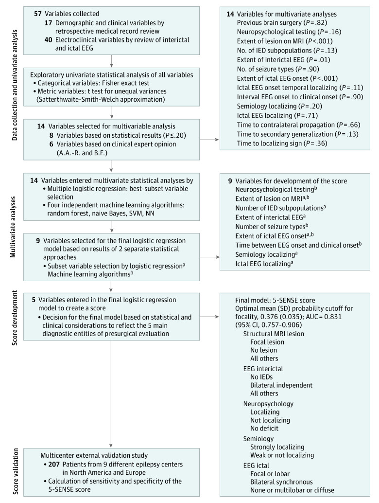
Design, setting, and participants: This was a monocentric cohort study for score development followed by multicenter validation with patient selection intervals between February 2002 to October 2018 and May 2002 to December 2019. The minimum follow-up period was 1 year. Patients with drug-resistant epilepsy undergoing SEEG at the Montreal Neurological Institute were analyzed to identify a focal seizure-onset zone. Selection criteria were 2 or more seizures in electroencephalography and availability of complete neuropsychological and neuroimaging data sets. For validation, patients from 9 epilepsy centers meeting these criteria were included. Analysis took place between May and July 2021.

Main outcomes and measures: Based on SEEG, patients were grouped as focal and nonfocal seizure-onset zone. Demographic, clinical, electroencephalography, neuroimaging, and neuropsychology data were analyzed, and a multiple logistic regression model for developing a score to predict SEEG focality was created and validated in an independent sample.

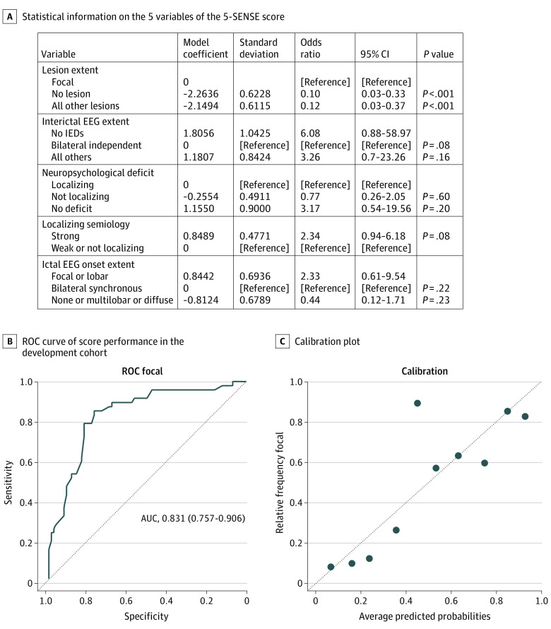
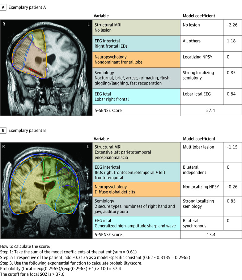
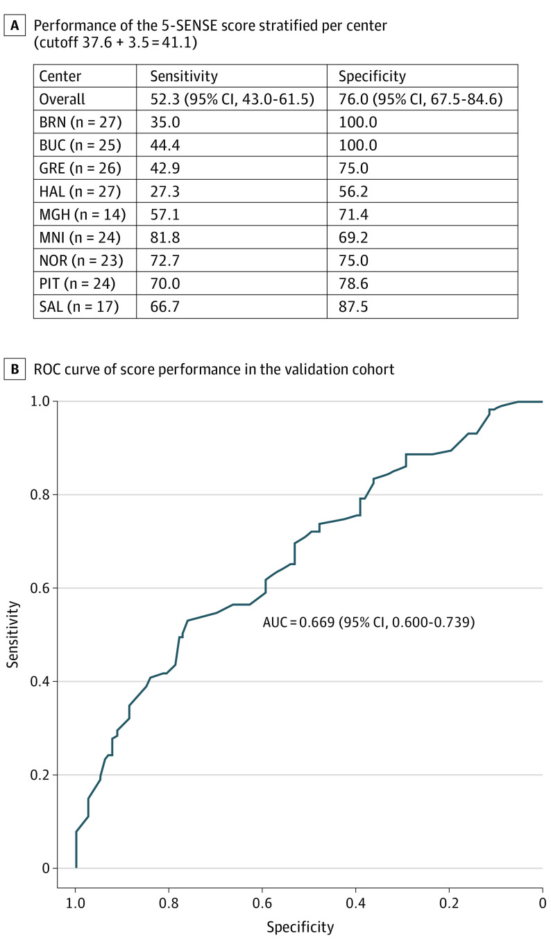
Results: A total of 128 patients (57 women [44.5%]; median [range] age, 31 [13-58] years) were analyzed for score development and 207 patients (97 women [46.9%]; median [range] age, 32 [16-70] years) were analyzed for validation. The score comprised the following 5 predictive variables: focal lesion on structural magnetic resonance imaging, absence of bilateral independent spikes in scalp electroencephalography, localizing neuropsychological deficit, strongly localizing semiology, and regional ictal scalp electroencephalography onset. The 5-SENSE score had an optimal mean (SD) probability cutoff for identifying a focal seizure-onset zone of 37.6 (3.5). Area under the curve, specificity, and sensitivity were 0.83, 76.3% (95% CI, 66.7-85.8), and 83.3% (95% CI, 72.30-94.1), respectively. Validation showed 76.0% (95% CI, 67.5-84.0) specificity and 52.3% (95% CI, 43.0-61.5) sensitivity.

Conclusions and relevance: High specificity in score development and validation confirms that the 5-SENSE score predicts patients where SEEG is unlikely to identify a focal seizure-onset zone. It is a simple and useful tool for assisting clinicians to reduce unnecessary invasive diagnostic burden on patients and overutilization of limited health care resources.


Conflict of interest statement

Conflict of Interest Disclosures: Dr Astner-Rohracher reported personal fees from Eisai (travel support and speaker honoraria) outside the submitted work. Dr Zimmermann reported grants from Federal State of Salzburg during the conduct of the study. Mr Avigdor is supported by the Canadian Open Neuroscience Platform. Dr Abdallah reported PhD studentship from Savoy Foundation. Dr Gotman reported grants from Canadian Institutes of Health Research during the conduct of the study. Dr Leitinger reported a travel grant from UCB Pharma and a speaker honorarium from Eisai outside of the submitted work. Dr Schuele reported personal fees from SK Life Science, Greenwhich Biosciences, Neurelis, Sunovion, Eisai, Epilog, and Monteris and grants from National Institute on Deafness and Other Communication Disorders and National Institute of Neurologic Disorders and Stroke outside the submitted work. Dr Trinka reported personal fees from UCB, Eisai, Marinus Pharmaceuticals, LivaNova, NewBridge, Hikma, EVER Pharma, Argenix, GL Pharma, GlaxoSmithKline, Biogen, Actavis, GW Pharmaceuticals, Sanofi and Novartis outside the submitted work; grants from Bial, Boehringer Ingelheim, GW Pharmaceuticals, Eisai, and Bayer outside the submitted work; and his institution has received grants from Biogen, UCB Pharma, Eisai, Red Bull, Merck, Bayer, the European Union, FWF Osterreichischer Fond zur Wissenschaftsforderung, undesministerium für Wissenschaft und Forschung, and Jubilaumsfond der Österreichischen Nationalbank outside the submitted work. Dr Urban reported personal fees from NeuroPace. Dr Frauscher reported a salary award from Fonds de Recherche du Québec and grants from Montreal Neurological Institute during the conduct of the study; personal fees from Eisai and UCB outside the submitted work; grants from Eisai outside the submitted work; and her research program is supported by the Canadian Institutes of Health Research, the Canadian Foundation for Innovation, the Natural Sciences and Engineering Research Council of Canada, the Savoy Epilepsy Foundation, the Hewitt Foundation, and start-up funding of the Montreal Neurological Institute. No other disclosures were reported.

Figures

Figure 1.
Figure 1.. Flowchart of Variable Selection and Score Development
AUC indicates area under the curve; EEG, electroencephalography; IED, interictal epileptiform discharge; MRI, magnetic resonance imaging; NN, neural network; SVM, support vector machine. aLogistic regression. bMachine learning algorithms.
Figure 2.
Figure 2.. The 5-SENSE Score for Prediction of SEEG Focality and Its Performance
The 5-SENSE score includes extent of the lesion on magnetic resonance imaging, extent of the ictal discharge, extent of interictal epileptiform discharges (IEDs), strength of localizing semiology, and localizing neuropsychological deficit. The area under the curve (AUC) demonstrates good discrimination (0.831 [95% CI, 0.757-0.906]) and good calibration. Sensitivity and specificity were 83.3% (95% CI, 72.3-94.1) and 76.3% (95% CI, 66.7-85.8), respectively. For classifying predicted probabilities into the focal and nonfocal categories, the optimal mean (SD) probability cutoff was 37.6 (3.5). EEG indicates electroencephalography; ROC, receiver operating characteristic curve.
Figure 3.
Figure 3.. Illustration of the Use of the 5-SENSE Score in 2 Exemplary Patients
A, A male patient in his late 30s with seizure onset during his late teen years. On high-resolution magnetic resonance imaging (MRI), there was no visible lesion. He experienced brief, nocturnal seizures with arrest and grimacing, followed by a flush, shaking of his head, and giggling, with immediate recuperation afterward. Ictal scalp electroencephalography (EEG) revealed a lobar onset over the right frontal head region. Interictal EEG showed spikes in the same area. His neuropsychological profile showed a mild nondominant frontal lobe disturbance. On stereo EEG (SEEG), a focal right orbitofrontal seizure-onset zone (SOZ) was discovered with subsequent resection. Histology revealed a focal cortical dysplasia (FCD) type 2b; the patient was completely seizure free following surgery. His 5-SENSE score for predicting a focal EEG results was 57.4. B, A male patient in his early 30s with seizure onset in his early 20s, following bacterial meningoencephalitis with empyema and residual extensive left parietotemporal encephalomalacia on MRI. Ictal scalp EEG showed a generalized discharge at seizure onset; interictally, there were independent epileptiform discharges over the right frontocentrotemporal as well as left frontotemporal regions, with right-sided predominance. He experienced 2 different seizure types with strongly localizing aura, consisting of numbness of his right hand and jaw or an auditory aura. Neuropsychology showed diffuse global deficits. SEEG revealed a widespread SOZ involving the left insula as well as the frontotemporoparietal cortex and immediate involvement of the left supplementary motor area (SMA); hence, no surgery was proposed. The patient’s 5-SENSE score precited a nonfocal SEEG result (13.4; cutoff for focality >37.6). IED indicates interictal epileptiform discharge; NPSY, neuropsychology.
Figure 4.
Figure 4.. Performance of the 5-SENSE Score in the Validation Cohort
The area under the curve (AUC) demonstrates good discrimination (0.655 [95% CI, 0.581-0.730]) and good calibration. Sensitivity and specificity were 52.3% (95% CI, 43.0-61.5) and 76.0% (95% CI, 67.5-84.6), respectively. For classifying predicted probabilities into the focal and nonfocal categories, the optimal mean (SD) probability cutoff was 41.1 (3.5). BRN indicates Masaryk University, Brno, Czech Republic; BUC, Carol Davila University of Medicine and Pharmacy Bucharest, Romania; GRE, Grenoble Institute of Neurosciences Centre Hospitalier Universitaire Grenoble Alpes, Grenoble, France; HAL, Dalhousie University and Hospital, Halifax, Canada; MGH, Massachusetts General Hospital, Boston; MNI, Montreal Neurological Institute and Hospital, Montreal, Canada; NOR, Northwestern University, Chicago, Illinois; PIT, University of Pittsburgh, Pittsburgh, Pennsylvania; ROC, receiver operating characteristic curve; SAL, Christian Doppler Clinic, Paracelsus Medical University Hospital, Salzburg, Austria.

Comment in

References

    1. Wiebe S, Blume WT, Girvin JP, Eliasziw M; Effectiveness and Efficiency of Surgery for Temporal Lobe Epilepsy Study Group . A randomized, controlled trial of surgery for temporal-lobe epilepsy. N Engl J Med. 2001;345(5):311-318. doi:10.1056/NEJM200108023450501 - DOI - PubMed
    1. Dwivedi R, Ramanujam B, Chandra PS, et al. . Surgery for drug-resistant epilepsy in children. N Engl J Med. 2017;377(17):1639-1647. doi:10.1056/NEJMoa1615335 - DOI - PubMed
    1. Lüders HO, Najm I, Nair D, Widdess-Walsh P, Bingman W. The epileptogenic zone: general principles. Epileptic Disord. 2006;8(suppl 2):S1-S9. - PubMed
    1. Rosenow F, Lüders H. Presurgical evaluation of epilepsy. Brain. 2001;124(Pt 9):1683-1700. doi:10.1093/brain/124.9.1683 - DOI - PubMed
    1. Frauscher B. Localizing the epileptogenic zone. Curr Opin Neurol. 2020;33(2):198-206. doi:10.1097/WCO.0000000000000790 - DOI - PubMed

Publication types