. 2022 Feb;474(2):217-229.
doi: 10.1007/s00424-021-02639-7. Epub 2021 Dec 6.

Proteolytic activation of the epithelial sodium channel (ENaC) by factor VII activating protease (FSAP) and its relevance for sodium retention in nephrotic mice

Affiliations

Proteolytic activation of the epithelial sodium channel (ENaC) by factor VII activating protease (FSAP) and its relevance for sodium retention in nephrotic mice

Ferruh Artunc et al. Pflugers Arch. 2022 Feb.

Abstract

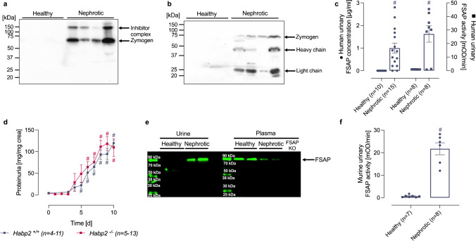
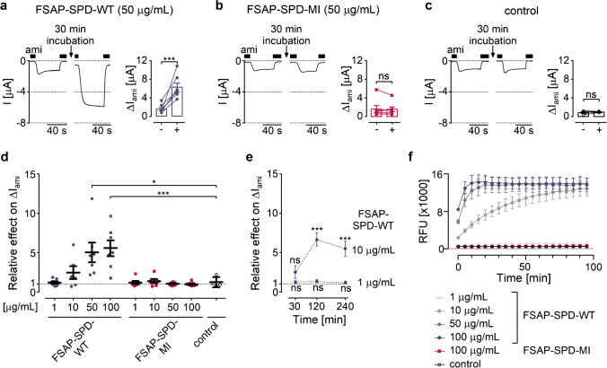
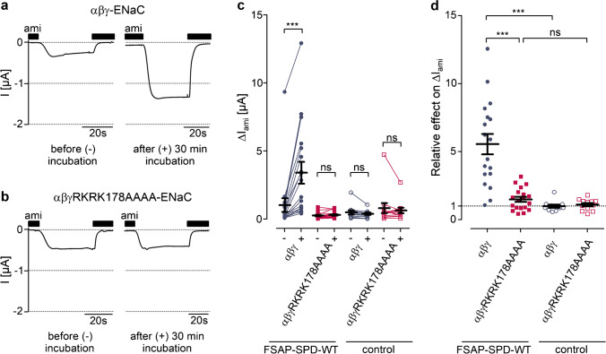
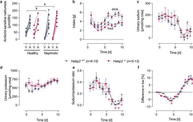
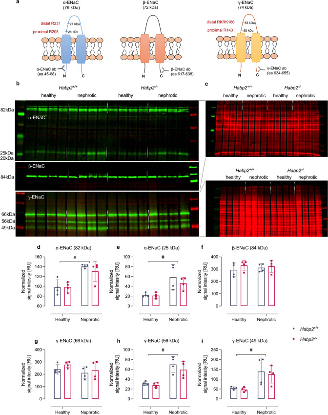
Proteolytic activation of the epithelial sodium channel (ENaC) by aberrantly filtered serine proteases is thought to contribute to renal sodium retention in nephrotic syndrome. However, the identity of the responsible proteases remains elusive. This study evaluated factor VII activating protease (FSAP) as a candidate in this context. We analyzed FSAP in the urine of patients with nephrotic syndrome and nephrotic mice and investigated its ability to activate human ENaC expressed in Xenopus laevis oocytes. Moreover, we studied sodium retention in FSAP-deficient mice (Habp2-/-) with experimental nephrotic syndrome induced by doxorubicin. In urine samples from nephrotic humans, high concentrations of FSAP were detected both as zymogen and in its active state. Recombinant serine protease domain of FSAP stimulated ENaC-mediated whole-cell currents in a time- and concentration-dependent manner. Mutating the putative prostasin cleavage site in γ-ENaC (γRKRK178AAAA) prevented channel stimulation by the serine protease domain of FSAP. In a mouse model for nephrotic syndrome, active FSAP was present in nephrotic urine of Habp2+/+ but not of Habp2-/- mice. However, Habp2-/- mice were not protected from sodium retention compared to nephrotic Habp2+/+ mice. Western blot analysis revealed that in nephrotic Habp2-/- mice, proteolytic cleavage of α- and γ-ENaC was similar to that in nephrotic Habp2+/+ animals. In conclusion, active FSAP is excreted in the urine of nephrotic patients and mice and activates ENaC in vitro involving the putative prostasin cleavage site of γ-ENaC. However, endogenous FSAP is not essential for sodium retention in nephrotic mice.

Keywords: Epithelial sodium channel (ENaC); FSAP-HABP2; Factor VII activating protease; Nephrotic syndrome; Serine protease.


Conflict of interest statement

The authors declare no competing interests.

Figures

Fig. 1
Fig. 1
Urinary excretion of FSAP in nephrotic syndrome. a Western blot from human urine samples (n = 4 healthy, n = 4 nephrotic) under non-reducing conditions using a rabbit polyclonal antibody. In nephrotic samples, FSAP is detected at 64 kDa as zymogen and at 150 kDa as part of a inhibitor complex. b Western blot of the same samples (n = 4 healthy, n = 4 nephrotic) as used in (A) under reducing conditions using a mix of two monoclonal antibodies. In addition to the detection of FSAP zymogen as single chain (64 kDa), both the light (27 kDa) and heavy chain (50 kDa) are detected which requires previous cleavage at the activation bond R311. Both chains dissociate under reducing conditions. c Quantitation of urinary FSAP concentration and activity in human nephrotic urine samples. Activity was measured with pro-uPA as substrate after immunocapture of FSAP. Results are quantified as uPA chromogenic substrate turnover in mOD min−1. d Proteinuria in wildtype (Habp2+/+) and FSAP-deficient mice (Habp2−/−) after induction of experimental nephrotic syndrome by doxorubicin. e Western blot for FSAP expression from plasma and urine of Habp2+/+ mice (n = 2). FSAP is detected at 64 kDa in its zymogen form in plasma samples and nephrotic urine. Compared to healthy plasma, FSAP expression appears to be reduced most likely due to urinary loss. The antibody does not recognize this band in the plasma from Habp2−/− mice proving the specificity of the antibody. f FSAP activity in mouse urine from Habp2+/+ mice as determined with pro-uPA as substrate. Results are quantified as uPA chromogenic substrate turnover in mOD min−1. #Significant difference between healthy and nephrotic samples
Fig. 2
Fig. 2
Stimulation of ENaC-mediated whole-cell currents by recombinant serine protease domain of FSAP. ac Representative whole-cell current traces recorded in oocytes expressing human αβγENaC before (left panels) and after (middle panels) 30-min incubation in ND96 containing FSAP-SPD-WT (50 µg mL−1 or 1.67 µM; a) or FSAP-SPD-MI (50 µg mL−1; b) or in protease-free ND96 (control; c). Data obtained from similar experiments as shown in left and middle panels are summarized in corresponding right panels. Amiloride-sensitive currents (ΔIami) were determined before ( −) and after ( +) incubation with FSAP-SPD-WT (a), FSAP-SPD-MI (b), or control solution (c). Measurements performed in the same oocyte are connected by a line (n = 4–7). d Summary of data from the same experiments as shown in ac and from additional experiments in which different protease concentrations were used as indicated. Incubation time (30 min) was the same in all experiments. The relative effect on ΔIami was calculated for each oocyte as the ratio of ΔIami measured after and before the incubation period (n = 4–7). Each data point corresponds to one individual oocyte. e Effect of 1 (0.03 µM) or 10 µg mL−1 (0.33 µM) FSAP-SPD-WT on ΔIami for different incubation times (30, 120, or 240 min). Significance is indicated for comparison with baseline ΔIami measured before incubation with the protease (n = 5–7). f Time course of proteolytic activity in the indicated incubation solutions detected using the fluorogenic substrate Boc-QAR-AMC (RFU = relative fluorescence unit; n = 10–11). *p < 0.05; †p < 0.01; ns non-significant; paired t-test (ac, e) or one-way ANOVA with Bonferroni post hoc test (d). Error bars, S.E
Fig. 3
Fig. 3
Mutation of ENaC at its putative prostasin cleavage site prevents its proteolytic activation by recombinant serine protease domain of FSAP. a, b Representative whole-cell current traces recorded in oocytes expressing human wild-type ENaC (αβγ-ENaC; a) or coexpressing wild-type α- and β-ENaC with mutant γ-ENaC (αβγRKRK178AAAA-ENaC; b) before (left panels) and after (right panels) 30 min incubation in a solution containing FSAP-SPD-WT (20 µg mL−1). c Summary of data obtained from similar experiments as shown in a, b and from additional experiments in which protease-free ND96 was used as control. ΔIami was determined before ( −) and after ( +) incubation in the indicated incubation solution. Measurements performed in the same oocyte are connected by a line (N = 2–3, n = 11–19). d Summary of the same data as shown in c normalized as relative effect of the indicated incubation solution on ΔIami (N = 2–3, n = 11–19). ‡p < 0.001; ns non-significant; paired t-test (c) or one-way ANOVA with Bonferroni post hoc test (d). Error bars, S.E. N indicates the number of different batches of oocytes; n indicates the numbers of individual oocytes measured
Fig. 4
Fig. 4
Impact of FSAP deficiency on ENaC activation and sodium retention in experimental nephrotic syndrome. a Natriuretic response to vehicle (injectable water, 5 µl g−1 bw) or amiloride (10 µg g−1 bw i.p.) in healthy and nephrotic Habp2+/+ and Habp2−/− mice. Urine was collected for 6 h after injection and all mice underwent vehicle and amiloride injection sequentially (at day − 14/ − 13 and day 7/8, respectively). bf Course of food and fluid intake, urinary sodium and potassium excretion and its ratio in spot urine samples and body weight taken in the morning after induction of nephrotic syndrome in Habp2+/+ and Habp2−/− mice. Note: Due to a variance of one day in the onset of proteolytic ENaC activation in experimental nephrotic syndrome, the data in ce were fit to the day of lowest urinary sodium (day 8) and to the day of lowest bodyweight (day 4) (f), which results in an x error depicted in the corresponding graphs. #Significant difference between healthy and nephrotic state. *Significant difference between the genotypes
Fig. 5
Fig. 5
Renal expression of ENaC subunits in experimental nephrotic syndrome. a Localization of the immunogenic sequences of the used antibodies against murine α-, β-, and γ-ENaC. In α- and γ-ENaC, the proximal and distal cleavage sites (designated from the N-terminus, respectively) are depicted. The antibody against N-terminal α-ENaC is supposed to detect full-length α-ENaC at 79 kDa (699 aa) and two N-terminal fragments with a mass of 27 kDa (231 aa) and 24 kDa (205 aa). The antibody against C-terminal β-ENaC is supposed to detect full-length β-ENaC at 72 kDa (638 aa). The antibody against C-terminal γ-ENaC is supposed to detect full-length γ-ENaC at 74 kDa (655 aa) and C-terminal fragments with a mass of 58 kDa (512 aa) after proximal cleavage and at 53 kDa (469 aa) after distal cleavage, respectively. Mass values are calculated from the amino acid sequences (omitting any N-glycosylations). b Western blots showing the expression of ENaC subunits in a plasma membrane preparation of kidney cortex from Habp2+/+ and Habp2−/− mice. α- and β-ENaC expression were analyzed in native samples on a 4–15% gradient gel after stripping, γ-ENaC expression was analyzed after deglycosylation of the same samples on an 8% gel. The higher molecular mass for α- and β-ENaC stems from N-glycosylation. c Total protein stain for control of loading and blotting. di Densitometry of the obtained bands normalized for total protein content of each lane (n = 4 each). #Significant difference between healthy and nephrotic state (two-way ANOVA)

References

    1. Artunc F, Nasir O, Amann K, Boini KM, Haring HU, Risler T, Lang F. Serum- and glucocorticoid-inducible kinase 1 in doxorubicin-induced nephrotic syndrome. Am J Physiol Renal Physiol. 2008;295:F1624–1634. doi: 10.1152/ajprenal.00032.2008. - DOI - PubMed
    1. Artunc F, Worn M, Schork A, Bohnert BN. Proteasuria—the impact of active urinary proteases on sodium retention in nephrotic syndrome. Acta Physiol (Oxf) 2019;225:e13249. doi: 10.1111/apha.13249. - DOI - PubMed
    1. Bohnert BN, Artunc F. Induction of nephrotic syndrome in mice by retrobulbar injection of doxorubicin and prevention of volume retention by sustained release aprotinin. J Vis Exp. 2018;135:e57642. doi: 10.3791/57642. - DOI - PMC - PubMed
    1. Bohnert BN, Daiminger S, Worn M, Sure F, Staudner T, Ilyaskin AV, Batbouta F, Janessa A, Schneider JC, Essigke D, Kanse S, Haerteis S, Korbmacher C, Artunc F. Urokinase-type plasminogen activator (uPA) is not essential for epithelial sodium channel (ENaC)-mediated sodium retention in experimental nephrotic syndrome. Acta Physiol (Oxf) 2019;227:e13286. doi: 10.1111/apha.13286. - DOI - PubMed
    1. Bohnert BN, Daniel C, Amann K, Voelkl J, Alesutan I, Lang F, Heyne N, Haring HU, Artunc F. Impact of phosphorus restriction and vitamin D-substitution on secondary hyperparathyroidism in a proteinuric mouse model. Kidney Blood Press Res. 2015;40:153–165. doi: 10.1159/000368491. - DOI - PubMed

Publication types

MeSH terms

LinkOut - more resources