. 2022:2416:133-156.
doi: 10.1007/978-1-0716-1908-7_10.

Generation of Pericytic-Vascular Progenitors from Tankyrase/PARP-Inhibitor-Regulated Naïve (TIRN) Human Pluripotent Stem Cells

Affiliations

Generation of Pericytic-Vascular Progenitors from Tankyrase/PARP-Inhibitor-Regulated Naïve (TIRN) Human Pluripotent Stem Cells

Ludovic Zimmerlin et al. Methods Mol Biol. 2022.

Abstract

Tankyrase/PARP inhibitor-regulated naïve human pluripotent stem cells (TIRN-hPSC) represent a new class of human stem cells for regenerative medicine that can differentiate into multi-lineage progenitors with improved in vivo functionality. Chemical reversion of conventional, primed hPSC to a TIRN-hPSC state alleviates dysfunctional epigenetic donor cell memory, lineage-primed gene expression, and potentially disease-associated aberrations in their differentiated progeny. Here, we provide methods for the reversion of normal or diseased patient-specific primed hPSC to TIRN-hPSC and describe their subsequent differentiation into embryonic-like pericytic-endothelial "naïve" vascular progenitors (N-VP). N-VP possess improved vascular functionality, high epigenetic plasticity, maintain greater genomic stability, and are more efficient in migrating to and re-vascularizing ischemic tissues than those generated from primed isogenic hPSC. We also describe detailed methods for the ocular transplantation and quantitation of vascular engraftment of N-VP into the ischemia-damaged neural retina of a humanized mouse model of ischemic retinopathy. The application of TIRN-hPSC-derived N-VP will advance vascular cell therapies of ischemic retinopathy, myocardial infarction, and cerebral vascular stroke.

Keywords: Differentiation; Human pluripotent stem cell; Ischemic retinopathy; Naïve pluripotency; PARP; Pericyte; Tankyrase inhibition; Vascular progenitors; Vascular regeneration.


Figures

Fig. 1
Fig. 1
Generation of embryonic pericytic-endothelial VP from primed hPSC. (a) Primed H9 hESC were differentiated to vascular mesoderm. CD31+ CD146+ CXCR4+ VP were further FACS-purified (red gate), and culture-expanded in EGM-2 medium into populations that contained distinct (b) endothelial CD144+ CD105+ CD34+ (purple-gated) and pericytic CD140b+ CD34 CD105lo (green-gated) populations. Expanded VP were assayed for endothelial function with (c) Ac-Dil-LDL uptake assays, and (d) In vivo Matrigel plug chimeric vascular network formation in NOD/SCID mice. Sections were stained with anti-human CD31 antibodies (brown). (e) Primed cord blood-hiPSC-derived CD31+ CD146+ VP formed assembled vascular tubes in collagen gel. (f) These tubes were imaged with transmission electron microscopy, which revealed cooperating endothelial-pericytic-like cells with bifurcations. L: lumen, n: nuclei
Fig. 2
Fig. 2
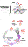
(a) Waddington landscape model for TIRN reversion (red) of primed hiPSC (blue), adapted from [2]. Epigenetic obstacles to VP differentiation may be overcome with molecular reversion to a TIRN epiblast-like state (red) with a developmentally more primitive epigenetic configuration. (b) Schema of paradigm for generating cGMP-grade, HLA-defined TIRN-hiPSC (from reprogrammed CD34+ hematopoietic progenitors) with improved multi-lineage vascular, neural, and retinal pigmented epithelial (RPE) differentiation for comprehensive regeneration of ischemic neural retina
Fig. 3
Fig. 3
Stepwise transition of isogenic primed hPSC to Tankyrase/PARP inhibitor-regulated naïve (TIRN)-hPSC culture conditions, and subsequent vascular directed differentiation protocol to N-VP. (a) Schematic of stepwise protocol for LIF-3i TIRN reversion of primed hPSC. (b) Schematic of APEL vascular progenitor (VP) differentiation system. AA: Activin A; B: BMP4; CHIR: CHIR99021; V: VEGF. Differentiated CD31-expressing N-VP are enriched with magnetic-activated cell sorting (MACS)
Fig. 4
Fig. 4
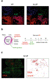
In vitro differentiation of N-VP from TIRN-hPSC and in vivo engraftment into a humanized murine model of ischemic retinopathy. (a) Representative images of primed VP versus N-VP from a fibroblast-hiPSC after 1 passage (1 week) in EGM-2 medium post-CD31+ MACS purification, demonstrating higher human CD31 (hCD31) and Ki-67 proliferation antigen expression. (b) Humanized murine model of ischemic retinal disease for quantifying N-VP engraftment (schematic adapted from [2]). Left panel, I/R location at anterior chamber and site of human cell injections into the vitreous body. Right panel, timeline of in vivo engraftment analysis. Immunodeficient NOG mice with retinal ischemia-reperfusion (I/R) injury can be leveraged for testing human N-VP therapies by injection directly into the vitreous body. Intraocular pressure (IOP) is elevated to 120 mm Hg for 90 min in the anterior chamber, followed by vascular reperfusion, which results in retinal vasculature loss, and apoptotic death of retinal neurons ~7 days following I/R injury. Although this model does not exactly simulate the sequence of events of diabetic retinopathy, it produces the same pathology seen in all retinopathies: acellular capillaries and ischemic neuronal degeneration. (c) Representative stitched image of a whole-mount retina demonstrating survival of HNA+ N-VP cells in the superficial vascular layers of the I/R damaged retina at 7 days following intra-vitreal injection
Fig. 5
Fig. 5
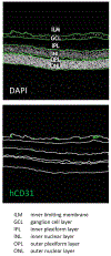
Method of human CD31+ engraftment quantitation in the vasculature of the neural layers (ILM, GCL, IPL, INL, OPL, ONL) of ischemia-injured mouse retinae. Shown is a representative example of ROI defining the neural retinal layers evaluated for human chimeric vascularization with murine blood vessels. A method of regional separation of neural retinal layers was employed for quantification of human CD31+ (hCD31) or alternatively CD34+ cells via automated counts (Analyze particles function) using the Fiji distribution of ImageJ software

Similar articles

Cited by

References

    1. Linask KK, Han M, Bravo-Valenzuela NJ (2014) Changes in vitelline and utero-placental hemodynamics: implications for cardiovascular development. Front Physiol 5:390. 10.3389/fphys.2014.00390 - DOI - PMC - PubMed
    1. Park TS, Zimmerlin L, Evans-Moses R et al. (2020) Vascular progenitors generated from tankyrase inhibitor-regulated naive diabetic human iPSC potentiate efficient revascularization of ischemic retina. Nat Commun 11(1):1195. 10.1038/s41467-020-14764-5 - DOI - PMC - PubMed
    1. Park TS, Bhutto I, Zimmerlin L et al. (2014) Vascular progenitors from cord blood-derived induced pluripotent stem cells possess augmented capacity for regenerating ischemic retinal vasculature. Circulation 129(3):359–372. 10.1161/CIRCULATI0NAHA.113.003000 - DOI - PMC - PubMed
    1. Park TS, Zimmerlin L, Zambidis ET (2013) Efficient and simultaneous generation of hematopoietic and vascular progenitors from human induced pluripotent stem cells. Cytometry A 83(1):114–126. 10.1002/cyto.a.22090 - DOI - PMC - PubMed
    1. Zambidis ET, Sinka L, Tavian M et al. (2007) Emergence of human angiohematopoietic cells in normal development and from cultured embryonic stem cells. Ann N Y Acad Sci 1106:223–232. 10.1196/annals.1392.010 - DOI - PMC - PubMed

Publication types

Substances

LinkOut - more resources