. 2021 Dec 6;19(12):e3001463.
doi: 10.1371/journal.pbio.3001463. eCollection 2021 Dec.

Heterophilic and homophilic cadherin interactions in intestinal intermicrovillar links are species dependent

Affiliations

Heterophilic and homophilic cadherin interactions in intestinal intermicrovillar links are species dependent

Michelle E Gray et al. PLoS Biol. .

Abstract

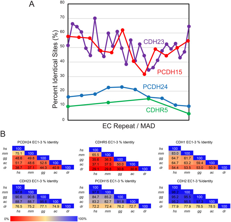
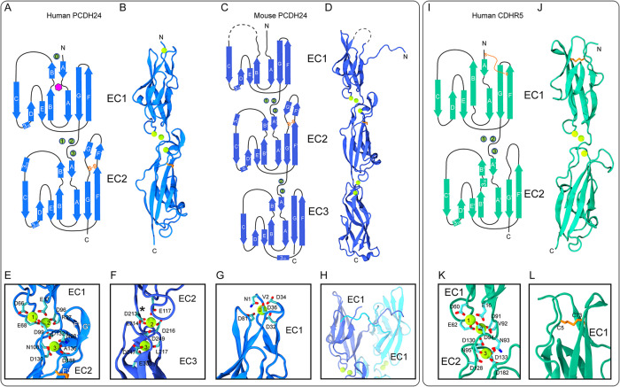
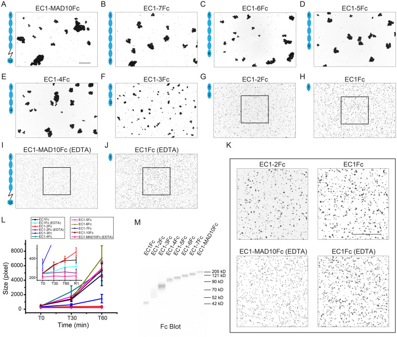
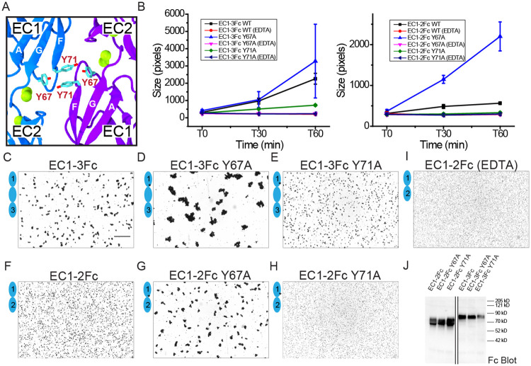
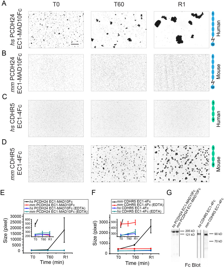
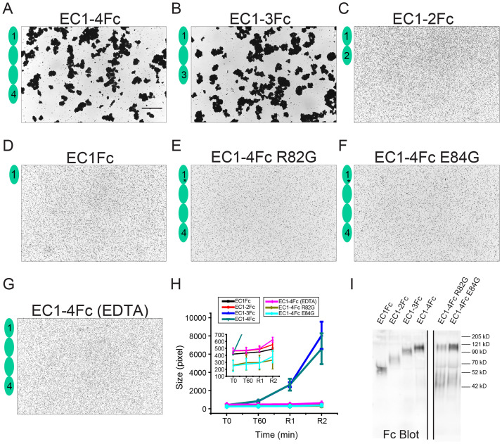
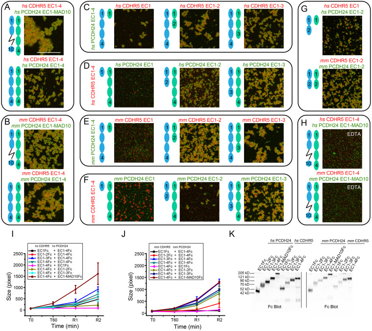
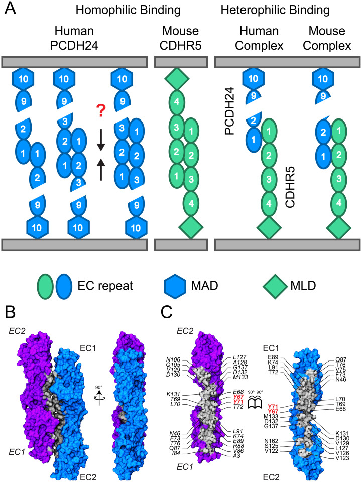
Enterocytes are specialized epithelial cells lining the luminal surface of the small intestine that build densely packed arrays of microvilli known as brush borders. These microvilli drive nutrient absorption and are arranged in a hexagonal pattern maintained by intermicrovillar links formed by 2 nonclassical members of the cadherin superfamily of calcium-dependent cell adhesion proteins: protocadherin-24 (PCDH24, also known as CDHR2) and the mucin-like protocadherin (CDHR5). The extracellular domains of these proteins are involved in heterophilic and homophilic interactions important for intermicrovillar function, yet the structural determinants of these interactions remain unresolved. Here, we present X-ray crystal structures of the PCDH24 and CDHR5 extracellular tips and analyze their species-specific features relevant for adhesive interactions. In parallel, we use binding assays to identify the PCDH24 and CDHR5 domains involved in both heterophilic and homophilic adhesion for human and mouse proteins. Our results suggest that homophilic and heterophilic interactions involving PCDH24 and CDHR5 are species dependent with unique and distinct minimal adhesive units.


Conflict of interest statement

The authors have declared that no competing interests exist.

Figures

Fig 1
Fig 1. Comparison of sequence conservation across species.
(A) Percent identity across the extracellular domains of PCDH24 (EC1-MAD10), CDH23 (EC1-MAD28), CDHR5 (EC1-4), and PCDH15 (EC1-MAD12) plotted against EC repeat/MAD number. Overall, the cadherins of the inner-ear tip link, CDH23 and PCDH15, have higher identity across a variety of species, while the intermicrovillar-link cadherins, PCDH24 and CDHR5, have poor sequence conservation across species (S1 and S2 Tables and S1 and S2 Figs). N- and C-termini are more conserved in CDH23 and PCDH15 than the middle region of these proteins. An opposite trend is observed for PCDH24 and CDHR5. (B) Percent identity of the first 3 EC repeats of PCDH24, CDHR5, CDH23, PCDH15, CDH1, and CDH2 demonstrate that PCDH24 and CDHR5 sequences are not highly conserved across several species (Homo sapiens, hs; Mus musculus, mm; Gallus gallus, gg; Anolis carolinesis, ac; Danio rerio, dr). However, the N-terminal repeats of CDH23, PCDH15, CDH1, and CDH2 are highly conserved. Sequences were obtained from NCBI (S3 Table and Methods). CDH1, Cadherin-1; CDH2, Cadherin-2; CDH23, Cadherin-23; CDHR2, cadherin-related family member 2; CDHR5, cadherin-related family member 5; EC, extracellular cadherin; MAD, membrane adjacent domain; PCDH15, protocadherin-15; PCDH24, protocadherin-24.
Fig 2
Fig 2. Structures of human (hs) PCDH24 EC1-2, mouse (mm) PCDH24 EC1-3, and hs CDHR5 EC1-2.
(A) Topology of hs PCDH24 EC1-2. The EC1 and EC2 repeats have a typical cadherin fold with 7 β-strands. A disulfide bond in EC2 is highlighted in orange. Calcium ions are shown as green circles. N-linked glycosylation at site p.N9 is highlighted by a magenta hexagon. (B) A ribbon representation of hs PCDH24 EC1-2 with calcium ions in green. (C) Topology of the mm PCDH24 EC1-3 shown as in (A). The dashed line indicates a loop not resolved. (D) A ribbon representation of mm PCDH24 EC1-3 as in (B). The structure is similar to hs PCDH24 EC1-2, but the N-terminus projects away from EC1. A N-terminal calcium ion was not present. (E) Detail of a linker between EC1 and EC2 of hs PCDH24 I showing canonical calcium-binding sites. Some sidechain and backbone atoms are not shown for clarity. (F) Detail of the mm PCDH24 EC2-3 linker, which lacks several canonical calcium-binding residues and thus only has 2 calcium ions bound. Shown as in (E). An asterisk (*) indicates the position where a calcium residue would be found in the canonical linker. (G) Detail of the N-terminal calcium bound at the tip of EC1 in the hs PCDH24 EC1-2 I structure. A similar calcium-binding motif is seen at the tip of CDH23. Residues coordinating calcium are shown in stick with backbone atoms omitted. (H) Detail of the N-terminus of mm PCDH24 EC1-3 (blue) showing its interaction with another protomer (cyan) in the asymmetric unit. (I) Topology of hs CDHR5 EC1-2 shown as in (A). (J) A ribbon representation of hs CDHR5 EC1-2 with calcium ions in green. (K) Detail of the linker between EC1 and EC2 of hs CDHR5 showing canonical calcium-binding sites. Shown as in (I, J). (L) Detail of the disulfide bond in EC1 in the hs CDHR5 EC1-2 structure. A similar disulfide bond is seen in PCDH15 EC1. CDH23, Cadherin-23; CDHR5, cadherin-related family member 5; PCDH15, protocadherin-15; PCDH24, protocadherin-24.
Fig 3
Fig 3. Homophilic binding assays of hs PCDH24.
(A-H) Protein G beads coated with the Fc-tagged full-length hs PCDH24 extracellular domain (A) and its C-terminal truncation versions (B-H). Each image shows bead aggregation observed after 60 min in the presence of 2 mM CaCl2. (I, J) Protein G beads coated with the Fc-tagged full-length hs PCDH24 extracellular domain (I) and EC1 (J) in the presence of 2 mM EDTA, shown as in (A-H). (K) Detail of binding assay results for EC1-2Fc, EC1Fc, EC1-MAD10Fc with EDTA, and EC1Fc with EDTA. Aggregation is still present for the shortest constructs. Bar– 500 μm. (L) Aggregate size for full-length and truncated versions of hs PCDH24 extracellular domains at the start of the experiment (T0) and after 30 and 60 min (T30, T60). Inset shows aggregate size of the shortest constructs compared to the EDTA control of EC1-MAD10Fc. An additional 1-min rocking step is denoted by R1. Error bars are standard error of the mean (n indicated in S4 Table; S1 Data). (M) Western blot shows expression and secretion of full-length and truncated versions of the Fc-tagged hs PCDH24 extracellular domain (S1 Raw Images). PCDH24, protocadherin-24.
Fig 4
Fig 4. Homophilic binding assays of hs PCDH24 mutants.
(A) Location of Y67 and Y71 residues at the largest antiparallel interface observed in the crystal structure hs PCDH24 EC1-2 II. Calcium ions are shown as green spheres, and EC1 strands are labeled. (B) Aggregate size for Fc-tagged hs PCDH24 EC1-3 (left) and EC1-2 (right) WT and mutants at the start of the experiment (T0) and after 30 and 60 min (T30, T60). Error bars are standard error of the mean (n indicated in S4 Table; S2 Data). (C-H) Protein G beads coated with hs PCDH24 EC1-3Fc WT (C), EC1-3Fc Y67A (D), EC1-3Fc Y71A (E), EC1-2Fc WT (F), EC1-2Fc Y67A (G), and EC1-2Fc Y71A (H). Each image shows bead aggregation observed after 60 min in the presence of 2 mM CaCl2. (I) Protein G beads coated with hs PCDH24 EC1-2Fc WT in the presence of 2 mM EDTA, shown as in (C-H). Bar– 500 μm. (J) Western blot shows expression and secretion of Fc-tagged hs PCDH24 EC1-3 and EC1-2 WT and mutants (S1 Raw Images). PCDH24, protocadherin-24; WT, wild type.
Fig 5
Fig 5. Comparison of homophilic binding assays of hs and mm PCDH24 and CDHR5.
(A-D) Protein G beads coated with Fc-tagged full-length hs PCDH24 (A), full-length mm PCDH24 (B), full-length hs CDHR5 (C), and full-length mm CDHR5 (D) cadherin extracellular domains, including MAD10 for PCDH24. None of the CDHR5 fragments included the MLD. Images show the aggregation observed at the start of the experiment (T0), after 60 min (T60) followed by rocking for 1 min (R1) in the presence of 2 mM CaCl2. Bar– 500 μm. (E) Aggregate size (S3 Data) for Fc-tagged full-length hs and mm PCDH24 extracellular domains in the presence of 2 mM CaCl2 and 2 mM EDTA at the start of the experiment (T0), after 60 min (T60) followed by rocking for 1 min (R1). Inset shows aggregate size of Fc-tagged full-length mm PCDH24 extracellular domain compared to the EDTA control of Fc-tagged full-length hs and mm PCDH24 extracellular domains. (F) Aggregate size (S4 Data) for Fc-tagged full-length hs and mm CDHR5 cadherin extracellular domains in the presence of 2 mM CaCl2 and 2 mM EDTA shown as in (E). Inset shows aggregate size of the Fc-tagged full-length hs CDHR5 cadherin extracellular domain compared to the EDTA control of Fc-tagged full-length hs and mm CDHR5 cadherin extracellular domain. Error bars in (E) and (F) are standard error of the mean (n indicated in S4 Table). (G) Western blot shows expression and secretion of Fc-tagged full-length hs and mm PCDH24 and CDHR5 cadherin extracellular domains (S1 Raw Images). CDHR5, cadherin-related family member 5; MLD, mucin-like domain; PCDH24, protocadherin-24.
Fig 6
Fig 6. Homophilic binding assays of mm CDHR5.
(A-D) Protein G beads coated with the Fc-tagged full-length mm CDHR5 cadherin extracellular domain (A) and its C-terminal truncation versions (B-D). Each image shows bead aggregation observed after 60 min followed by rocking for 2 min in the presence of 2 mM CaCl2. (E, F) Protein G beads coated with mutants mm CDHR5 EC1-4Fc R82G (E) and EC1-4Fc E84G (F) observed after 60 min followed by rocking for 2 min in the presence of 2 mM CaCl2. (G) Protein G beads coated with the full-length mm CDHR5 cadherin extracellular domain in the presence of 2 mM EDTA shown as in (A). Bar– 500 μm. (H) Aggregate size for full-length, truncated versions and mutants of mm CDHR5 cadherin extracellular domains at the start of the experiment (T0), after 60 min (T60) followed by rocking for 1 min (R1) and 2 min (R2). Inset shows aggregate size of EC1Fc, EC1-2Fc, the EDTA control of EC1-4Fc and EC1-4Fc mutants R82G and E84G. Error bars are standard error of the mean (n indicated in S4 Table; S5 Data). (I) Western blot shows expression and secretion of Fc-tagged full-length, truncated, and mutant versions of mm CDHR5 cadherin extracellular domains (S1 Raw Images). CDHR5, cadherin-related family member 5.
Fig 7
Fig 7. Heterophilic binding assays of hs and mm PCDH24 and CDHR5.
(A, B) Images from binding assays of Fc-tagged hs PCDH24 (full-length extracellular domain and EC1–4Fc) mixed with the Fc-tagged full-length hs CDHR5 cadherin extracellular domain (A) and Fc-tagged mm PCDH24 (full-length extracellular domain and EC1-4Fc) mixed with Fc-tagged full-length mm CDHR5 cadherin extracellular domain (B). Green fluorescent Protein A beads are coated with Fc-tagged PCDH24 fragments and red fluorescent Protein A beads are coated with Fc-tagged CDHR5 in all panels. All images are details of complete views shown in S18 Fig. Images show bead aggregation observed after 60 min followed by rocking for 2 min in the presence of 2 mM CaCl2. (C-F) Images from binding assays of truncations of Fc-tagged hs CDHR5 mixed with hs PCDH24 EC1-4Fc (C), truncations of Fc-tagged hs PCDH24 mixed with hs CDHR5 EC1-4Fc (D), truncations of Fc-tagged mm CDHR5 mixed with mm PCDH24 EC1-4Fc (E), and truncations of Fc-tagged mm PCDH24 mixed with mm CDHR5 EC1-4Fc (F). Images show bead aggregation observed after 60 min followed by rocking for 2 min in the presence of 2 mM CaCl2. (G) Images from binding assays of minimum EC repeats required for heterophilic adhesion of hs and mm PCDH24 and CDHR5. The minimum units for heterophilic adhesion for the human proteins are CDHR5 EC1Fc and PCDH24 EC1-2Fc. The minimum units for heterophilic adhesion for the mouse proteins are CDHR5 EC1-2Fc and PCDH24 EC1-2Fc. Images show bead aggregation observed after 60 min followed by rocking for 2 min in the presence of 2 mM CaCl2. (H) Protein A beads coated with full-length hs and mm PCDH24 and CDHR5 cadherin extracellular domains (including MAD10 for PCDH24 and without the MLD for CDHR5) in the presence of 2 mM EDTA shown as in (A, B). Bar– 500 μm. (I) Aggregate size (S6 Data) for Fc-tagged full-length and truncated constructs of hs PCDH24 and hs CDHR5 cadherin extracellular domains (including MAD10 for PCDH24 when indicated and without the MLD for CDHR5) at the start of the experiment (T0), after 60 min (T60) followed by rocking for 1 min (R1) and 2 min (R2). (J) Aggregate size (S7 Data) for Fc-tagged full-length and truncated constructs of mm PCDH24 and mm CDHR5 as in (I). Error bars in I and J are standard error of the mean (n indicated in S4 Table). (K) Western blot shows expression and secretion of Fc-tagged full-length and truncated versions of hs and mm PCDH24 and CDHR5 cadherin extracellular domains (including MAD10 for PCDH24 when indicated and without the MLD for CDHR5; S1 Raw Images). CDHR5, cadherin-related family member 5; EC, extracellular cadherin; MLD, mucin-like domain; PCDH24, protocadherin-24.
Fig 8
Fig 8. Binding modes for PCDH24 and CDHR5 extracellular domains.
(A) Schematics of homophilic and heterophilic adhesion mediated by human and mouse PCDH24 and CDHR5. Homophilic adhesion that results in small bead aggregates can be mediated by hs PCDH24 EC1 and EC1-2, while homophilic adhesion that results in large bead aggregates requires hs PCDH24 EC1-3. Homophilic adhesion mediated by mm CDHR5 requires EC1-3. Heterophilic adhesion is mediated by PCDH24 EC1-2 and CDHR5 EC1 when using human proteins and by PCDH24 EC1-2 and CDHR5 EC1-2 when using mouse proteins. (B) Crystallographic antiparallel interface in hs PCDH24 EC1-2 II. Molecular surface representation of 2 hs PCDH24 EC1-2 monomers in the crystal with the interaction interface formed by overlapped antiparallel EC1-2 repeats. Two perpendicular views are shown. (C) Interaction surface exposed with interfacing residues listed and shown in silver. CDHR5, cadherin-related family member 5; EC, extracellular cadherin; MAD, membrane adjacent domain; MLD, mucin-like domain; PCDH24, protocadherin-24.

Similar articles

Cited by

References

    1. Mukherjee S., Vaishnava S., Hooper L. V. Multi-layered regulation of intestinal antimicrobial defense. Cell Mol Life Sci. 2008;65:3019–3027. doi: 10.1007/s00018-008-8182-3 - DOI - PMC - PubMed
    1. Selsted M.E., Ouellette A.J. Mammalian defensins in the antimicrobial immune response. Nat Immunol. 2005;6:551–557. doi: 10.1038/ni1206 - DOI - PubMed
    1. Shifrin D.A., Tyska M.J. Ready…aim…fire into the lumen: a new role for enterocyte microvilli in gut host defense. Gut Microbes. 2012;3:460–2. doi: 10.4161/gmic.21247 - DOI - PMC - PubMed
    1. Maroux S., Coudrier E., Feracci H., Gorvel J.-P., Louvard D. Molecular organization of the intestinal brush border. Biochimie. 1988;70:1297–1306. doi: 10.1016/0300-9084(88)90198-8 - DOI - PubMed
    1. Mooseker M.S. Organization, chemistry, and assembly of the cytoskeletal apparatus of the intestinal brush border. Annu Rev Cell Biol. 1985;1:209–41. doi: 10.1146/annurev.cb.01.110185.001233 - DOI - PubMed

Publication types

MeSH terms

LinkOut - more resources