. 2021 Dec 21;12(6):e0305921.
doi: 10.1128/mBio.03059-21. Epub 2021 Dec 7.

The Retropepsin-Type Protease APRc as a Novel Ig-Binding Protein and Moonlighting Immune Evasion Factor of Rickettsia

Affiliations

The Retropepsin-Type Protease APRc as a Novel Ig-Binding Protein and Moonlighting Immune Evasion Factor of Rickettsia

Pedro Curto et al. mBio. .

Abstract

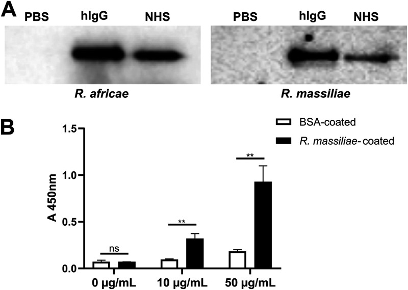
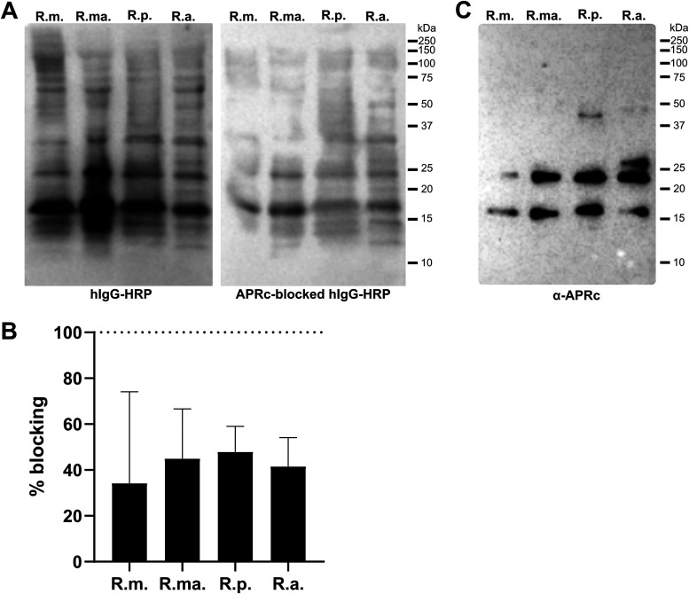
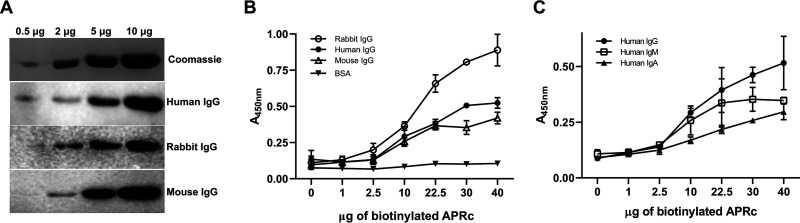
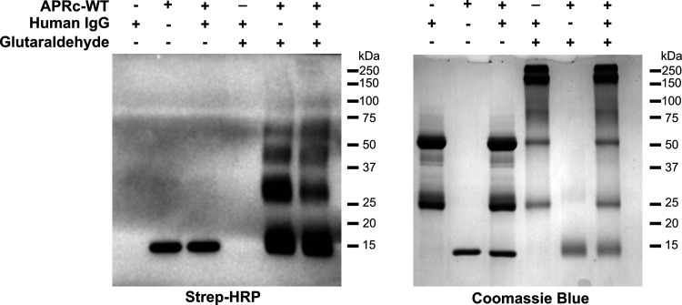
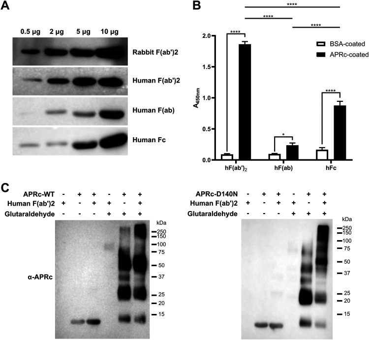
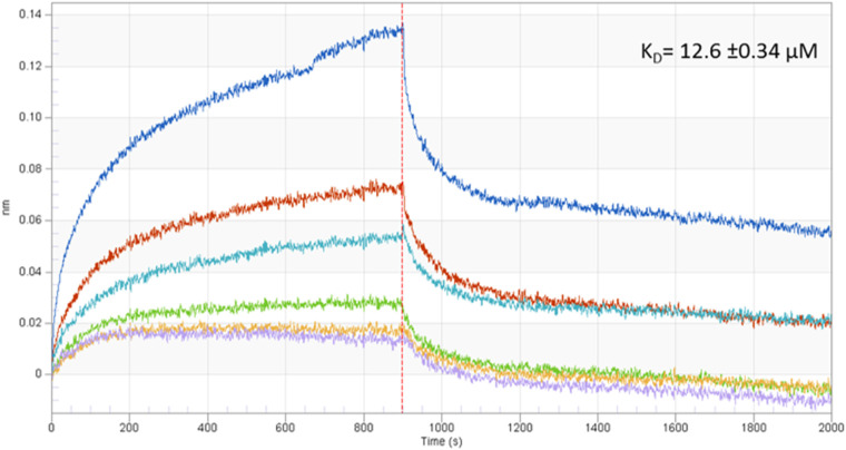
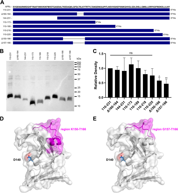
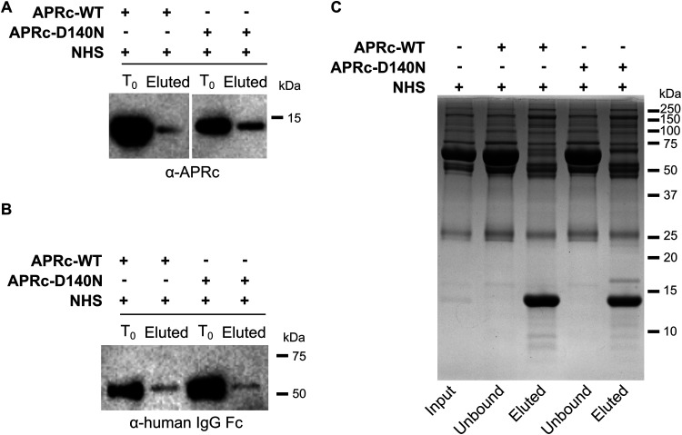
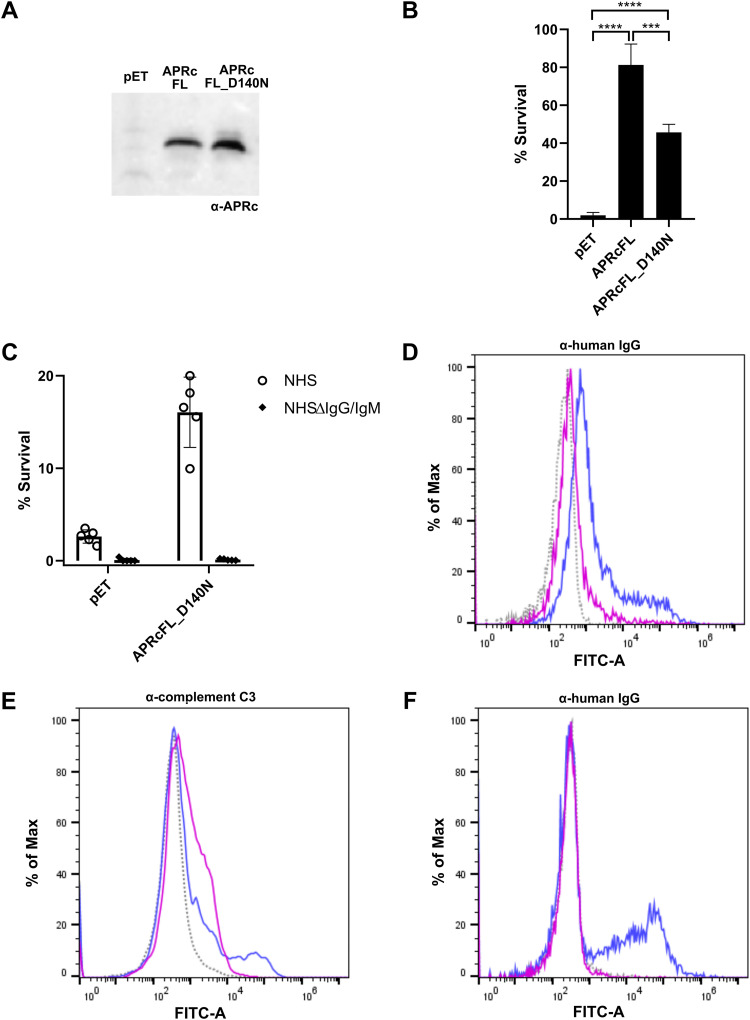
Rickettsiae are obligate intracellular Gram-negative bacteria transmitted by arthropod vectors. Despite their reduced genomes, the function(s) of the majority of rickettsial proteins remains to be uncovered. APRc is a highly conserved retropepsin-type protease, suggested to act as a modulator of other rickettsial surface proteins with a role in adhesion/invasion. However, APRc's function(s) in bacterial pathogenesis and virulence remains unknown. This study demonstrates that APRc targets host serum components, combining nonimmune immunoglobulin (Ig)-binding activity with resistance to complement-mediated killing. We confirmed nonimmune human IgG binding in extracts of different rickettsial species and intact bacteria. Our results revealed that the soluble domain of APRc is capable of binding to human (h), mouse, and rabbit IgG and different classes of human Ig (IgG, IgM, and IgA) in a concentration-dependent manner. APRc-hIgG interaction was confirmed with total hIgG and normal human serum. APRc-hIgG displayed a binding affinity in the micromolar range. We provided evidence of interaction preferentially through the Fab region and confirmed that binding is independent of catalytic activity. Mapping the APRc region responsible for binding revealed the segment between amino acids 157 and 166 as one of the interacting regions. Furthermore, we demonstrated that expression of the full-length protease in Escherichia coli is sufficient to promote resistance to complement-mediated killing and that interaction with IgG contributes to serum resistance. Our findings position APRc as a novel Ig-binding protein and a novel moonlighting immune evasion factor of Rickettsia, contributing to the arsenal of virulence factors utilized by these intracellular pathogens to aid in host colonization. IMPORTANCE Many Rickettsia organisms are pathogenic to humans, causing severe infections, like Rocky Mountain spotted fever and Mediterranean spotted fever. However, immune evasion mechanisms and pathogenicity determinants in rickettsiae are far from being resolved. We provide evidence that the highly conserved rickettsial retropepsin-type protease APRc displays nonimmune immunoglobulin (Ig)-binding activity and participates in serum resistance. APRc emerges then as a novel Ig-binding protein from Gram-negative bacteria and the first to be identified in Rickettsia. Bacterial surface proteins capable of Ig binding are known to be multifunctional and key players in immune evasion. We demonstrate that APRc is also a novel moonlighting protein, exhibiting different actions on serum components and acting as a novel evasin. This work strengthens APRc as a virulence factor in Rickettsia and its significance as a potential therapeutic target. Our findings significantly contribute to a deeper understanding of the virulence strategies used by intracellular pathogens to subvert host immune responses.

Keywords: APRc; Rickettsia; aspartic protease; evasin; immune evasion; nonimmune immunoglobulin-binding; retropepsin; serum resistance.


Figures

FIG 1
FIG 1
Nonimmune IgG binding at the surface of Rickettsia species. (A) PFA-fixed Rickettsia (R. africae and R. massiliae) bacteria were incubated with human IgG (hIgG), 50% NHS, or PBS as control for 2 h at 37°C. After incubation, bacteria were washed twice with PBS, and proteins that bound at the surface of Rickettsia were then eluted with PBS containing 1 M NaCl and analyzed by Western blotting with HRP-labeled anti-human IgG Fc antibody. (B) PFA-fixed R. massiliae bacteria were applied as a coating onto 96-well plates at 4.5 × 107 bacteria per well. BSA at 1 μg/well was used as a negative control. Nonimmune IgG binding was then evaluated by incubation with HRP-labeled rabbit IgG at different concentrations. Binding was detected at 450 nm. Data represent mean values ± standard deviations from three replicates. Significance was determined by an unpaired t test using GraphPad Prism 8 (ns, not significant; **, P < 0.01).
FIG 2
FIG 2
APRc as a potential nonimmune IgG-binding protein at the surface of Rickettsia. (A) Total protein extracts (12 μg) from different Rickettsia species (R. montanensis [R.m.], R. massiliae [R.ma.], R. parkeri [R.p.], and R. africae [R.a.]) were assayed for the presence of nonimmune IgG-binding proteins. Rickettsial protein extracts were evaluated by far-Western blotting with HRP-labeled human IgG (left) or HRP-labeled human IgG previously blocked with purified recombinant soluble domain APRc (APRc110–231His) catalytically inactive at a ratio of 1:550 m/m (hIgG to APRc) for 3 h at room temperature (right). (B) Quantification of far-Western blotting for the protein bands with molecular weights of approximately 24 and 16 kDa in rickettsial extracts probed with HRP-labeled human IgG versus APRc-blocked HRP-labeled human IgG. Percentage of blocking was determined as follows: 100 − [(band intensity for APRc-blocked HRP-labeled human IgG/band intensity for HRP-labeled human IgG) × 100]. Data represent mean values ± standard deviations from three independent replicates. (C) Total protein extracts (12 μg) from different Rickettsia species (R. montanensis [R.m.], R. massiliae [R.ma.], R. parkeri [R.p.], and R. africae [R.a.]) were evaluated by Western blotting with an anti-APRc antibody.
FIG 3
FIG 3
APRc binds IgGs from different origins as well as different classes of Igs. (A) Different amounts of recombinant and purified APRc were assessed for their ability to bind IgGs from different mammalian origins (human IgG, rabbit IgG, and mouse IgG) by far-Western blotting. (B) The ability of APRc to bind IgGs from different origins was evaluated by ELISA. BSA and IgGs from rabbit IgG, human IgG, and mouse IgG were applied as a coating onto 96-well plates at 1 μg/well and incubated with different concentrations of biotinylated APRc. Binding was then detected with HRP-conjugated streptavidin at 450 nm. Data represent mean values ± standard deviations from three replicates. (C) Binding of APRc to different classes of immunoglobulins was assessed by ELISA. Human immunoglobulins from different classes (IgG, IgM, and IgA) were applied as a coating onto 96-well plates at 1 μg/well and incubated with different concentrations of biotinylated APRc. Binding was then detected with HRP-conjugated streptavidin at 450 nm. Data represent mean values ± standard deviations from three replicates.
FIG 4
FIG 4
APRc-hIgG binding stabilizes the APRc oligomeric state. Recombinant biotinylated APRc110–231 was incubated with human IgG at equimolar ratios (166.85 pmol) in PBS, pH 7.4, for 4 h at 37°C. After incubation, samples were treated with glutaraldehyde, or water as a control, and incubated for 4 min at 37°C. The reaction was stopped by adding 1 M Tris-HCl, pH 8.0, and the products of incubation were resolved by SDS-PAGE under reducing conditions and assessed by Western blotting with HRP-streptavidin (left) and Coomassie blue staining as evidence for loading (right). WT, wild type.
FIG 5
FIG 5
APRc binds to IgG preferentially through the Fab region. (A) Different amounts of recombinant soluble domain of APRc were assessed for their ability to bind rabbit HRP-F(ab′)2 and human HRP-F(ab′)2, HRP-F(ab′), and HRP-Fc domain by far-Western blotting. (B) The ability of APRc to bind to the different human IgG fragments was evaluated by ELISA. BSA and APRc110–231His were applied as a coating onto 96-well plates at 1 μg/well and incubated with 9.09 pmol/well of HRP-labeled human F(ab′)2, F(ab′), and Fc. Binding was detected upon incubation with HRP substrate at 450 nm. Data represent mean values ± standard deviations from three replicates. Significance was determined by two-way ANOVA followed by Tukey multiple-comparison test using GraphPad Prism 8 (*, P < 0.05; ****, P < 0.0001). (C) Differences in migration of APRc wild type (WT) and mutant in the presence of human F(ab′)2. Recombinant soluble domains of wild-type (APRc-WT; left panel) and mutant (APRc-D140N; right panel) APRc110–231His were incubated with human F(ab′)2 at an equimolar ratio (166.85 pmol), for 4 h at 37°C. After incubation, samples were treated with glutaraldehyde, or water as a control, and incubated for 4 min at 37°C. The reaction was stopped by adding 1 M Tris-HCl, pH 8.0, and the products of incubation were resolved by SDS-PAGE under reducing conditions and assessed by Western blotting with an anti-APRc antibody.
FIG 6
FIG 6
Binding affinity between APRc and human IgG. The binding affinity between APRc and human IgG was determined using biolayer interferometry (BLI). Human IgG was bound to anti-human IgG Fc-capture (AHC) biosensors and incubated with several dilutions, 0.42 μM (purple), 0.85 μM (yellow), 1.70 μM (green), 3.40 μM (light blue), 6.80 μM (orange), and 13.60 μM (dark blue), of APRc110–231His. The real-time binding response (nm) is plotted against time for different concentrations of APRc. Data were analyzed using the Data Analysis software (version 9.0; FortéBio) and a 1:1 binding interaction model with global fitting.
FIG 7
FIG 7
Binding to human IgG involves APRc domain residues in the loop region 157 to 166. (A) Amino acid sequence of the APRc soluble domain and schematic representation of the different truncated forms comprising deletions at each terminal as well as internal deletions, as follows: APRc110–231His, APRc(Δ160–164)110–231His, APRc144–231His, APRc110–173His, APRc110–189His, APRc110–218His, APRc110–225His, APRc(Δ150–166)110–231His, and APRc(Δ157–166)100–231His. (B and C) The soluble domain and the truncated forms of APRc were recombinantly expressed in E. coli. After expression, cells were harvested and normalized to an OD600 of 3, and equivalent amounts of total protein extracts were loaded onto SDS-PAGE gels and transferred to PVDF membranes. (B) Representative far-Western blot analysis with HRP-labeled rabbit IgG. (C) Densitometric analysis of IgG binding to APRc soluble domain and the respective truncated forms. The band intensity of each protein construct was normalized for the Coomassie blue staining of each corresponding lane. Data represent the mean ± standard deviation (SD) from 6 independent biological replicates, and the ratios were normalized for the APRc soluble domain construct, APRc110–231His. Significance was determined using a one-way ANOVA followed by Dunnett multiple-comparison test using GraphPad Prism 8 (ns, not significant; *, P < 0.05; **, P < 0.01). (D and E) Cartoon and surface representation of APRc (PDB ID, 5C9F) colored in white with alpha-helix and wide loop region 150 to 166 shown in magenta, with side chains of residues represented in line mode (D). (E) Only wide loop region 157 to 166 is highlighted in magenta for comparison. The catalytic aspartate (Asp140) is highlighted in blue and shown in stick mode. Evident surface exposure of the region 150 to 166 corroborates its major contribution to IgG complex formation. Images were generated using PyMOL (PyMOL molecular graphics system version 1.2r2; DeLano Scientific, LLC).
FIG 8
FIG 8
APRc binds IgG in human serum samples and targets additional serum components. The nonimmune APRc-IgG interaction in serum samples was further evaluated by immunoprecipitation (A) and pulldown (B) assays. (A) For the immunoprecipitation assay, the dimeric form of APRc (APRc110–231His) and the corresponding active site mutant [APRc110–231(D140N)His] were independently incubated with NHS (15× diluted in PBS), followed by incubation with protein A Mag Sepharose slurry. Magnetic beads were then washed, and protein elution was carried out by denaturation with SDS sample buffer diluted 6× in PBS. Detection of APRc in input (T0) and eluted samples (Eluted) was carried out by Western blotting with anti-APRc antibody. (B) For the pulldown analysis, the dimeric form of APRc (APRc110–231His) and the corresponding active site mutant [APRc110–231(D140N)His] were independently incubated with His Mag Sepharose Ni bead slurry. Upon binding, magnetic beads were incubated with NHS (15× diluted in PBS). After incubation, the beads were washed and protein was then eluted and denatured with SDS sample buffer diluted 6× in PBS. Detection of immunoglobulins in input (T0) and eluted samples (Eluted) was carried out by Western blotting with anti-human IgG Fc. (C) SDS-PAGE analysis followed by Coomassie blue staining of the samples from the pulldown assays: NHS sample without APRc (input), samples collected after sedimentation of the His Mag Sepharose Ni beads (unbound), and eluted samples (eluted).
FIG 9
FIG 9
APRc protects E. coli from complement-mediated killing, and interaction with IgG contributes to serum resistance. (A) Western blot analysis with anti-APRc antibody of total protein extracts from BL21 Star(DE3) E. coli strain expressing the empty vector backbone pET28a (pET) or the plasmid encoding untagged full-length APRc wild type (APRcFL) and the corresponding catalytic mutant (APRcFL_D140N). (B) The serum-sensitive BL21 Star(DE3) E. coli strain expressing the empty vector (pET) or the plasmid encoding APRcFL and APRcFL_D140N was independently incubated 1:1 with PBS or in PBS containing 40% NHS for 1 h. After incubation, the samples were serially diluted, plated onto LB agar plates, and incubated overnight at 37°C. The average number of CFU per milliliter was calculated from the replicate plate counts. The data are presented as survival rate, which was calculated as the percentage of the original cell number at T0 (considered 100% survival). Data represent the mean ± SD from 4 independent biological replicates. Significance was determined using a one-way ANOVA followed by Tukey multiple-comparison test using GraphPad Prism 8 (***, P < 0.001; ****, P < 0.0001). (C) BL21 Star(DE3) E. coli strain expressing the empty vector (pET) or the plasmid encoding APRcFL_D140N was independently incubated 1:1 with PBS or in PBS containing 40% NHS or 40% NHSΔIgG/IgM for 1 h. After incubation, samples were treated as described for panel B. Data represent the mean ± SD from 4 independent biological replicates. (D) Flow cytometry analysis to query for human IgG deposition at the surface of E. coli cells. PFA-fixed E. coli expressing the untagged full-length APRc catalytic mutant (APRcFL_D140N) was incubated with HBSS (gray, dotted trace), HBSS containing 40% NHS (blue trace), and HBSS containing 40% NHSΔIgG/IgM (pink trace) for 1 h, followed by detection using anti-human IgG (Fc-specific)-FITC antibody. (E and F) Flow cytometry analysis querying for complement C3 (E) and IgG (F) deposition at the surface of R. massiliae. PFA-fixed R. massiliae was incubated with HBSS (gray, dotted trace), HBSS containing 50% NHS (blue trace), and HBSS containing 50% NHSΔIgG/IgM (pink trace) for 1 h, followed by detection using anti-complement C3 and secondary detection with goat anti-rabbit FITC-conjugated antibody or anti-human IgG (Fc-specific)-FITC antibody, respectively.

Similar articles

Cited by

References

    1. Driscoll TP, Verhoeve VI, Guillotte ML, Lehman SS, Rennoll SA, Beier-Sexton M, Rahman MS, Azad AF, Gillespie JJ. 2017. Wholly Rickettsia! Reconstructed metabolic profile of the quintessential bacterial parasite of eukaryotic cells. mBio 8:e00859-17. doi:10.1128/mBio.00859-17. - DOI - PMC - PubMed
    1. Blanton LS. 2019. The rickettsioses: a practical update. Infect Dis Clin North Am 33:213–229. doi:10.1016/j.idc.2018.10.010. - DOI - PMC - PubMed
    1. Fang R, Blanton LS, Walker DH. 2017. Rickettsiae as emerging infectious agents. Clin Lab Med 37:383–400. doi:10.1016/j.cll.2017.01.009. - DOI - PubMed
    1. Álvarez-Hernández G, Roldán JFG, Milan NSH, Lash RR, Behravesh CB, Paddock CD. 2017. Rocky Mountain spotted fever in Mexico: past, present, and future. Lancet Infect Dis 17:e189–e196. doi:10.1016/S1473-3099(17)30173-1. - DOI - PubMed
    1. Chan YG, Riley SP, Martinez JJ. 2010. Adherence to and invasion of host cells by spotted fever group rickettsia species. Front Microbiol 1:139. doi:10.3389/fmicb.2010.00139. - DOI - PMC - PubMed

Publication types

LinkOut - more resources