. 2021 Dec 21;12(6):e0325421.
doi: 10.1128/mbio.03254-21. Epub 2021 Dec 7.

Plasma Membrane Anchoring and Gag:Gag Multimerization on Viral RNA Are Critical Properties of HIV-1 Gag Required To Mediate Efficient Genome Packaging

Affiliations

Plasma Membrane Anchoring and Gag:Gag Multimerization on Viral RNA Are Critical Properties of HIV-1 Gag Required To Mediate Efficient Genome Packaging

Alice Duchon et al. mBio. .

Abstract

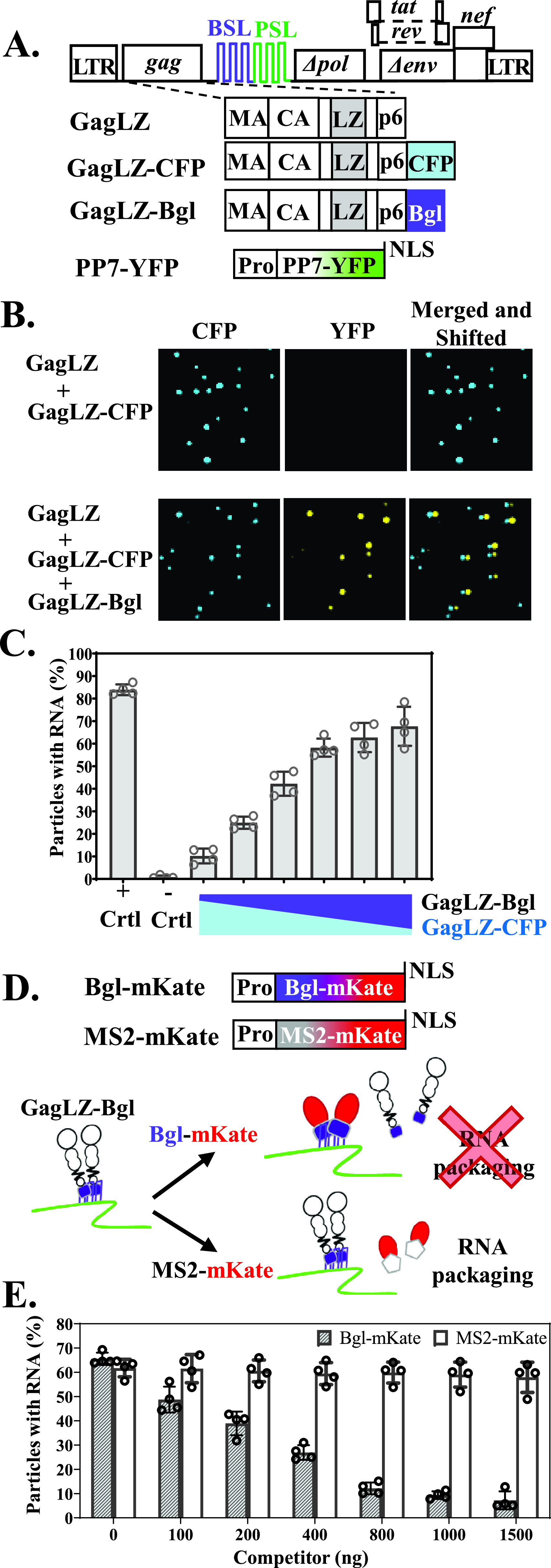
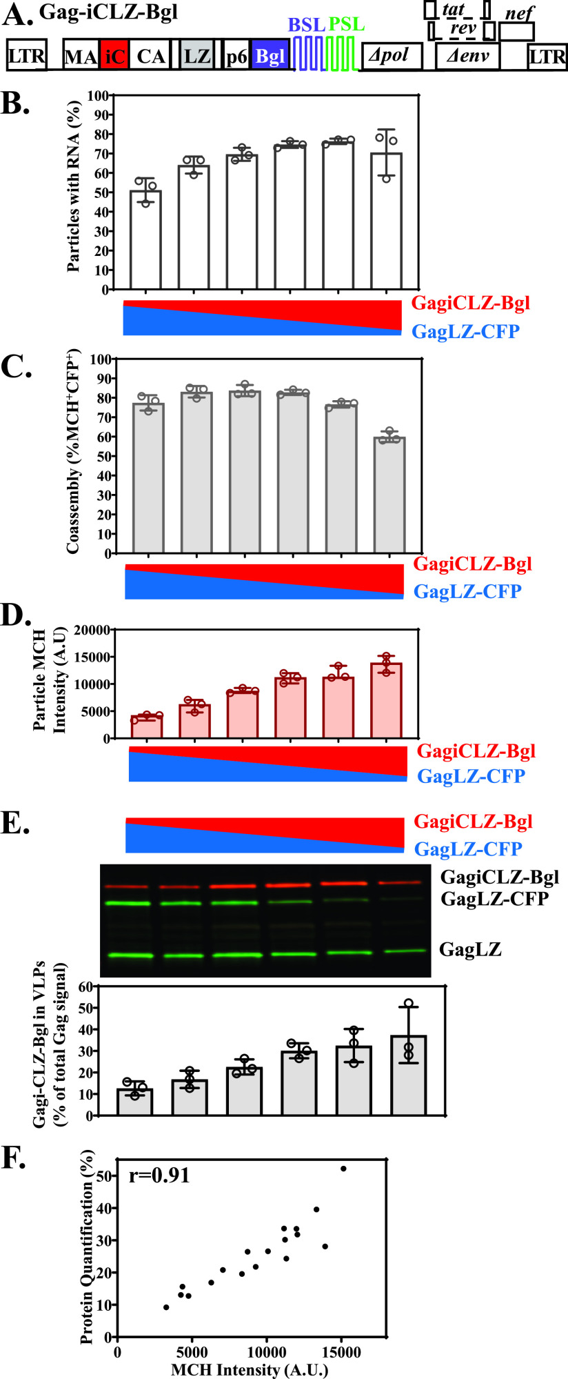
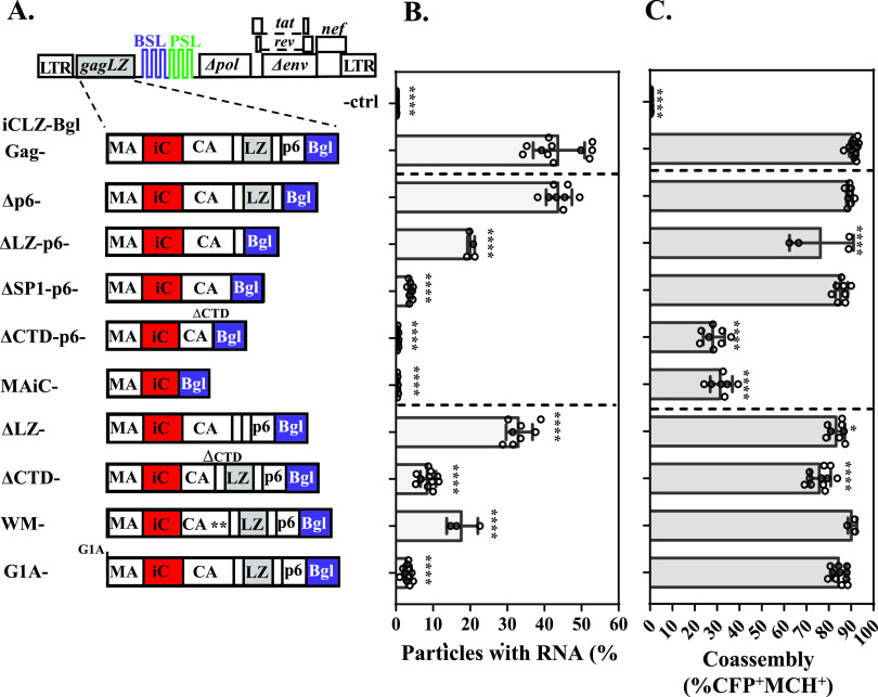
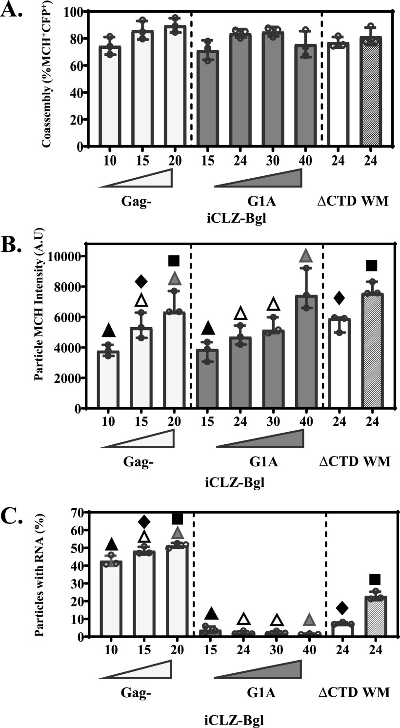
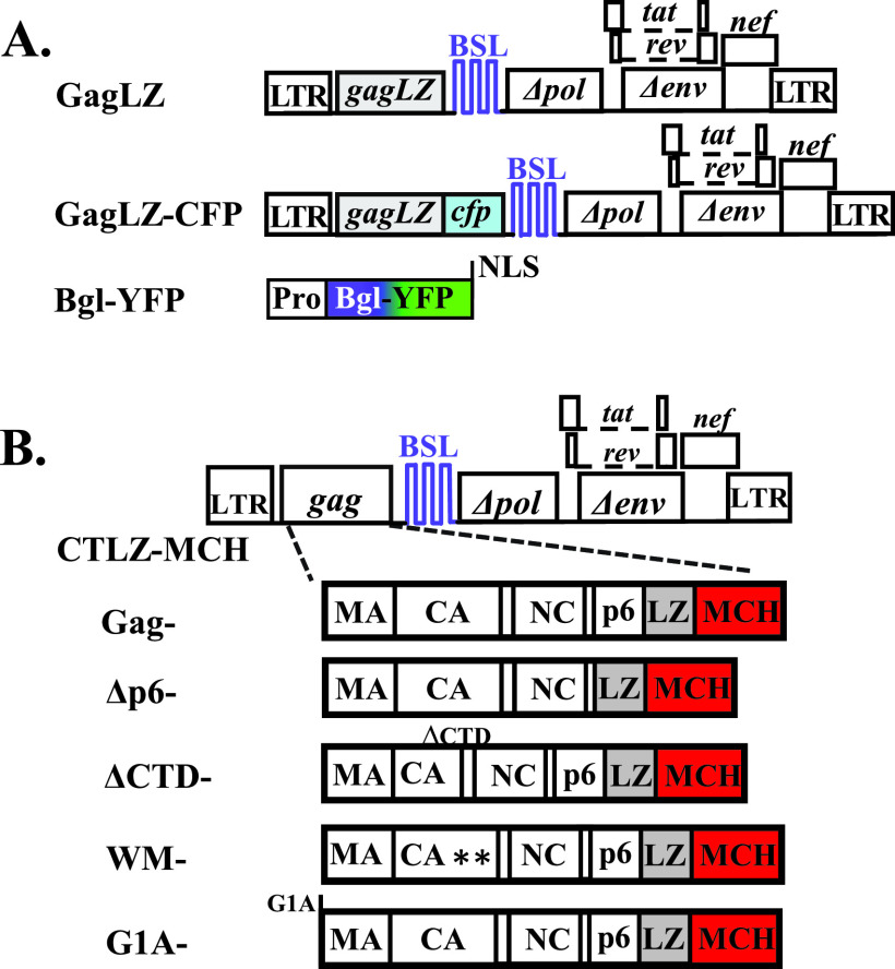
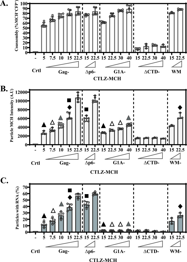
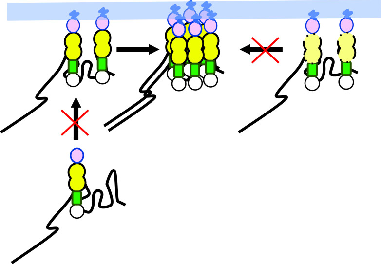
Human immunodeficiency virus type 1 (HIV-1) Gag selects and packages the HIV RNA genome during virus assembly. However, HIV-1 RNA constitutes only a small fraction of the cellular RNA. Although Gag exhibits a slight preference to viral RNA, most of the cytoplasmic Gag proteins are associated with cellular RNAs. Thus, it is not understood how HIV-1 achieves highly efficient genome packaging. We hypothesize that besides RNA binding, other properties of Gag are important for genome packaging. Many Gag mutants have assembly defects that preclude analysis of their effects on genome packaging. To bypass this challenge, we established complementation systems that separate the particle-assembling and RNA-binding functions of Gag: we used a set of Gag proteins to drive particle assembly and an RNA-binding Gag to package HIV-1 RNA. We have developed two types of RNA-binding Gag in which packaging is mediated by the authentic nucleocapsid (NC) domain or by a nonviral RNA-binding domain. We found that in both cases, mutations that affect the multimerization or plasma membrane anchoring properties of Gag reduce or abolish RNA packaging. These mutant Gag can coassemble into particles but cannot package the RNA genome efficiently. Our findings indicate that HIV-1 RNA packaging occurs at the plasma membrane and RNA-binding Gag needs to multimerize on RNA to encapsidate the viral genome. IMPORTANCE To generate infectious virions, HIV-1 must package its full-length RNA as the genome during particle assembly. HIV-1 Gag:RNA interactions mediate genome packaging, but the mechanism remains unclear. Only a minor portion of the cellular RNA is HIV-1 RNA, and most of the RNAs associated with cytoplasmic Gag are cellular RNAs. However, >94% of the HIV-1 virions contain viral RNA genome. We posited that, besides RNA binding, other properties of Gag contribute to genome packaging. Using two complementation systems, we examined features of Gag that are important for genome packaging. We found that the capacities for Gag to multimerize and to anchor at the plasma membrane are critical for genome packaging. Our results revealed that Gag needs to multimerize on viral RNA at the plasma membrane in order to package RNA genome.

Keywords: Gag; Gag:RNA interactions; RNA; genome packaging; human immunodeficiency virus; membrane targeting; multimerization.


Figures

FIG 1
FIG 1
NC-independent packaging system and experimental approach. (A) General structures of constructs used to express GagLZ, GagLZ-CFP, GagLZ-Bgl, and PP7-YFP. A nuclear localization signal (NLS) is fused to the C terminus of YFP. All constructs contain a leucine zipper motif (LZ; shown in gray) in place of NC and two sets of stem-loop sequences, BSL (purple) and PSL (green), inserted into pol, which are recognized by BglG protein and PP7 coat protein, respectively. CFP is shown in blue, BglG in purple, and YFP in green. LTR, long terminal repeat. (B) Representative images of particles generated by GagLZ and GagLZ-CFP (top) and by GagLZ, GagLZ-CFP, and GagLZ-Bgl (bottom). Images from CFP and YFP channels are shown individually or merged and shifted by 4 pixels to visualize colocalized signals. (C) Proportions of RNA packaging when different amounts of RNA-binding Gag were used. The sample labeled Gag is the positive control generated from transfection with wild-type Gag and Gag-CFP. With the titration, GagLZ plasmid was maintained at half, whereas plasmids encoding GagLZ-CFP and GagLZ-Bgl together constitute the remaining half of the Gag-encoding plasmid. PP7-YFP was cotransfected in all samples. Proportions of particles with RNA are shown for each condition. The ratios of Gag/Gag-CFP in the positive control (+ Crtl), GagLZ/GagLZ-CFP in the negative control (- Crtl), and GagLZ/(GagLZ-CFP + GagLZ-Bgl) of the titration experiments were 1:1. In the titrations, the percentages of transfected GagLZ-Bgl plasmid among the Gag-coding plasmids were 2.5, 5, 10, 20, 25, and 40%. (D) Schematics of Bgl-mKate and MS2-mKate, both containing a C-terminal NLS. Diagram depicting competitive (Bgl-mKate) versus noncompetitive (MS2-mKate) RNA-binding proteins and their impact on genome packaging. Bgl-mKate competes with GagLZ-Bgl for binding to BSL that is present in the viral RNA, resulting in a loss of packaged RNA (top). MS2-mKate cannot recognize BSL present in the viral RNA and should not impact RNA packaging through GagLZ-Bgl:BSL interactions (bottom). (E) A mixture of GagLZ/GagLZ-CFP/GagLZ-Bgl at a 2:1:1 ratio along with PP7-YFP was transfected into cells with increasing amounts of competing (Bgl-mKate) or noncompeting (MS2-mKate), 0 to 1,500 ng. This corresponds to a molar ratio ranging from 1:1 (100 ng) to 1:14 (1,500 ng) GagLZ-Bgl:Bgl-mKate competitor. The percentages of particles containing RNA are depicted for each Bgl-mKate (gray bars) and MS2-YFP (white bars) condition. Results of four independent experiments (open circles) are indicated as means ± standard deviations (error bars).
FIG 2
FIG 2
Visualizing the RNA-binding Gag allows for correlation of MCH particle intensity with protein incorporation into viral particles. (A) General structure of Gag-iCLZ-Bgl. An internal mCherry (iC) was inserted between MA and CA domains (red). Other abbreviations are the same as in Fig. 1. (B) The percentage of particles containing PP7-YFP-labeled RNA. Of the Gag-encoding plasmids, GagLZ (77%) was transfected into 293T cells with increasing and decreasing amounts of Gag-iCLZ-Bgl (4 to 20%) and GagLZ-CFP (19 to 3%), respectively. The percentage of Gag-iCLZ-Bgl used in the titrations were 4, 7, 10, 15, 18, and 20%. (C) The percentage of Gag-iCLZ-Bgl coassembled into particles was calculated throughout the titration. The mean ± standard deviation from three independent experiments is indicated. (D) Median MCH particle intensity in arbitrary units (A.U.) of coassembled particles is depicted as a median of three independent trials ± 95% confidence interval (95% CI). (E) Representative Western blot image of viral lysate probed with anti-p24 (green) and anti-MCH (red) antibodies. The positions of bands corresponding to Gag-iCLZ-Bgl, GagLZ-CFP, and GagLZ are indicated on the right. The percentage of Gag-iCLZ-Bgl relative to total Gag species packaged into particles as quantified by densitometry of Western blots from three independent experiments is shown below as the mean ± standard deviation. Individual values from independent experiments are shown as open circles in each panel. (F) Correlation plot of the percentage of Gag-iCLZ-Bgl protein incorporated into particles versus the corresponding median MCH particle intensity for each experimental data point shown in panels D and E. The Pearson correlation coefficient is 0.91.
FIG 3
FIG 3
Mutational analysis to determine features in HIV-1 Gag important for genome packaging and coassembly. (A) Schematic of deletion and point mutations made in Gag-iCLZ-Bgl. Amino acids 184 and 185 in CA of the WM mutant are denoted by two asterisks. GagLZ, GagLZ-CFP, and PP7-YFP alone (negative control) or with the indicated Gag-iCLZ-Bgl mutants were transfected into 293T cells, and viral particles were analyzed for RNA packaging efficiency (B) and coassembly (C). Dashed lines separate negative control (-ctrl) and Gag-iCLZ-Bgl from serial truncation mutants (middle) and single domain or point mutants (bottom). In these experiments, the ratios of GagLZ/GagLZ-CFP/Gag-iCLZ-Bgl constructs were 2:1:1 in all samples except in the negative control (-ctrl), in which GagiCLZ-Bgl was not used and the ratio of GagLZ/GagLZ-CFP was 1:1. All samples were compared with results from Gag-iCLZ-Bgl. Statistical significance was evaluated by a one-way ANOVA with Bonferroni’s multiple-comparison posttest and indicated as follows: *, P < 0.05; ****, P < 0.0001. Individual values from 3 to 11 independent trials are indicated by open circles. Bars indicate means ± standard deviations.
FIG 4
FIG 4
Loss in RNA packaging is independent of mutant Gag incorporation. (A) Coassembly of Gag-iCLZ-Bgl and GagLZ-CFP are indicated as a percentage of total viral particles; bars represent the means ± standard deviations. (B) Median MCH intensity of coassembled particles from three independent experiments with 95% CI. Paired symbols indicate median MCH intensities of similar values. Diamonds and squares denote conditions under which Gag- and ΔCTD- or WM-iCLZ-Bgl particles display comparable MCH intensity, respectively. Filled, open, and gray triangles correspond to conditions in which Gag- and G1A-iCLZ-Bgl particles exhibit comparable MCH intensities. (C) The proportion of RNA-containing viral particles from cells transfected with GagLZ, GagLZ-CFP, PP7-YFP and various amounts of Gag-, G1A-, ΔCTD-, or WM-iCLZ-Bgl is shown as the mean ± standard deviation. One-way ANOVA with Bonferonni’s multiple-comparisons posttest was used. (C) Symbols indicate statistical analysis of conditions in which MCH particle intensity are similar. Each paired symbol combination represents P < 0.0001. (A to C) Titrations of Gag- and G1A- are separated from each other and ΔCTD- and WM-iCLZ-Bgl by dashed lines. The percentage of each iCLZ-Bgl plasmid transfected into cells is indicated on the x axis. Individual values from three independent experiments are shown as open circles.
FIG 5
FIG 5
NC-dependent packaging system. (A) General structures of constructs used to express GagLZ, GagLZ-CFP, and Bgl-YFP. (B) NC-containing Gag constructs. Parent construct in which each Gag-CTLZ-MCH mutant was made is depicted at top, followed by schematics of Gag-CTLZ-MCH and mutants. A LZ domain (gray) and a mCherry fluorescent protein (MCH) were fused to the C terminus of Gag. Point mutations made at amino acids 184 and 185 in CA of the WM mutant are denoted by asterisks. All constructs contain a set of stem-loop sequences, BSL (purple), inserted into pol which are recognized by Bal-YFP.
FIG 6
FIG 6
NC-dependent RNA packaging requires functional Gag plasma membrane anchoring and multimerization. (A to C) GagLZ, GagLZ-CFP, Bgl-YFP alone (negative control [- Ctrl]) or along with Gag-CTLZ-MCH mutants were transfected into cells and viral particles analyzed for coassembly of Gag proteins (A), MCH particle intensity (B), and RNA packaging (C). The mean ± standard deviation (A and C) or median with 95% CI (B) is shown. Individual values from three to five independent experiments are indicated by open circles. The percentage of each Gag-CTLZ-MCH among the total Gag-encoding plasmids transfected into cells is indicated on the x axis. Gag-, Δp6-, G1A, ΔCTD-, and WM -CTLZ-MCH samples are separated by dashed lines. One-way ANOVA with Bonferroni’s multiple comparisons posttest was used. Symbols indicate statistical analysis of conditions in which MCH particle intensity are similar. RNA packaging efficiencies between Gag-CTLZ-MCH and mutants in filled triangles (P < 0.01), diamond (P < 0.001), empty and gray triangles (P < 0.0001), square (not significant [ns]).
FIG 7
FIG 7
Proposed model for HIV-1 RNA packaging. Wild-type Gag proteins bind viral RNA (black line) at the plasma membrane (shown as light blue area) (top left). To package RNA, Gag proteins must multimerize on viral RNA to form stable complex (top center). Gag with multimerization defects (top right) and Gag with membrane anchoring defects (bottom) cannot form stable Gag:viral RNA complex, resulting in genome packaging defects. Two copies of viral RNA are shown in the stable complex; however, for the BglG-mediated genome packaging, the number of RNA genomes in a particle is not known.

Similar articles

Cited by

References

    1. Freed EO. 2015. HIV-1 assembly, release and maturation. Nat Rev Microbiol 13:484–496. doi:10.1038/nrmicro3490. - DOI - PMC - PubMed
    1. Kuzembayeva M, Dilley K, Sardo L, Hu WS. 2014. Life of psi: how full-length HIV-1 RNAs become packaged genomes in the viral particles. Virology 454-455:362–370. doi:10.1016/j.virol.2014.01.019. - DOI - PMC - PubMed
    1. Rein A. 2019. RNA packaging in HIV. Trends Microbiol 27:715–723. doi:10.1016/j.tim.2019.04.003. - DOI - PMC - PubMed
    1. Nikolaitchik OA, Dilley KA, Fu W, Gorelick RJ, Tai SH, Soheilian F, Ptak RG, Nagashima K, Pathak VK, Hu WS. 2013. Dimeric RNA recognition regulates HIV-1 genome packaging. PLoS Pathog 9:e1003249. doi:10.1371/journal.ppat.1003249. - DOI - PMC - PubMed
    1. Schur FK, Obr M, Hagen WJ, Wan W, Jakobi AJ, Kirkpatrick JM, Sachse C, Kräusslich HG, Briggs JA. 2016. An atomic model of HIV-1 capsid-SP1 reveals structures regulating assembly and maturation. Science 353:506–508. doi:10.1126/science.aaf9620. - DOI - PubMed

Publication types

Substances