. 2021 Dec 6;9(1):e1119.
doi: 10.1212/NXI.0000000000001119. Print 2022 Jan.

Stress Signal ULBP4, an NKG2D Ligand, Is Upregulated in Multiple Sclerosis and Shapes CD8+ T-Cell Behaviors

Affiliations

Stress Signal ULBP4, an NKG2D Ligand, Is Upregulated in Multiple Sclerosis and Shapes CD8+ T-Cell Behaviors

Ana Carmena Moratalla et al. Neurol Neuroimmunol Neuroinflamm. .

Abstract

Background and objectives: We posit the involvement of the natural killer group 2D (NKG2D) pathway in multiple sclerosis (MS) pathology via the presence of specific NKG2D ligands (NKG2DLs). We aim to evaluate the expression of NKG2DLs in the CNS and CSF of patients with MS and to identify cellular stressors inducing the expression of UL16-binding protein 4 (ULBP4), the only detectable NKG2DL. Finally, we evaluate the impact of ULBP4 on functions such as cytokine production and motility by CD8+ T lymphocytes, a subset largely expressing NKG2D, the cognate receptor.

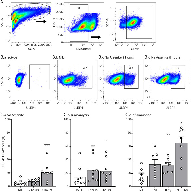
Methods: Human postmortem brain samples and CSF from patients with MS and controls were used to evaluate NKG2DL expression. In vitro assays using primary cultures of human astrocytes and neurons were performed to identify stressors inducing ULBP4 expression. Human CD8+ T lymphocytes from MS donors and age/sex-matched healthy controls were isolated to evaluate the functional impact of soluble ULBP4.

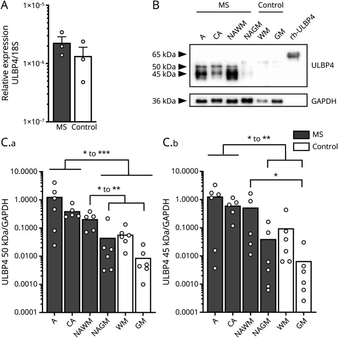
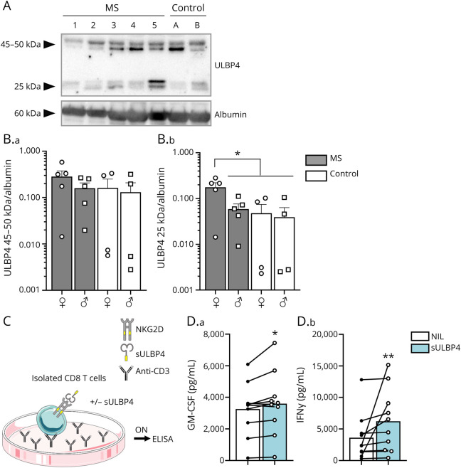
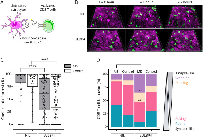
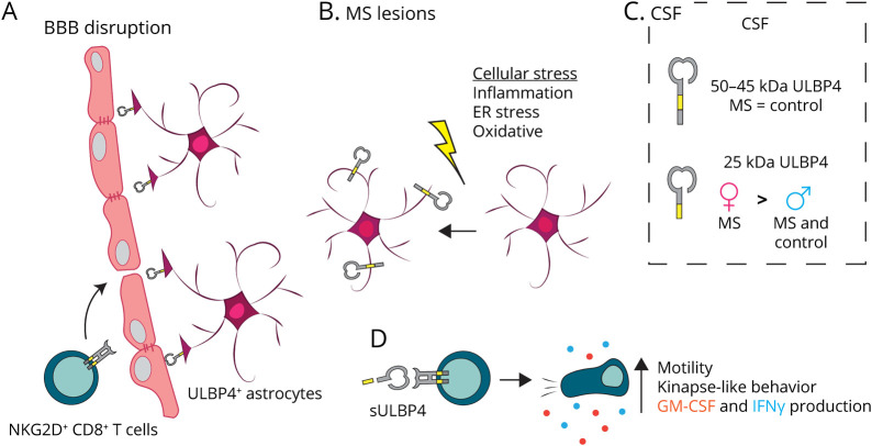
Results: We detected mRNA coding for the 8 identified human NKG2DLs in brain samples from patients with MS and controls, but only ULBP4 protein expression was detectable by Western blot. ULBP4 levels were greater in patients with MS, particularly in active and chronic active lesions and normal-appearing white matter, compared with normal-appearing gray matter from MS donors and white and gray matter from controls. Soluble ULBP4 was also detected in CSF of patients with MS and controls, but a smaller shed/soluble form of 25 kDa was significantly elevated in CSF from female patients with MS compared with controls and male patients with MS. Our data indicate that soluble ULBP4 affects various functions of CD8+ T lymphocytes. First, it enhanced the production of the proinflammatory cytokines GM-CSF and interferon-γ (IFNγ). Second, it increased CD8+ T lymphocyte motility and favored a kinapse-like behavior when cultured in the presence of human astrocytes. CD8+ T lymphocytes from patients with MS were especially altered by the presence of soluble ULBP4 compared with healthy controls.

Discussion: Our study provides new evidence for the involvement of NKG2D and its ligand ULBP4 in MS pathology. Our results point to ULBP4 as a viable target to specifically block 1 component of the NKG2D pathway without altering immune surveillance involving other NKG2DL.


Figures

Figure 1
Figure 1. ULBP4 Expression Is Elevated in Brain Lesions From Patients With MS
(A) Relative mRNA expression of ULBP4 in postmortem brain samples from untreated patients with MS and controls (epilepsy) (Ctl) expressed as 2-ΔCt compared with 18S. Each dot represents 1 donor (n = 3 per group). Data are shown as mean + SEM. (B and C) Western blot analysis of ULBP4 in postmortem brain lysates from patients with MS and controls (Ctl). MS samples were characterized as active (A) MS lesion, chronic active (CA) MS lesion, NAWM, and NAGM. Control samples were dissected from either white or gray matter (WM/GM). (B) Representative Western blot showing ULBP4 and GAPDH detection in samples from 1 MS donor and 1 control as well as the positive control rhULBP4. (C) Quantification of 50 kDa (C.a) and 45 kDa (C.b) ULBP4 bands relative to GAPDH levels. Each dot represents 1 donor. Data are shown as mean for 5–6 patients with MS (5 untreated SPMS and 1 fingolimod-treated transitional RRMS-SPMS) and 6 controls. Comparison between different groups is indicated with lines: Kruskal-Wallis test and uncorrected Dunn test; *p < 0.05, **p < 0.01, and ***p < 0.001. CA = chronic active; GAPDH = glyceraldehyde 3-phosphate dehydrogenase; MS = multiple sclerosis; NAGM = normal-appearing gray matter; NAWM = normal-appearing white matter; rhULBP4 = recombinant human ULBP4; RRMS = relapsing-remitting MS; SPMS = secondary progressive MS; ULBP4 = UL16-binding protein 4.
Figure 2
Figure 2. ULBP4 Is Mainly Expressed by Astrocytes in Brain Tissue From Patients With MS and Controls
(A) Representative images for the costaining of ULBP4 (cyan) and GFAP (magenta) in paraffin-embedded sections from untreated MS donors (NAGM and WM lesion) and controls (no brain related disease); n = 4 for each group. Nuclei were stained with 4',6-diamidino-2-phenylindole (blue). Corresponding isotype controls are shown. Yellow arrows indicate ULBP4+GFAP+ cells, and red arrows indicate ULBP4-negative GFAP+ cells. Scale bars = 25 μM. (B) Representative enlarged image showing colocalization of GFAP and ULBP4 in the brain section. Scale bars = 10 µM. GFAP = glial fibrillary acidic protein; MS = multiple sclerosis; NAGM = normal-appearing gray matter; WM = white mater; ULBP4 = UL16-binding protein 4.
Figure 3
Figure 3. Several Types of Cellular Stress Increase the Proportion of ULBP4-Expressing Astrocytes
(A–C) Flow cytometry analysis of ULBP4 expression by human astrocytes. (A) Gating strategy from 1 representative donor. Cell debris, doublets, and dead cells were excluded, and GFAP+ cells were selected for analysis. (B) Representative dot plots showing isotype control (B.a) or ULBP4 detection on living GFAP+ gated cells. Astrocytes were either kept under normal culture conditions (NIL) (B.b) or exposed to sodium arsenite (Na Arsenite) for 2 (B.c) and 6 hours (B.d). Percentage of ULBP4+ cells is indicated. (C) Quantification of ULBP4 expression on GFAP+ cells after exposure to sodium arsenite (C.a), tunicamycin (C.b), or proinflammatory cytokines (C.c). Each dot represents 1 donor. Data are shown as mean or mean +SEM, n = 7–9. Friedman test and Dunn multiple comparison test comparing NIL vs Na Arsenite or DMSO vs tunicamycin; 1-way analysis of variance comparing NIL vs cytokine *p < 0.05, **p < 0.01 ***p < 0.001. DMSO = dimethylsulfoxide; GFAP = Glial fibrillary acidic protein; MS = multiple sclerosis; IFNγ = interferon-γ; TNF = tumor necrosis factor; ULBP4 = UL16-binding protein 4.
Figure 4
Figure 4. Soluble ULBP4 Is Elevated in CSF From Female Patients With MS and Enhances Proinflammatory Properties of CD8+ T Lymphocytes
(A and B) Western blot analysis of ULBP4 and albumin levels in CSF from patients with MS and controls (Ctl). (A) Representative Western blot including 5 untreated MS donors (1–5) and 2 controls (A and B) illustrating expression of ULBP4 and albumin. (B) Quantification of 45–50 kDa (B.a) and 25 kDa (B.b) bands of ULBP4 relative to albumin according to sex (♀: female; ♂: male) and disease (MS vs control). Each dot represents 1 donor. Mean ± SEM, n = 5 female untreated MS, n = 5 male untreated MS, n = 4 female controls, and n = 4 male controls. (C and D) Production of GM-CSF and IFNγ by CD8+ T lymphocytes stimulated with or without rhULBP4. (C) Diagram illustrating culture conditions: isolated CD8+ T lymphocytes were stimulated overnight with plate-bound anti-CD3 in the presence or absence of sULBP4 before collecting supernatants for ELISA measurements. (D) Quantification of secreted GM-CSF (D.a) and IFNγ (D.b) levels (pg/mL) in the presence (blue bars) or absence (white bars) of rhULBP4. Each dot represents 1 donor. Mean, n = 10 per group. Paired t test for GM-CSF production and Wilcoxon test for IFNγ production comparing NIL vs sULBP4; *p < 0.05 and **p < 0.01. GM-CSF = granulocyte-macrophage colony-stimulating factor; MS = multiple sclerosis; rhULBP4 = recombinant human ULBP4; sULBP4 = soluble UL16-binding protein 4; ULBP4 = UL16-binding protein 4.
Figure 5
Figure 5. Soluble ULBP4 Enhances the Motility of CD8+ T Lymphocytes
(A–D) Time-lapse imaging of activated CD8+ T lymphocytes cocultured with human astrocytes in the presence or absence of sULBP4. (A) Diagram illustrating culture conditions. CD8+ T lymphocytes were activated overnight on anti-CD3 coated plates in the presence of anti-CD28 and interleukin-15, CFSE-labeled, and then added to astrocytes in the presence or absence of sULBP4 and imaged for 2 hours. (B) Three-dimensional time-lapse view at T = 0, T = 1, and T = 2 hours of activated CD8+ T lymphocytes (green) cocultured with astrocytes (magenta). (C and D) Analysis of the motility and behavior of CD8+ T lymphocytes from 3 untreated patients with MS and 3 sex- and age-matched healthy controls on coculture with astrocytes in the presence or absence of sUBLP4. Presented data are pooled from 3 independent experiments. (C) Coefficient of arrest (%) of individual CD8+ T lymphocytes; each dot represents 1 cell. Kruskal-Wallis test comparing sULBP4 vs NIL for each donors' group ****p < 0.0001. (D) Proportion of CD8+ T lymphocytes exhibiting the scanning, dancing, poking, and round behaviors when cocultured with astrocytes in the presence or absence of sULBP4. One way analysis of variance comparing NIL vs sULBP4 for each donors' group, *p < 0.05, **p < 0.01. CFSE = carboxyfluorescein succinimidyl ester; MS = multiple sclerosis; sULBP4 = soluble UL16-binding protein 4; ULBP4 = UL16-binding protein 4.
Figure 6
Figure 6. Proposed Involvement of the NKG2D Pathway in MS Pathology
(A) CD8+ T lymphocytes can enter the CNS of patients with MS in part due to disrupted BBB. All infiltrating CD8+ T lymphocytes express NKG2D and thus can interact with ULBP4-expressing astrocytes including those having end feet in close proximity to the BBB. (B) Various cellular stresses (inflammation, ER stress, and oxidative stress) present in the brain of patients with MS can upregulate ULBP4 expression by astrocytes, as supported by our in vitro data. Reactive astrocytes, which are abundantly present in MS lesion, represent the predominant cell type expressing ULBP4 in the brain of patients with MS. (C) Soluble forms of ULBP4 are detectable in CSF; a soluble 25-kDa ULBP4 form is significantly elevated in CSF from female patients with MS compared with groups (male patients with MS and controls of both sexes). (D) Soluble ULBP4 can affect immune cell functions. On contact with sULBP4, CD8+ T lymphocytes increase their secretion of GM-CSF and IFNγ. Moreover, addition of sULBP4 to CD8+ T lymphocytes cocultured with astrocytes increases their motility and favors kinapse-like behaviors, which are more dynamic and may lead to enhanced displacement within the tissue. BBB = blood-brain barrier; ER = endoplasmic reticulum; MS = multiple sclerosis; sULBP4 = soluble UL16-binding protein 4.

Similar articles

Cited by

References

    1. Lassmann H. Multiple sclerosis pathology. Cold Spring Harb Perspect Med. 2018;8(3):a02893. - PMC - PubMed
    1. Escartin C, Galea E, Lakatos A, et al. . Reactive astrocyte nomenclature, definitions, and future directions. Nat Neurosci. 2021;24(3):312-325. - PMC - PubMed
    1. Brambilla R. The contribution of astrocytes to the neuroinflammatory response in multiple sclerosis and experimental autoimmune encephalomyelitis. Acta Neuropathol. 2019;137(5):757-783. - PMC - PubMed
    1. Wensveen FM, Jelenčić V, Polić B. NKG2D: a master regulator of immune cell responsiveness. Front Immunol. 2018;9:441. - PMC - PubMed
    1. Babic M, Romagnani C. The role of natural killer group 2, member D in chronic inflammation and autoimmunity. Front Immunol. 2018;9:1219. - PMC - PubMed

Publication types

MeSH terms

Substances

Grants and funding