. 2021 Dec 14;118(50):e2114856118.
doi: 10.1073/pnas.2114856118.

Sleep promotes the formation of dendritic filopodia and spines near learning-inactive existing spines

Affiliations

Sleep promotes the formation of dendritic filopodia and spines near learning-inactive existing spines

Avital Adler et al. Proc Natl Acad Sci U S A. .

Abstract

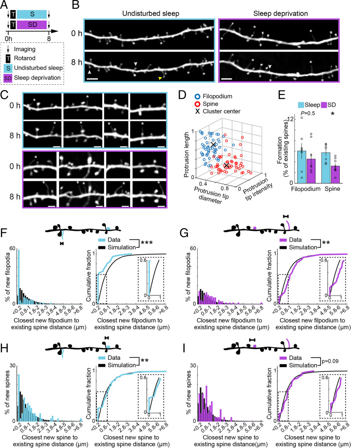
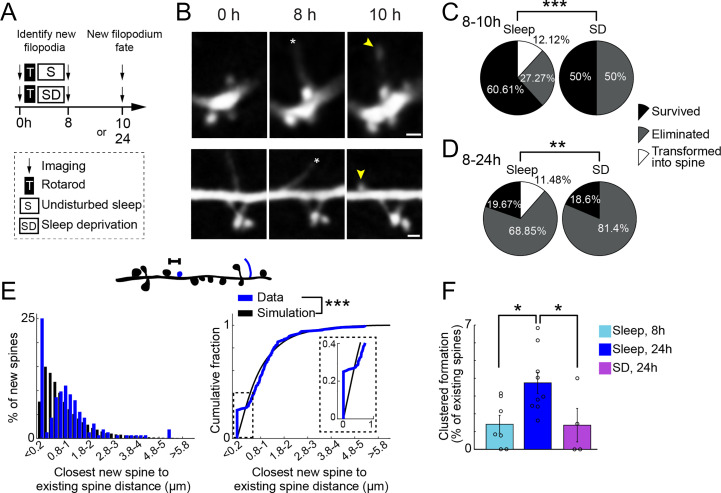
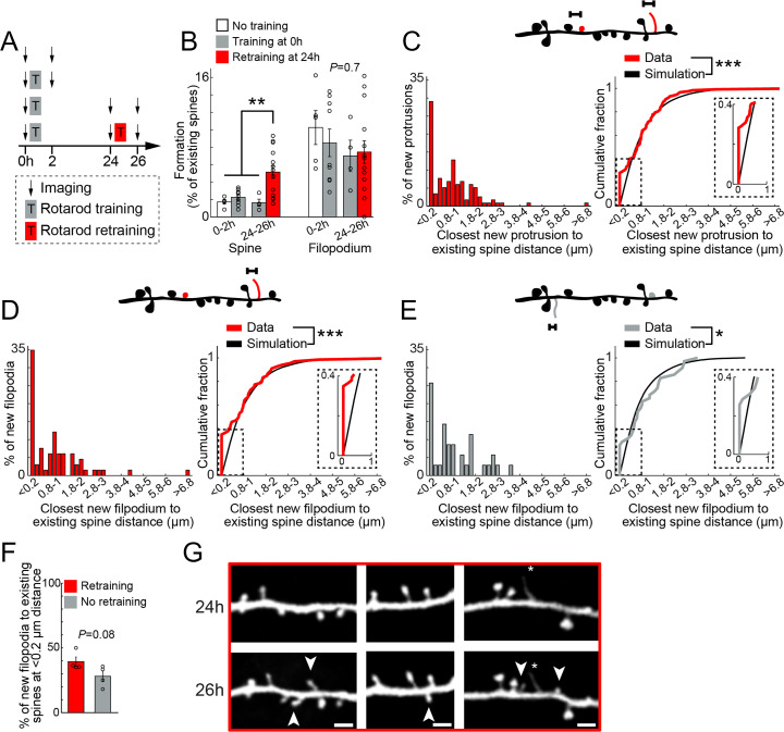
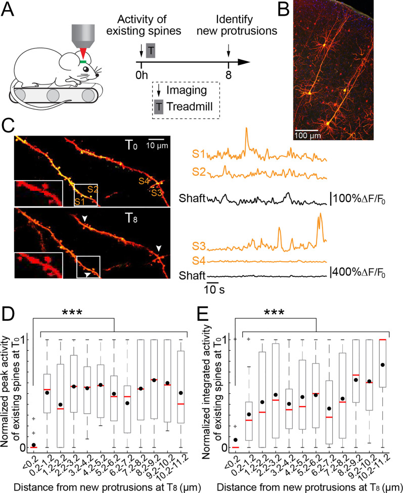
Changes in synaptic connections are believed to underlie long-term memory storage. Previous studies have suggested that sleep is important for synapse formation after learning, but how sleep is involved in the process of synapse formation remains unclear. To address this question, we used transcranial two-photon microscopy to investigate the effect of postlearning sleep on the location of newly formed dendritic filopodia and spines of layer 5 pyramidal neurons in the primary motor cortex of adolescent mice. We found that newly formed filopodia and spines were partially clustered with existing spines along individual dendritic segments 24 h after motor training. Notably, posttraining sleep was critical for promoting the formation of dendritic filopodia and spines clustered with existing spines within 8 h. A fraction of these filopodia was converted into new spines and contributed to clustered spine formation 24 h after motor training. This sleep-dependent spine formation via filopodia was different from retraining-induced new spine formation, which emerged from dendritic shafts without prior presence of filopodia. Furthermore, sleep-dependent new filopodia and spines tended to be formed away from existing spines that were active at the time of motor training. Taken together, these findings reveal a role of postlearning sleep in regulating the number and location of new synapses via promoting filopodial formation.

Keywords: clustering; dendritic spines; filopodia; sleep.


Conflict of interest statement

The authors declare no competing interest.

Figures

Fig. 1.
Fig. 1.
Sleep promotes clustered formation of new filopodia and spines with existing spines within 8 h after motor training. (A) Schematic of experimental paradigm. After imaging and training, mice were either left undisturbed or subjected to SD. They were then imaged again to assess the effect of SD. (B) Example of new spines (white arrowheads), filopodia (asterisks), and filopodium transition into spine (yellow arrowhead) in a L5 pyramidal cell dendrite 8 h following rotarod training and sleep (Left) or rotarod training and SD (Right) (Scale bar: 5 µm). (C) Examples of new filopodia (asterisks) in L5 pyramidal cell dendrites 8 h following rotarod training and sleep (Top) or rotarod training and SD (Bottom) (Scale bar: 2 µm). (D) Characteristics of new protrusions following rotarod motor training in sleep and sleep-deprived animals. For each new protrusion (circle), its head intensity (normalized to adjacent shaft), head diameter, and length are plotted. Protrusions defined as spines and filopodia based on these morphological characteristics are color coded (red and blue respectively). X marks the center of each cluster based on K-means algorithm. A total of 51 new spines and 49 new filopodia from eight animals. Data from sleep and sleep-deprived animals were not different and were pooled together. (E) The percentage of dendritic filopodia and spines formed in sleep (blue) and sleep-deprived (purple) mice. Black circles: individual animals. A total of 7 and 8 mice in sleep and sleep-deprivation groups, respectively. (F) Top: schematic of distances measured on individual dendritic segments between new filopodium (blue) and existing spines (black). Left: distribution of closest distances measured (blue) and simulated (black, Materials and Methods) in animals left undisturbed following motor training. Right: Cumulative sum of closest distances measured (blue) saturate faster than simulated (black), showing new filopodia cluster with existing spines. Inset: enlargement of 0 to 1 µm distance. A total of 80 new filopodia in 42 dendrites from seven animals. (G) Similar as in F for animals subjected to SD. Following SD, new filopodia do not cluster with existing spines and are further away from existing spines than expected by chance. A total of 72 new filopodia in 44 dendrites from eight animals. (H) Similar as in F for new spines in animals with undisturbed sleep. Following sleep, new spines cluster with existing spines. A total of 71 new spines in 42 dendrites from seven animals. (I) Similar as in F for new spines in animals subjected to SD. Following SD, new spines do not cluster with existing spines and are randomly distributed. A total of 47 new spines in 44 dendrites from eight animals. Percent formation was tested using Mann–Whitney U test. Cumulative sums were tested using Kolmogorov–Smirnov test. *P < 0.05, **P < 0.01, ***P < 0.001.
Fig. 2.
Fig. 2.
Sleep-dependent clustered formation of new filopodia contributes to clustered new spine formation 24 h after training. (A) Schematic of experimental paradigm. The fate of new filopodia identified at 8 h was determined at either 10 h or 24 h. (B) Examples of new filopodia (asterisks) in L5 pyramidal cell dendrites 8 h following rotarod training and sleep that were transformed into new spines at 10 h (yellow arrowhead) (Scale bar: 1 µm). (C) New filopodia fate. Sleep and sleep-deprived animals were imaged again at 10 h to assess the fate of new filopodia (survived, eliminated, or transformed into spine). Only in sleep animals new filopodia were transformed into new spines. Sleep; 33 new filopodia in four mice. SD; 16 new filopodia in four mice. (D) Similar as in C for filopodia fate at 24 h. Only in sleep animals new filopodia were transformed into new spines. Sleep; 61 new filopodia in 4 mice. SD; 43 new filopodia in 4 mice. (E) Top: schematic of distances measured on individual dendritic segments between new spines (blue) and existing spines (black). Left: distribution of closest distances measured (blue) and simulated (black, Materials and Methods) in animals left undisturbed 24 h following motor training. Right: Cumulative sum of closest distances measured (blue) saturate faster than simulated (black), showing new spines cluster with existing spines 24 h following motor training. Inset: enlargement of 0 to 1 µm distance. A total of 164 new spines in 82 dendrites from nine animals. (F) The percentage of clustered new spines over existing spines formed 24 h following training in mice left undisturbed (blue) is larger than that formed 8 h following training in mice left undisturbed (light blue), as well as that formed 24 h following training in mice with SD (purple); 7, 9, and 4 mice, respectively. Distribution of new filopodia fate was tested using χ2 test. Cumulative sum was tested using Kolmogorov–Smirnov test. Percent clustered formation was tested using Kruskal–Wallis test followed by Tukey–Kramer for multiple comparison. *P < 0.05, **P < 0.01, ***P < 0.001.
Fig. 3.
Fig. 3.
Retraining promotes rapid spine formation without intermediate filopodium transformation. (A) Schematic of experimental paradigm. Animals either did not receive rotarod training, were trained once (training), or twice (retraining). Animals were imaged before and 2 h following training and retraining. Some animals were imaged in all four time points. (B) The percentage of dendritic spines and filopodia formed over 2 h in response to training (Left, gray versus white) and retraining (Right, red versus gray). Black circles: individual animals. Five and nine mice for no training and training, respectively. A total of 4 and 14 mice for no retraining and retraining, respectively. Retraining but not initial training led to rapid new spine formation within 2 h. (C) Top: schematic of distances measured on individual dendritic segments between retraining-induced new spines and filopodia (red) and existing spines (black). Left: distribution of closest distances measured in retraining animals. Right: Cumulative sum of closest distances measured (red) saturate faster than simulated (black, Materials and Methods), showing new protrusions following retraining cluster with existing spines. Inset: enlargement of 0 to 1 µm distance. A total of 117 new protrusions in 48 dendrites from five animals. (D) Similar as in C for new filopodia in retraining animals. Following retraining, new filopodia cluster with existing spines. A total of 66 new filopodia in 48 dendrites from five animals. (E) Similar as in C for new filopodia in animals receiving a single training session (no retraining). Similar to retraining, also in control new filopodia cluster with existing spines. A total of 35 new filopodia in 34 dendrites from four animals. (F) The percentage of new filopodia that are ≤ 0.2 µm from existing spines is similar in animals that did (red, 4 mice) or did not (gray, 4 mice) receive retraining. Black circles: individual animals. (G) Examples of retraining-induced new spines (arrowheads) at 26 h without prior filopodia presences at 24 h (Scale bar: 2 µm). Percent formation was tested using Kruskal–Wallis test followed by Tukey–Kramer for multiple comparison. Cumulative sums were tested using Kolmogorov–Smirnov test. New filopodia at ≤ 0.2 µm were tested using Mann–Whitney U test. *P < 0.05, **P < 0.01, ***P < 0.001.
Fig. 4.
Fig. 4.
Clustered new protrusions are formed near inactive existing spines. (A) Schematic of experimental paradigm. Ca2+ activity of existing spines was imaged in response to running on the treadmill before training. At 8 h later, the animals were imaged again, and training-induced new spines and filopodia were identified. (B) L5 pyramidal neurons were sparsely labeled with both GCaMP7s and tdTomato in M1. (C) Left: example of two dendrites from the same L5 pyramidal neuron at times 0 h (Top) and 8 h (Bottom). White rectangle box is enlarged on the Left. Arrowheads point to three new spines. Right: ΔF/F0 of single spines marked on the image and the dendritic shafts. (D) Normalized peak activity of existing spines at time 0 h in response to forward running on the treadmill as a function of their distance from where new spines and filopodia were formed 8 h later. Existing spines that are clustered (≤ 0.2 µm) with new protrusions are less active. Black dots indicate the mean. Red lines indicate the median. Gray box shows the 25th and the 75th percentile, and the whiskers extend to the most extreme data point not considered outliers. Gray pluses show individual outliers. A total of 219 existing spines relative to 98 new protrusions from seven mice. (E) Similar as in D showing the normalized integrated activity of existing spines at time 0 h. Existing spines that are clustered (≤ 0.2 µm) with new protrusions are less active. A total of 219 existing spines relative to 98 new protrusions from seven mice. Activity of existing spines as a function of distance from new protrusions was tested using Kruskal–Wallis test followed by Tukey–Kramer test for multiple comparisons. ***P < 0.001.

References

    1. Bhatt D. H., Zhang S., Gan W. B., Dendritic spine dynamics. Annu. Rev. Physiol. 71, 261–282 (2009). - PubMed
    1. Yuste R., Bonhoeffer T., Morphological changes in dendritic spines associated with long-term synaptic plasticity. Annu. Rev. Neurosci. 24, 1071–1089 (2001). - PubMed
    1. Buonomano D. V., Merzenich M. M., Cortical plasticity: From synapses to maps. Annu. Rev. Neurosci. 21, 149–186 (1998). - PubMed
    1. Yang G., Pan F., Gan W. B., Stably maintained dendritic spines are associated with lifelong memories. Nature 462, 920–924 (2009). - PMC - PubMed
    1. Xu T., et al. , Rapid formation and selective stabilization of synapses for enduring motor memories. Nature 462, 915–919 (2009). - PMC - PubMed

Publication types

LinkOut - more resources