

Licochalcone H Induces Cell Cycle Arrest and Apoptosis in Human Skin Cancer Cells by Modulating JAK2/STAT3 Signaling
- PMID: 34873073
- PMCID: PMC8724845
- DOI: 10.4062/biomolther.2021.149
Licochalcone H Induces Cell Cycle Arrest and Apoptosis in Human Skin Cancer Cells by Modulating JAK2/STAT3 Signaling
Abstract
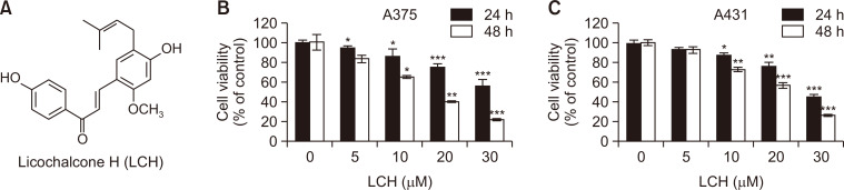
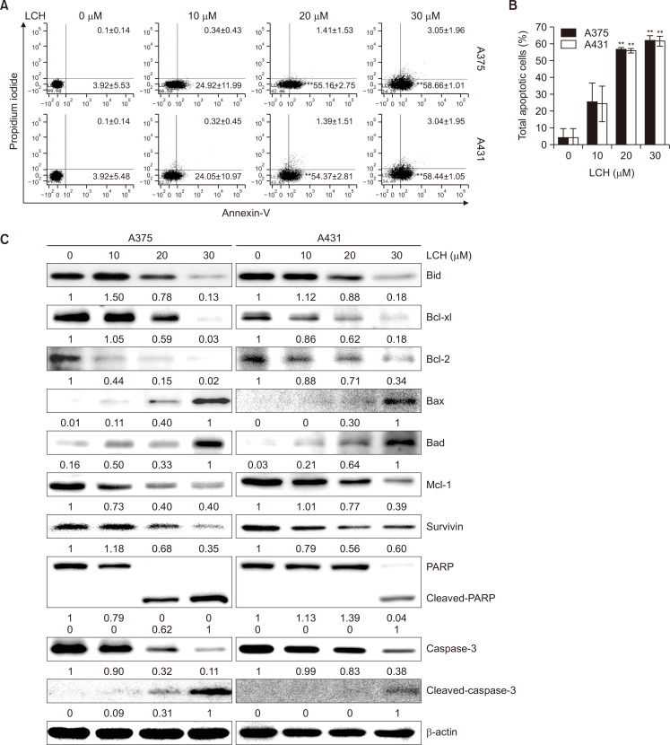
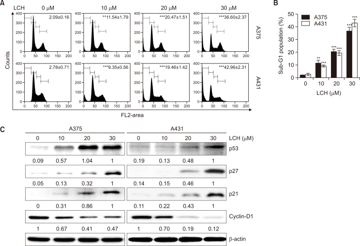
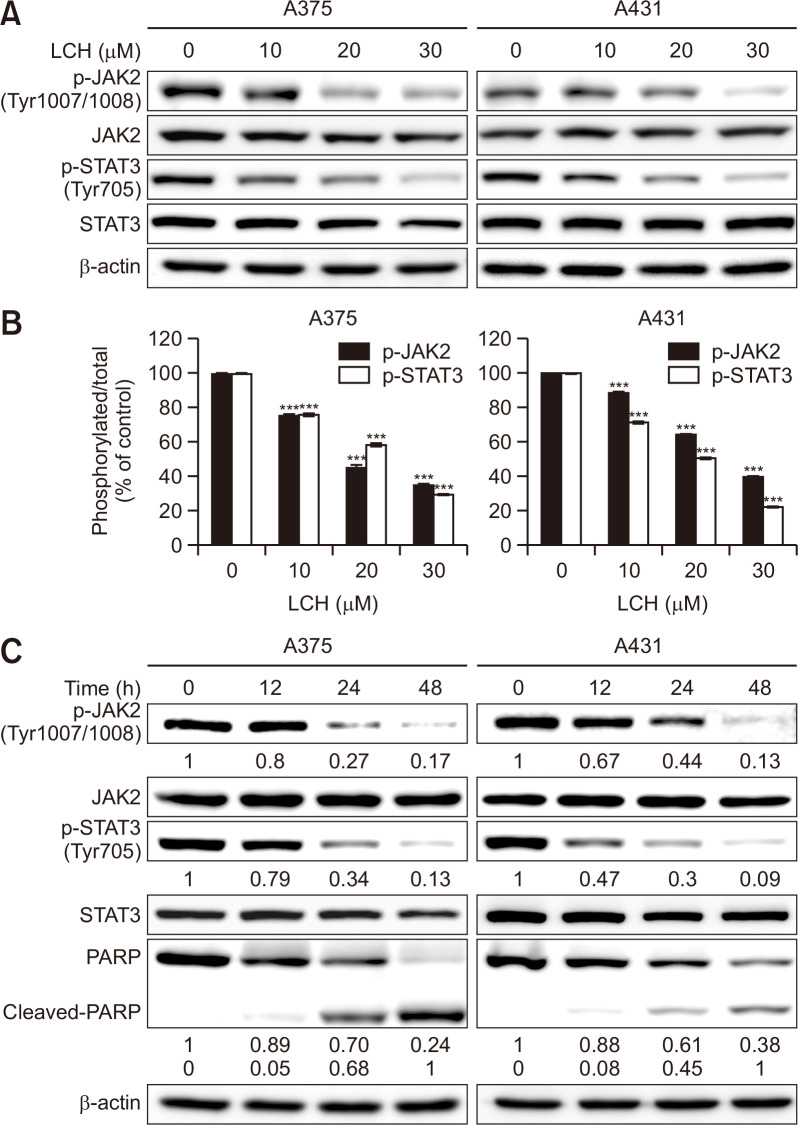
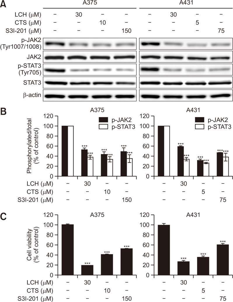
Licochalcone H (LCH) is a phenolic compound synthetically derived from licochalcone C (LCC) that exerts anticancer activity. In this study, we investigated the anticancer activity of LCH in human skin cancer A375 and A431 cells. The 3-(4,5-dimethylthiazol- 2-yl)-5-(3-carboxymethoxyphenyl)-2-(4-sulfophenyl)-2H-tetrazolium (MTS) cell viability assay was used to evaluate the antiproliferative activity of LCH. Cell cycle distribution and the induction of apoptosis were analyzed by flow cytometry. Western blotting assays were performed to detect the levels of proteins involved in cell cycle progression, apoptosis, and the JAK2/STAT3 signaling pathway. LCH inhibited the growth of cells in dose- and time-dependent manners. The annexin V/propidium iodide double staining assay revealed that LCH induced apoptosis, and the LCH-induced apoptosis was accompanied by cell cycle arrest in the G1 phase. Western blot analysis showed that the phosphorylation of JAK2 and STAT3 was decreased by treatment with LCH. The inhibition of the JAK2/STAT3 signaling pathway by pharmacological inhibitors against JAK2/STAT3 (cryptotanshinone (CTS) and S3I-201) simulated the antiproliferative effect of LCH suggesting that LCH induced apoptosis by modulating JAK2/STAT3 signaling.
Keywords: Apoptosis; Human skin cancer; JAK2; Licochalcone H; STAT3.
Conflict of interest statement
There are no conflicts of interest.
Figures
Similar articles
-
JAK2 regulation by licochalcone H inhibits the cell growth and induces apoptosis in oral squamous cell carcinoma.Phytomedicine. 2019 Jan;52:60-69. doi: 10.1016/j.phymed.2018.09.180. Epub 2018 Sep 18. Phytomedicine. 2019. PMID: 30599913
-
Cryptotanshinone induces cell cycle arrest and apoptosis through the JAK2/STAT3 and PI3K/Akt/NFκB pathways in cholangiocarcinoma cells.Drug Des Devel Ther. 2017 Jun 15;11:1753-1766. doi: 10.2147/DDDT.S132488. eCollection 2017. Drug Des Devel Ther. 2017. PMID: 28670110 Free PMC article.
-
Inhibition of JAK2/STAT3 Signaling Pathway Suppresses Proliferation of Burkitt's Lymphoma Raji Cells via Cell Cycle Progression, Apoptosis, and Oxidative Stress by Modulating HSP70.Med Sci Monit. 2018 Sep 8;24:6255-6263. doi: 10.12659/MSM.910170. Med Sci Monit. 2018. PMID: 30194286 Free PMC article.
-
Licochalcone H Synthesized by Modifying Structure of Licochalcone C Extracted from Glycyrrhiza inflata Induces Apoptosis of Esophageal Squamous Cell Carcinoma Cells.Cell Biochem Biophys. 2020 Mar;78(1):65-76. doi: 10.1007/s12013-019-00892-3. Epub 2019 Nov 9. Cell Biochem Biophys. 2020. PMID: 31707583
-
Anticancer effects of licochalcones: A review of the mechanisms.Front Pharmacol. 2023 Jan 23;14:1074506. doi: 10.3389/fphar.2023.1074506. eCollection 2023. Front Pharmacol. 2023. PMID: 36755942 Free PMC article.
Cited by
-
Targeting STAT3 and NF-κB Signaling Pathways in Cancer Prevention and Treatment: The Role of Chalcones.Cancers (Basel). 2024 Mar 8;16(6):1092. doi: 10.3390/cancers16061092. Cancers (Basel). 2024. PMID: 38539427 Free PMC article. Review.
-
MS-5, a Naphthalene Derivative, Induces Apoptosis in Human Pancreatic Cancer BxPC-3 Cells by Modulating Reactive Oxygen Species.Biomol Ther (Seoul). 2023 Jan 1;31(1):68-72. doi: 10.4062/biomolther.2022.127. Epub 2022 Nov 16. Biomol Ther (Seoul). 2023. PMID: 36380602 Free PMC article.
-
Licochalcone H Targets EGFR and AKT to Suppress the Growth of Oxaliplatin -Sensitive and -Resistant Colorectal Cancer Cells.Biomol Ther (Seoul). 2023 Nov 1;31(6):661-673. doi: 10.4062/biomolther.2023.155. Biomol Ther (Seoul). 2023. PMID: 37899744 Free PMC article.
-
Licochalcone D Inhibits Skin Epidermal Cells Transformation through the Regulation of AKT Signaling Pathways.Biomol Ther (Seoul). 2023 Nov 1;31(6):682-691. doi: 10.4062/biomolther.2023.162. Biomol Ther (Seoul). 2023. PMID: 37899745 Free PMC article.
-
SIRT7-Mediated MVP Desuccinylation Facilitates Tongue Squamous Cell Carcinoma Progression by Activating JAK2/STAT3 Pathway.Cell Biol Int. 2025 Sep;49(9):1184-1196. doi: 10.1002/cbin.70048. Epub 2025 Jun 30. Cell Biol Int. 2025. PMID: 40586636 Free PMC article.
References
LinkOut - more resources
Full Text Sources
Research Materials
Miscellaneous
