. 2021 Dec 6;11(1):23494.
doi: 10.1038/s41598-021-02757-3.

Adaptive value of foot-and-mouth disease virus capsid substitutions with opposite effects on particle acid stability

Affiliations

Adaptive value of foot-and-mouth disease virus capsid substitutions with opposite effects on particle acid stability

Flavia Caridi et al. Sci Rep. .

Abstract

Foot-and-mouth disease virus (FMDV) is a picornavirus that exhibits an extremely acid sensitive capsid. This acid lability is directly related to its mechanism of uncoating triggered by acidification inside cellular endosomes. Using a collection of FMDV mutants we have systematically analyzed the relationship between acid stability and the requirement for acidic endosomes using ammonium chloride (NH4Cl), an inhibitor of endosome acidification. A FMDV mutant carrying two substitutions with opposite effects on acid-stability (VP3 A116V that reduces acid stability, and VP1 N17D that increases acid stability) displayed a rapid shift towards acid lability that resulted in increased resistance to NH4Cl as well as to concanamicyn A, a different lysosomotropic agent. This resistance could be explained by a higher ability of the mutant populations to produce NH4Cl-resistant variants, as supported by their tendency to accumulate mutations related to NH4Cl-resistance that was higher than that of the WT populations. Competition experiments also indicated that the combination of both amino acid substitutions promoted an increase of viral fitness that likely contributed to NH4Cl resistance. This study provides novel evidences supporting that the combination of mutations in a viral capsid can result in compensatory effects that lead to fitness gain, and facilitate space to an inhibitor of acid-dependent uncoating. Thus, although drug-resistant variants usually exhibit a reduction in viral fitness, our results indicate that compensatory mutations that restore this reduction in fitness can promote emergence of resistance mutants.


Conflict of interest statement

The authors declare no competing interests.

Figures

Figure 1
Figure 1
Double FMDV mutant with VP3 A116V and VP1 N17D does not follow the relation between uncoating pH and resistance to NH4Cl in FMDV. (a) Combinatorial mutants analyzed in the study. Symbols in the first column identify Each mutant, with the amino acid substitutions, the uncoating pH (pH50) and references included in the columns on the right. See text for details. (b) Relation between uncoating pH and sensitivity to endosomal neutralization by NH4Cl of FMDV mutants,. FMDV mutants are identified with the symbols explained in (a). BHK-21 cells treated or not with 25 mM NH4Cl were infected (MOI of 0.5 PFU/cell) with FMDV C-S8c1 (WT) or its variants. Virus yield obtained in samples treated with NH4Cl was determined at 8 h post-infection, and is expressed as a percentage of that obtained in samples not treated with the drug. Mean virus yield (n = 3) of each mutant was plotted as a function of its uncoating pH, estimated by pH50 value. (c) Location on the structure of FMDV C-S8c1 capsid of amino acid substitutions present in the mutants analyzed. An inside schematic view of a pentameric subunit is displayed. Only amino acid main chains are shown, for clarity. VP1 is green, VP2 is magenta, VP3 is cyan, and VP4 is yellow.
Figure 2
Figure 2
Double FMDV mutant VP3 A116V + VP1 N17D also displays increased resistance to the inhibitor of endosome acidification Concanamycin A. Analysis of the resistance of FMDV WT and FMDV VP3 A116V + VP1 N17D to Concanamycin A. BHK-21 cells treated or not with 100 nM Concanamycin A, were infected with FMDV FMDV VP3 A116V VP1 N17D, single mutants VP3 A116V, VP1 N17D or WT at a MOI of 0.5 PFU/cell. Virus yield at 8 h post-infection was determined by standard plaque assay. Two tailed Student’s t-test P values between double mutant and the rest of viruses were corrected for multiple comparisons using Bonferroni’s method (**P < 0.005; n.s. non-significant). Data represent the means ± SDs (n = 2–3).
Figure 3
Figure 3
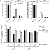
Acid-lability shift of FMDV VP3 A116V + VP1 N17D population after a single passage in the presence of NH4Cl. Acid sensitivity profiles of WT (a), VP3 A116V (b), VP1 N17D (c), and double mutant VP3 A116V + VP1 N17D (d) after a single amplification in the presence of NH4Cl. Viruses were grown in the presence or absence of 25 mM NH4Cl (MOI of 0.5 PFU/cell) for 8 h, and the acid sensitivity of the progeny populations was analyzed. For this purpose, equal number of PFU from each population were treated with PBS at pH 7.3, 6.6 and 6.3 for 30 min (abscissae). The pH was neutralized, and the remaining PFU in each sample was determined in BHK-21 cells. Infectivity was calculated as the percentage of PFU recovered at each different pH relative to that obtained at pH 7.3 (ordinate). Two tailed Student’s t-test P values between control and NH4Cl-treated populations were corrected for multiple comparisons using the Sidak–Bonferroni method (**P < 0.005). Data represent the means ± SDs (n = 3).
Figure 4
Figure 4
Viral populations carrying VP3 A116V and VP1 N17D replacements display increased resistance to NH4Cl but not to GuHCl. (a) Analysis of the resistance of FMDV WT and FMDV VP3 A116V + VP1 N17D to NH4Cl and GuHCl. BHK-21 cells treated or not with 25 mM NH4Cl or 4 mM GuHCl, were infected with FMDV WT or FMDV VP3 A116V VP1 N17D at a MOI of 0.5 PFU/cell. Virus yield at 8 h post-infection was determined by standard plaque assay. Two tailed Student’s t-test P values between control and drug-treated samples were corrected for multiple comparisons using the Sidak–Bonferroni method (*P < 0.05). Data represent the means ± SDs (n = 3–6). (b) Plaque assay of FMDV WT and FMDV VP3 A116V + VP1 N17D in the absence or presence of inhibitors. Viral progeny obtained from transfection of viral RNA from infectious clones was titrated in in semi-solidum medium in the absence of drug or the presence of 25 mM NH4Cl, 4 mM GuHCl. The number of lysis plaques produced in each condition was determined by crystal violet staining (30 h post-infection) and is expressed as the percentage of PFU developed in presence of the drug relative to that obtained in absence of the drug (n = 3–6). (c) Effect of MOI on the resistance against NH4Cl and GuHCl. Monolayers of BHK-21 cells treated or not with 25 mM NH4Cl, 4 mM GuHCl or no inhibitor (control) were infected with FMDV WT or FMDV VP3 A116V + VP1 N17D, at a MOI of 0.1, 0.01 or 0.001 PFU/cell. Virus yield produced in the presence of each drug (8 h post-infection) was determined by standard plaque assay and is expressed as a percentage of that obtained in the absence of inhibitors (control) (n = 3). Two tailed Student’s t-test P values between control and drug-treated samples were corrected for multiple comparisons using the Sidak–Bonferroni method (**P < 0.005). Data represent the means ± SDs.
Figure 5
Figure 5
Fitness gain of VP3 A116V + VP1 N17D mutant. (a, b) Increase in fitness conferred by VP3 A116V + VP1 N17D replacements both in presence and in absence of NH4Cl. Competition experiments between WT and double mutant VP3 A116V + VP1 N17D during serial passage of a virus mixture, in the absence (a) or in the presence of 25 mM NH4Cl (b). An initial MOI of 0.1 PFU/cell (0.05 PFU/cell for each virus) was used for each infection. Approximate percentage of competing genomes during serial passages was determined by sequencing the capsid coding cDNA. Passage 0 denote the initial mixture of the viruses. Data represent the means ± SDs (n = 3). (c) Rapid fixation of VP1 T22N replacement (nucleotide substitution C3272A) in VP3 A116V + VP1 N17D mutant passaged in presence of 25 mM NH4Cl. The percentage of genomes carrying nucleotide substitution C3272A was calculated in three independent passage series (Experiments 1, 2 and 3).

Similar articles

References

    1. Mateu MG. Assembly, stability and dynamics of virus capsids. Arch. Biochem. Biophys. 2013;531:65–79. doi: 10.1016/j.abb.2012.10.015. - DOI - PubMed
    1. Acharya R, et al. The three-dimensional structure of foot-and-mouth disease virus at 2.9 A resolution. Nature. 1989;337:709–716. doi: 10.1038/337709a0. - DOI - PubMed
    1. Newman JF, Rowlands DJ, Brown F. A physico-chemical sub-grouping of the mammalian picornaviruses. J. Gen. Virol. 1973;18:171–180. doi: 10.1099/0022-1317-18-2-171. - DOI - PubMed
    1. Grubman MJ, Baxt B. Foot-and-mouth disease. Clin. Microbiol. Rev. 2004;17:465–493. doi: 10.1128/CMR.17.2.465-493.2004. - DOI - PMC - PubMed
    1. Vazquez-Calvo A, Saiz JC, McCullough KC, Sobrino F, Martin-Acebes MA. Acid-dependent viral entry. Virus Res. 2012;20:20. - PubMed

Publication types

Substances