. 2021 Dec 6;11(1):23440.
doi: 10.1038/s41598-021-02898-5.

Peptides derived from gp43, the most antigenic protein from Paracoccidioides brasiliensis, form amyloid fibrils in vitro: implications for vaccine development

Affiliations

Peptides derived from gp43, the most antigenic protein from Paracoccidioides brasiliensis, form amyloid fibrils in vitro: implications for vaccine development

Thyago R Cardim-Pires et al. Sci Rep. .

Abstract

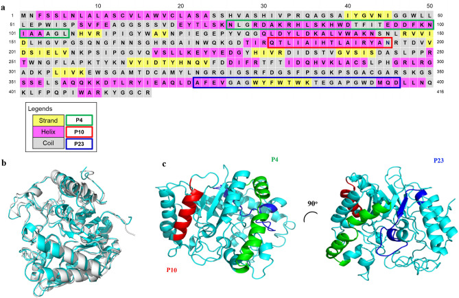
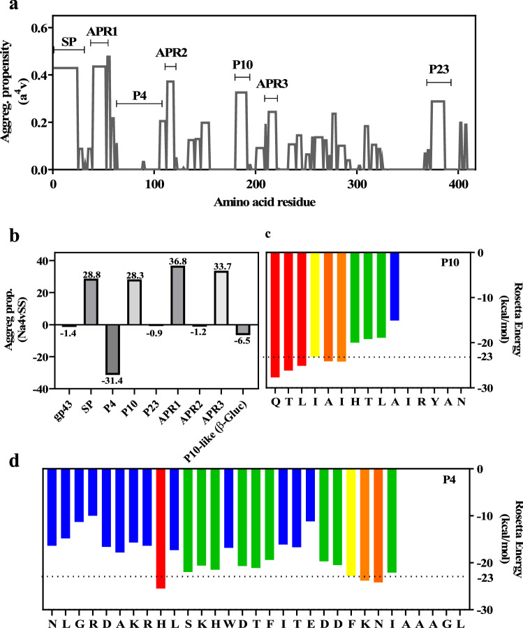
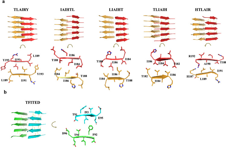
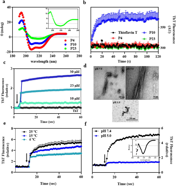
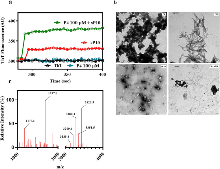
Fungal infection is an important health problem in Latin America, and in Brazil in particular. Paracoccidioides (mainly P. brasiliensis and P. lutzii) is responsible for paracoccidioidomycosis, a disease that affects mainly the lungs. The glycoprotein gp43 is involved in fungi adhesion to epithelial cells, which makes this protein an interesting target of study. A specific stretch of 15 amino acids that spans the region 181-195 (named P10) of gp43 is an important epitope of gp43 that is being envisioned as a vaccine candidate. Here we show that synthetic P10 forms typical amyloid aggregates in solution in very short times, a property that could hamper vaccine development. Seeds obtained by fragmentation of P10 fibrils were able to induce the aggregation of P4, but not P23, two other peptides derived from gp43. In silico analysis revealed several regions within the P10 sequence that can form amyloid with steric zipper architecture. Besides, in-silico proteolysis studies with gp43 revealed that aggregation-prone, P10-like peptides could be generated by several proteases, which suggests that P10 could be formed under physiological conditions. Considering our data in the context of a potential vaccine development, we redesigned the sequence of P10, maintaining the antigenic region (HTLAIR), but drastically reducing its aggregation propensity.


Conflict of interest statement

The authors declare no competing interests.

Figures

Figure 1
Figure 1
Primary sequence and predicted secondary structure elements of gp43 from Paracoccidioides brasiliensis strain B339 highlighting the three peptides studied here (P4, P10 and P23). (a) The primary structure was obtained at GenBank access number AAG36697.1. Secondary structure elements of gp43 were predicted by PSIPRED and are indicated by pink, yellow and grey cells where color refers to, respectively, α-helix, β-strand and random-coil structures. Green, red and blue outlines represent P4, P10 and P23 primary sequences, respectively. (b) The structural model of gp43 was generated by using AlphaFold software (cyan), which was aligned with the β-glucanase structure (gray). (c) Positions of P4, P10 and P23 in the structural model of gp43 are depicted in green, red and blue, respectively. Superposition of both structures in B was generated by PyMOL (The PyMOL Molecular Graphics System, Version 1.2r3pre, Schrödinger, LLC.).
Figure 2
Figure 2
Aggregation propensity analysis of entire gp43 and its derived peptides. (a) Aggrescan analysis of entire gp43 showing its aggregation propensity scores as a function of its primary sequence. SP: signal peptide (amino acids 1–35); APR1, 2 and 3: amyloid-prone regions 1, 2 and 3. Regions corresponding to P4, P10 and P23 are marked. (b) Aggrescan analysis of different segments of gp43 (SP, APR, P4, P10 and P23) as well as the corresponding sequence of P10 in β-glucanase from P. lutzii (QTLAAIRALANRYAK). (c and d) ZipperDB analysis of P10 and P4, respectively, showing the hexapeptides predicted to form steric zippers (Rosetta energy values bellow − 23 kcal/mol). Yellow, orange and red bars indicate the first residue of a hit hexapeptide (N to C terminal); green and blue bars indicate that the hexapeptide beginning at that residue does not fit into a steric zipper.
Figure 3
Figure 3
Hexapeptides derived from P10 and P4 (only a single stretch) form steric zippers in silico. P10 (a) and P4 (b) sequences were analyzed by Cordax (LOURO et al., 2020). The algorithm identified five hexapeptides in the sequence of P10 (TLIAIH, LIAIHT, IAIHTL, TLAIRY and HTLAIR) capable of forming steric zippers. Except for HTLAIR, each peptide forms two sheets (orange and red) composed of parallel β-strands (antiparallel in HTLAIR). In panel (a), the lower images show details of the interdigitation of the lateral chains of the amino acids in the steric zippers facing the interior of the bilayer. (b) Only one hexapeptide of P4 (TFITED; blue and green) adopts a steric zipper structure in an antiparallel fashion.
Figure 4
Figure 4
In solution, P10 undergoes aggregation forming amyloid fibrils. (a) Circular dichroism spectra of 100 μM of soluble P4, P10 and P23 in 5% TFE (see also the inset for more details of P23 secondary structure). (b) Kinetics of P10 aggregation (blue) at pH 7.4, 25 °C by measuring Thioflavin-T (ThT) binding. P4 and P23 do not form ThT-positive aggregates in solution under these conditions. In all experiments (except in panel 4C), the concentrations of peptides were 100 μM. (c) Aggregation of P10 (pH 7.4; 25 °C) shows concentration dependence. [P10] = 10, 25 and 50 μM, as indicated. (d) TEM images show the presence of mature amyloid fibrils composed of P10 only at pH 7.4 (25 °C), while at pH 5.0 amorphous aggregates predominate; the right-hand image at pH 7.4 is a zoom of the left-hand one. The inset of panel (f) shows the circular dichroism spectrum of the amyloid fibrils composed of P10, showing their β-sheet content. (e) Aggregation of P10 at pH 7.4 (100 μM) diminishes at low temperatures: Kinetics were performed at 4, 15 and 25 °C, as indicated. (f) Aggregation of P10 at 25 °C, 100 μM at pH 5.0 (blue) and 7.4 (black). The arrows in panels C, E and F indicate the moment when the peptides were added to the buffer. ThT binding was measured by exciting the samples at 450 nm and recording emission at 485 nm. Error bars in (b) and (e) are SD in three independent experiments. In (c), (e) and (f) the error bars are shorter than the size of the symbols.
Figure 5
Figure 5
Seeds composed of P10 (sP10) can seed P4 amyloidal aggregation. (a) P4 alone (blue, 100 μM) does not aggregate in solution unless P10 seeds are added (green; 5% seeds). In red is shown the ThT signal of the seeds alone in solution and in black is the ThT emission when free in solution. (b) TEM images of the amorphous aggregates formed by P4 alone (P4, upper left), P10 fibrils (P10, upper right), seeds of P10 (sP10; lower left) and aggregates formed when P4 is seeded by P10 (P4 + sP10; lower right). (c) Mass spectrometry analysis of P4 fibrils grown in the presence of P10 seeds showing the expected molecular masses of P10 (mass: 1,697) and P4 (mass: 3,426). These data suggest the incorporation of P4 into the P10 seeds giving rise to the appearance of hybrid fibrils. All experiments were performed at pH 7.4, 25 °C.

Similar articles

Cited by

References

    1. Cordova L. A., Torres J. Paracoccidioidomycosis in StatPearls. (StatPearls Publishing, 2021). - PubMed
    1. Sardi J, et al. In vitro Paracoccidioides brasiliensis biofilm and gene expression of adhesins and hydrolytic enzymes. Virulence. 2015;6:642–651. doi: 10.1080/21505594.2015.1031437. - DOI - PMC - PubMed
    1. Braz JD, et al. Gene expression of Paracoccidioides virulence factors after interaction with macrophages and fibroblasts. Mem. Inst. Oswaldo Cruz. 2021;26:e200592. doi: 10.1590/0074-02760200592. - DOI - PMC - PubMed
    1. Vicentini AP, et al. Binding of Paracoccidioides brasiliensis to laminin through surface glycoprotein gp43 leads to enhancement of fungal pathogenesis. Infect. Immun. 1994;62:1465–1469. doi: 10.1128/iai.62.4.1465-1469.1994. - DOI - PMC - PubMed
    1. Mendes-Giannini MJ, et al. Binding of extracellular matrix proteins to Paracoccidioides brasiliensis. Microbes Infect. 2006;8:1550–1559. doi: 10.1016/j.micinf.2006.01.012. - DOI - PubMed

Publication types

MeSH terms