. 2021 Dec 6;11(1):23476.
doi: 10.1038/s41598-021-02949-x.

Optimization in the expression of ASFV proteins for the development of subunit vaccines using poxviruses as delivery vectors

Affiliations

Optimization in the expression of ASFV proteins for the development of subunit vaccines using poxviruses as delivery vectors

Jaime Lopera-Madrid et al. Sci Rep. .

Abstract

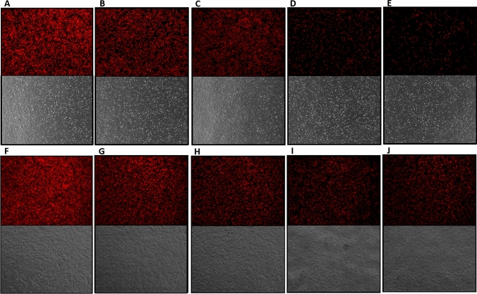
African swine fever virus (ASFV) causes a highly contagious hemorrhagic disease that affects domestic pig and Eurasian wild boar populations. To date, no safe and efficacious treatment or vaccine against ASF is available. Nevertheless, there are several reports of protection elicited by experimental vaccines based on live attenuated ASFV and some levels of protection and reduced viremia in other approaches such as DNA, adenovirus, baculovirus, and vaccinia-based vaccines. Current ASF subunit vaccine research focuses mainly on delivering protective antigens and antigen discovery within the ASFV genome. However, due to the complex nature of ASFV, expression vectors need to be optimized to improve their immunogenicity. Therefore, in the present study, we constructed several recombinant MVA vectors to evaluate the efficiency of different promoters and secretory signal sequences in the expression and immunogenicity of the p30 protein from ASFV. Overall, the natural poxvirus PrMVA13.5L promoter induced high levels of both p30 mRNA and specific anti-p30 antibodies in mice. In contrast, the synthetic PrS5E promoter and the S E/L promoter linked to a secretory signal showed lower mRNA levels and antibodies. These findings indicate that promoter selection may be as crucial as the antigen used to develop ASFV subunit vaccines using MVA as the delivery vector.


Conflict of interest statement

The authors declare no competing interests.

Figures

Figure 1
Figure 1
Generation and characterization of MVA-p30 viruses. Diagram of the MVA genome and DNA fragments used for homologous recombination to generate the recombinant MVA viruses. Hind III restriction endonuclease sites within the genome of MVA and the HA gene are indicated. Each expression cassette containing the p30 gene under the control of either the S E/L, pHyb, PrS5E, or PrMVA-13.5L promoters, and the S E/L promoter in frame with the C13L secretory signal are depicted. Also included in each cassette is the mCherry gene under the control of the p11 promoter and flanking regions to the HA gene, where the expression cassettes were inserted into the MVA genome.
Figure 2
Figure 2
Expression levels of p30 were analyzed at the mRNA level. DF-1 and Vero cells were infected with the indicated MVA-p30 virus, and after 24 h, total RNA was isolated and reverse transcribed. (A) indicates mRNAs from DF-1 cells and (B) from Vero cells. Fold p30 expression levels were calculated relative to the cells infected with MVA-GFP control and after normalization to the β-actin gene. Sample reactions, including MVA-GFP, were performed in triplicate and two independent repetitions. Statistically significant differences between MVA-p30 viruses are shown (P < 0.05, one-way ANOVA with Tukey’s multiple comparison test). For DF-1 cells: *P < 0.01; **P < 0.002; ***P < 0.0003. For Vero cells: **P < 0.0012; ***P < 0.0004; *****P < 0.0001.
Figure 3
Figure 3
Detection of p30 protein expression by immunoblotting assay. Monolayers of DF-1 and Vero cells were infected with each MVA-p30 virus at an MOI of 5, and then total protein extraction was performed at 24 h p.i., and extracted proteins were subjected to SDS-PAGE followed by western blot analysis. (A) indicates proteins from DF-1 cells and (B) from Vero cells. The order is as follows, MVA-GFP control (1), PrMVA-13.5 (2), pHyb (3), S E/L-C13L (4), PrS5E (5), and S E/L (6). A parallel blot incubated with a ß-actin specific monoclonal antibody served as loading control. Molecular masses of marker proteins are indicated. Full-length blots are presented in Supplementary Fig. 1.
Figure 4
Figure 4
Detection of p30 protein expression by immunofluorescence assay. Monolayers of DF-1 and Vero cells were infected with each MVA-p30 virus at an MOI of 1, and processed 24 h after infection. All images were acquired using a 10X objective and a green filter set at 70% intensity. Row A to E corresponds to DF-1 cells, and row F to J to Vero cells. Each column corresponds to: (A,F) MVA-PrMVA-13.5L-p30, (B,G) MVA-pHyb-p30, (C,H) MVA-S E/L-C13L-p30, (D,I) MVA-PrS5E-p30, and (E,J) MVA-S E/L-p30. Bright-field images are also included.
Figure 5
Figure 5
Comparison of fluorescence intensity of recombinant MVA viruses. DF-1 and Vero cells were infected with each recombinant virus at an MOI of 1, and at 24 h p.i., images were acquired using a 4 × objective and a Texas red filter set at 100% intensity. Row A to E corresponds to DF-1 cells, and row F to J to Vero cells. Each column corresponds to: (A,F) MVA-PrMVA-13.5L-p30, (B,G) MVA-pHyb-p30, (C,H) MVA-S E/L-C13L-p30, (D,I) MVA-PrS5E-p30, and (E,J) MVA-S E/L-p30. Bright-field images are also included.
Figure 6
Figure 6
Expression of mCherry mRNA. DF-1 and Vero cells were infected for 24 h with each recombinant virus. After total RNA was isolated and reverse transcribed, the fold expression of mCherry was calculated relative to the cells infected with MVA-GFP control and normalized to the β-actin gene. All reactions were performed in triplicate and two independent repetitions. Statistically significant differences between constructs are shown (P < 0.05, one-way ANOVA with Tukey’s multiple comparison test). For DF-1 cells: *P < 0.01; **P < 0.003; ***P < 0.001. For Vero cells: **P < 0.004; ***P < 0.0006; ****P < 0.0001.
Figure 7
Figure 7
Characterization of serum samples obtained from mice vaccinated with MVA-p30 constructs. BALB/c mice were primed and boosted (days 21 and 35) with either MVA-PrMVA-13.5L-p30, MVA-S E/L-C13L-p30, MVA-PrS5E-p30, or MVA-GFP. Purified p30 protein (baculovirus-expressed) were used as coating antigen for ELISA assays. Detection of specific anti-p30 antibodies at days 21, 28, and 42 p.i. for all animal groups are shown (n = 4 mice per group). Statistically significant differences between antibody titers are shown (P < 0.05, one-way ANOVA with Tukey’s multiple comparison test) *P < 0.05; **P < 0.005; ***P < 0.0005.

Similar articles

Cited by

References

    1. Parker J, Plowright W, Pierce MA. The epizootiology of African swine fever in Africa. Vet. Rec. 1969;85:668–674. - PubMed
    1. Anderson E, Hutchings G, Mukarati N, Wilkinson P. African swine fever virus infection of the bushpig (Potamochoerus porcus) and its significance in the epidemiology of the disease. Vet. Microbiol. 1998;62:1–15. doi: 10.1016/S0378-1135(98)00187-4. - DOI - PubMed
    1. Tulman ER, Delhon GA, Ku BK, Rock DL. Current Topics in Microbiology and Immunology. Springer; 2009. African swine fever virus; pp. 43–87. - PubMed
    1. Hamdy FM, Dardiri AH. Clinical and immunologic responses of pigs to African swine fever virus isolated from the Western Hemisphere. Am. J. Vet. Res. 1984;45:711–714. - PubMed
    1. Mebus CA, Dardiri AH. Western hemisphere isolates of African swine fever virus: Asymptomatic carriers and resistance to challenge inoculation. Am. J. Vet. Res. 1980;41:1867–1869. - PubMed

Publication types