Comparative Study
. 2021 Dec 6;11(1):23465.
doi: 10.1038/s41598-021-02972-y.

Comparative assessment of favipiravir and remdesivir against human coronavirus NL63 in molecular docking and cell culture models

Affiliations
Comparative Study

Comparative assessment of favipiravir and remdesivir against human coronavirus NL63 in molecular docking and cell culture models

Yining Wang et al. Sci Rep. .

Abstract

Human coronavirus NL63 (HCoV-NL63) mainly affects young children and immunocompromised patients, causing morbidity and mortality in a subset of patients. Since no specific treatment is available, this study aims to explore the anti-SARS-CoV-2 agents including favipiravir and remdesivir for treating HCoV-NL63 infection. We first successfully modelled the 3D structure of HCoV-NL63 RNA-dependent RNA polymerase (RdRp) based on the experimentally solved SARS-CoV-2 RdRp structure. Molecular docking indicated that favipiravir has similar binding affinities to SARS-CoV-2 and HCoV-NL63 RdRp with LibDock scores of 75 and 74, respectively. The LibDock scores of remdesivir to SARS-CoV-2 and HCoV-NL63 were 135 and 151, suggesting that remdesivir may have a higher affinity to HCoV-NL63 compared to SARS-CoV-2 RdRp. In cell culture models infected with HCoV-NL63, both favipiravir and remdesivir significantly inhibited viral replication and production of infectious viruses. Overall, remdesivir compared to favipiravir is more potent in inhibiting HCoV-NL63 in cell culture. Importantly, there is no evidence of resistance development upon long-term exposure to remdesivir. Furthermore, combining favipiravir or remdesivir with the clinically used antiviral cytokine interferon-alpha resulted in synergistic effects. These findings provided a proof-of-concept that anti-SARS-CoV-2 drugs, in particular remdesivir, have the potential to be repurposed for treating HCoV-NL63 infection.


Conflict of interest statement

The authors declare no competing interests.

Figures

Figure 1
Figure 1
Binding mode of favipiravir to the SARS-CoV-2 and HCoV-NL63 RdRp. (A) Favipiravir, bound to the SARS-CoV-2 RdRp (atom color ribbons) binding site, was depicted as surface representation with H-bond donor (purple) and acceptor (green). (B) Molecular interactions of favipiravir (atom color sticks) with the SARS-CoV-2 RdRp residues (yellow sticks). (C) 2D diagram of interactions between SARS-CoV-2 RdRp and favipiravir. (D) Favipiravir, bound to the HCoV-NL63 RdRp (atom color ribbons) binding site, was depicted as surface representation with H-bond donor (purple) and acceptor (green). (E) Molecular interactions of favipiravir (atom color sticks) with the HCoV-NL63 RdRp residues (yellow sticks). (F) 2D diagram of interactions between HCoV-NL63 RdRp and favipiravir.
Figure 2
Figure 2
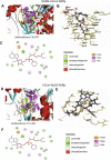
Binding mode of remdesivir to the SARS-CoV-2 and HCoV-NL63 RdRp. (A) Remdesivir, bound to the SARS-CoV-2 RdRp (atom color ribbons) binding site, was depicted as surface representation with H-bond donor (purple) and acceptor (green). (B) Molecular interactions of remdesivir (atom color sticks) and the SARS-CoV-2 RdRp residues (yellow sticks). (C) 2D diagram of interactions between SARS-CoV-2 RdRp and remdesivir. (D) Remdesivir, bound to the HCoV-NL63 RdRp (atom color ribbons) binding site, was depicted as surface representation with H-bond donor (purple) and acceptor (green). (E) Molecular interactions of remdesivir (atom color sticks) and the HCoV-NL63 RdRp residues (yellow sticks). (F) 2D diagram of interactions between HCoV-NL63 RdRp and remdesivir.
Figure 3
Figure 3
The root-mean-square deviations (RMSDs) of the backbone atoms and its ligands complex relative to their coordinates as a function of simulation time. (A) The average RMSDs of SARS-CoV-2 RdRp backbone atoms (Blue line) and its complexes with favipiravir (Green Line) or remdesivir (Red line) were plotted against the total simulation time. (B) The average RMSDs of HCoV-NL63 RdRp backbone atoms (Blue line) and its complexes with favipiravir (Green Line) or remdesivir (Red line) were plotted against the total simulation time.
Figure 4
Figure 4
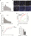
Antiviral effects of favipiravir against HCoV-NL63 in cell culture models. (A) Dose-dependent inhibition of HCoV-NL63 replication in LLC-MK2 cell line by favipiravir treatment. Intracellular viral RNA quantified by qRT-PCR was normalized to housekeeping gene GAPDH and presented relative to the control (CTR) (set as 1) (n = 6). (B) Immunofluorescence microscopy analysis of dsRNA, the intermediate of replicating HCoV-NL63 genomic RNA, upon treatment of different concentrations of favipiravir in LLC-MK2 and Caco-2 cell lines. Nuclei were visualized by DAPI (blue). (C) Dose-dependent inhibition of HCoV-NL63 replication by favipiravir in Caco-2 cell line (n = 6). (D) Caco-2 cells were infected with 0.1 MOI HCoV-NL63 and treated with different concentrations of favipiravir for 48 h. 50% effective concentration (EC50) curve was quantified by qRT-PCR, and 50% cytotoxic concentration (CC50) curve was determined by MTT assay. The left and right Y-axis of the graphs represent mean % inhibition of virus yield and cytotoxicity of the drug, respectively (n = 6–8). (E) Caco-2 cells were infected with 0.1 MOI HCoV-NL63, and then untreated or treated with 10 μM favipiravir for 5 days. Supernatant was collected every day to quantify secreted viruses by qRT-PCR, calculated as genomic copy numbers (n = 6). Standard curve for calculation of genomic copy numbers is included in Supplementary Fig. S3. (F) Caco-2 cells were infected with 0.1 MOI HCoV-NL63, and then untreated or treated with 1 or 100 μM favipiravir for 48 h. Virus titers from different groups was determined by TCID50 assay (n = 6). Data represent as mean ± SD. *P < 0.05; **P < 0.01; ***P < 0.001.
Figure 5
Figure 5
Antiviral effects of remdesivir against HCoV-NL63 in vitro. (A) Dose-dependent inhibition of HCoV-NL63 replication by remdesivir in LLC-MK2 cell line. Intracellular viral RNA quantified by qRT-PCR was normalized to housekeeping gene GAPDH and presented relative to the control (CTR) (set as 1) (n = 6–8). (B) Immunofluorescence microscopy analysis of dsRNA, the replicating HCoV-NL63 genomic RNA, upon treatment of different concentrations of remdesivir in LLC-MK2 and Caco-2 cell lines. Nuclei were visualized by DAPI (blue). (C) Dose-dependent inhibition of HCoV-NL63 replication by remdesivir on in Caco-2 cell line (n = 6). (D) Caco-2 cells were infected with 0.1 MOI HCoV-NL63 and treated with different concentrations of remdesivir for 48 h. 50% effective concentration (EC50) curve was quantified by qRT-PCR, and 50% cytotoxic concentration (CC50) curve was determined by MTT assay. The left and right Y-axis of the graphs represent mean % inhibition of virus yield and cytotoxicity of the drug, respectively (n = 6–8). (E) Caco-2 cells were infected with 0.1 MOI HCoV-NL63, and then untreated or treated with 1 μM remdesivir for 5 days. Supernatant was collected every day to quantify secreted viruses by qRT-PCR and calculated as genomic copy numbers (n = 6). Standard curve for calculation of genomic copy numbers is included in Supplementary Fig. S3. (F) Caco-2 cells were infected with HCoV-NL63 at an MOI of 0.1, then untreated or treated with 0.3 or 3 μM remdesivir for 48 h. Virus titers from different groups was determined by TCID50 assay (n = 6). Data represent as mean ± SD. *P < 0.05; **P < 0.01; ***P < 0.001. (G) Comparing the inhibitory potency of favipiravir and remdesivir in Caco-2 cells infected with HCoV-NL63. (H) HCoV-NL63 was serially passaged in Caco-2 cells exposed to no remdesivir (as control) or increasing concentrations of remdesivir for 20 passages. 1 μM remdesivir was used in passage 1–10, which was increased to 2 μM at the subsequent passages. The effect of remdesivir (1 μM) on HCoV-NL63 harvested at passage 5, 10, 15 and 20 was quantified using qRT-PCR. Data represent as mean ± SD. *P < 0.05; **P < 0.01; ***P < 0.001.
Figure 6
Figure 6
The effects of combining antivirals in Caco-2 cells infected with HCoV-NL63. The antiviral effects of favipiravir in combination with remdesivir (A), or IFN-α in combination with favipiravir (B) or remdesivir (C) respectively. The results were analyzed by the MacSynergyII model. The three-dimensional surface plot represents the differences (within 95% confidence interval) between actual experimental effects and theoretical additive effects of the combination at various concentrations (n = 4).

Similar articles

Cited by

References

    1. Li P, Liu J, Ma Z, Bramer WM, Peppelenbosch MP, Pan Q. Estimating global epidemiology of low-pathogenic human coronaviruses in relation to the COVID-19 context. J. Infect. Dis. 2020;222(4):695–696. doi: 10.1093/infdis/jiaa321. - DOI - PMC - PubMed
    1. Ma Z, Liu J, Pan Q. Overwhelming COVID-19 clinical trials: Call for prospective meta-analyses. Trends Pharmacol. Sci. 2020;41(8):501–503. doi: 10.1016/j.tips.2020.05.002. - DOI - PMC - PubMed
    1. Li P, Ikram A, Peppelenbosch MP, Ma Z, Pan Q. Systematically mapping clinical features of infections with classical endemic human coronaviruses. Clin. Infect. Dis. 2020;73:554–555. doi: 10.1093/cid/ciaa1386. - DOI - PMC - PubMed
    1. da Veiga ABG, Martins LG, Riediger I, Mazetto A, Debur MD, Gregianini TS. More than just a common cold: Endemic coronaviruses OC43, HKU1, NL63, and 229E associated with severe acute respiratory infection and fatality cases among healthy adults. J. Med. Virol. 2020;93:1002–1007. doi: 10.1002/jmv.26362. - DOI - PubMed
    1. van der Hoek L, Pyrc K, Jebbink MF, et al. Identification of a new human coronavirus. Nat. Med. 2004;10(4):368–373. doi: 10.1038/nm1024. - DOI - PMC - PubMed

Publication types

MeSH terms