Review
. 2022 Jun;23(6):760-771.
doi: 10.1111/mpp.13170. Epub 2021 Dec 6.

Extracellular vesicles: Their functions in plant-pathogen interactions

Affiliations
Review

Extracellular vesicles: Their functions in plant-pathogen interactions

Qingfeng Zhou et al. Mol Plant Pathol. 2022 Jun.

Abstract

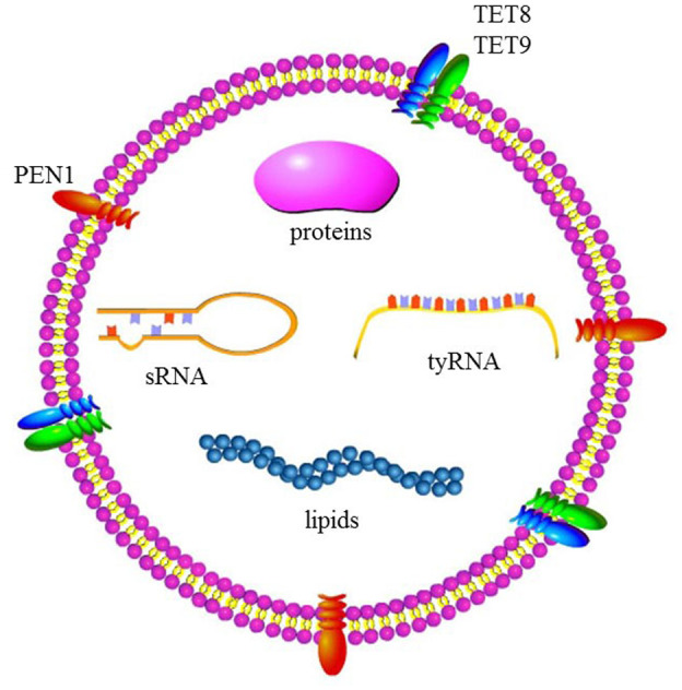
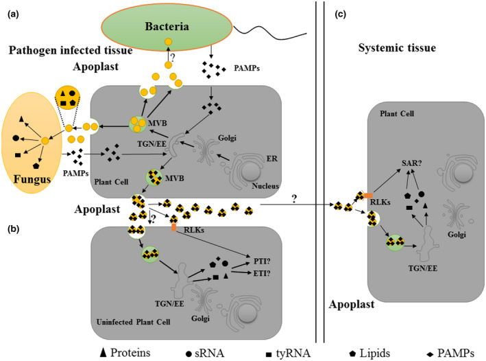
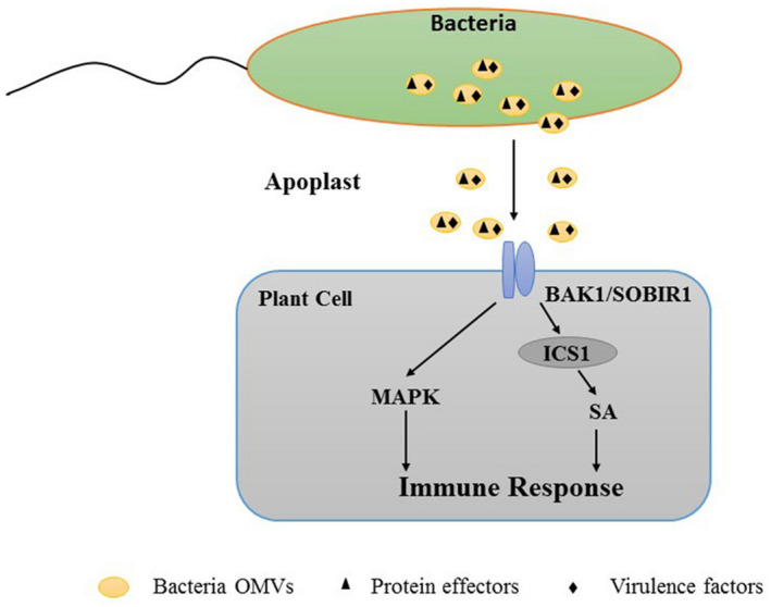
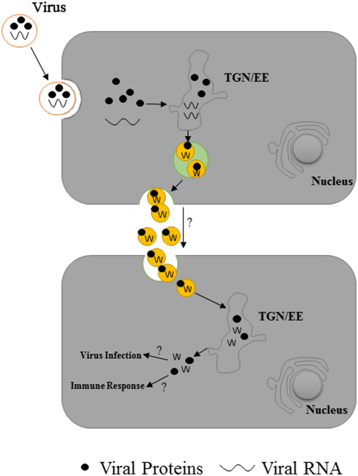
Extracellular vesicles (EVs) are rounded vesicles enclosed by a lipid bilayer membrane, released by eukaryotic cells and by bacteria. They carry various types of bioactive substances, including nucleic acids, proteins, and lipids. Depending on their cargo, EVs have a variety of well-studied functions in mammalian systems, including cell-to-cell communication, cancer progression, and pathogenesis. In contrast, EVs in plant cells (which have rigid walls) have received very little research attention for many decades. Increasing evidence during the past decade indicates that both plant cells and plant pathogens are able to produce and secrete EVs, and that such EVs play key roles in plant-pathogen interactions. Plant EVs contains small RNAs (sRNAs) and defence-related proteins, and may be taken up by pathogenic fungi, resulting in reduced virulence. On the other hand, EVs released by gram-negative bacteria contain a wide variety of effectors and small molecules capable of activating plant immune responses via pattern-recognition receptor- and BRI1-ASSOCIATED RECEPTOR KINASE- and SUPPRESSOR OF BIR1-mediated signalling pathways, and salicylic acid-dependent and -independent processes. The roles of EVs in plant-pathogen interactions are summarized in this review, with emphasis on important molecules (sRNAs, proteins) present in plant EVs.

Keywords: effector-triggered immunity; exosome; extracellular vesicles; pathogen-associated molecular patterns-triggered immunity; plant; plant disease; systemic acquired resistance.


Figures

FIGURE 1
FIGURE 1
Cargo of plant extracellular vesicles (EVs). PEN1, plant‐specific penetration 1; sRNA, small RNA; TET, tetraspanin; tyRNA, tiny RNA
FIGURE 2
FIGURE 2
Function of plant extracellular vesicles (EVs) in plant–microbe interactions. (a) Plant EVs can be taken up by pathogens and can inhibit the virulence of pathogens by releasing their cargos (small RNAs [sRNA], proteins, tiny RNAs [tyRNA], and lipids). Meanwhile, pathogen‐associated molecular patterns (PAMPs) from pathogens can be loaded into plant EVs in pathogen‐infected cells. (b) Plant EVs may be functionally related to the signal transmission of PAMP‐triggered immunity (PTI) and effector‐triggered immunity (ETI). First, PAMPs in plant EVs released by infected cells are recognized by receptor‐like kinases (RLKs) located on the membrane to activate PTI in adjacent uninfected cells. Second, plant EVs contain abundant proteins involved in PTI and ETI. When plant cells perceive pathogens, the contents of plant EVs secreted by these cells may change. These EVs can be taken up by adjacent pathogen‐free cells and induce them to produce PTI and ETI downstream signals. (c) Plant EVs may be functionally related to systemic acquired resistance (SAR) signal transmission. Plant EVs contain SAR‐related proteins and PAMPs. During SAR, EVs released by pathogen‐infected cells may be transported to systemic tissues via the apoplast to transmit SAR signals when they are taken up by target cells or induce an immune response by the recognition of PAMPs and RLKs in systemic tissues. ER, endoplasmic reticulum; MVB, multivesicular bodies endosomes; TGN/EE, trans‐Golgi network/early endosome
FIGURE 3
FIGURE 3
Bacterial outer membrane vesicles (OMVs), which contain protein effectors, can elicit plant protective immune responses. The immune co‐receptors BAK1 and SOBIR1 mediate the perception of, and the response to, OMVs. The OMVs induce immune responses via salicylic acid (SA)‐dependent/‐independent processes and MAPK pathways
FIGURE 4
FIGURE 4
Plant extracellular vesicles (EVs) are involved in the virus‐induced immune response. Plant EVs released by virus‐infected cells contain viral proteins and RNA. These EVs may facilitate the spread of viruses from infected cells to adjacent cells, or act as pathogen‐associated molecular pattern (PAMP) carriers to induce plant immune responses

References

    1. Akers, J.C. , Gonda, D. , Kim, R. , Carter, B.S. & Chen, C.C. (2013) Biogenesis of extracellular vesicles (EV): exosomes, microvesicles, retrovirus‐like vesicles, and apoptotic bodies. Journal of Neuro‐Oncology, 113, 1–11. - PMC - PubMed
    1. An, Q. , Ehlers, K. , Kogel, K.H. , van Bel, A.J. & Hückelhoven, R. (2006) Multivesicular compartments proliferate in susceptible and resistant MLA12‐barley leaves in response to infection by the biotrophic powdery mildew fungus. New Phytologist, 172, 563–576. - PubMed
    1. Anand, P.K. , Anand, E. , Bleck, C.K. , Anes, E. & Griffiths, G. (2010) Exosomal Hsp70 induces a pro‐inflammatory response to foreign particles including mycobacteria. PLoS One, 5, e10136. - PMC - PubMed
    1. Bahar, O. , Mordukhovich, G. , Luu, D.D. , Schwessinger, B. , Daudi, A. , Jehle, A.K. et al. (2016) Bacterial outer membrane vesicles induce plant immune responses. Molecular Plant‐Microbe Interaction, 29, 374–384. - PubMed
    1. Baldrich, P. , Rutter, B.D. , Karimi, H.Z. , Podicheti, R. , Meyers, B.C. & Innes, R.W. (2019) Plant extracellular vesicles contain diverse small RNA species and are enriched in 10‐ to 17‐nucleotide "tiny" RNAs. The Plant Cell, 31, 315–324. - PMC - PubMed

Publication types