Review
. 2021 Dec 1;14(12):dmm048785.
doi: 10.1242/dmm.048785. Epub 2021 Dec 7.

Current approaches and advances in the imaging of stroke

Affiliations
Review

Current approaches and advances in the imaging of stroke

Pragati Kakkar et al. Dis Model Mech. .

Abstract

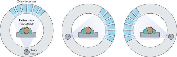
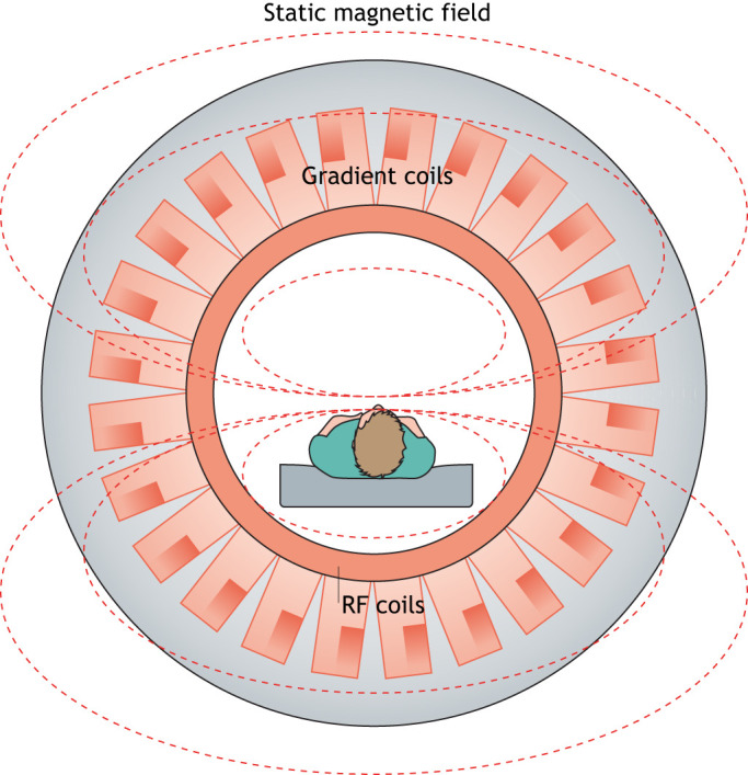
A stroke occurs when the blood flow to the brain is suddenly interrupted, depriving brain cells of oxygen and glucose and leading to further cell death. Neuroimaging techniques, such as computed tomography and magnetic resonance imaging, have greatly improved our ability to visualise brain structures and are routinely used to diagnose the affected vascular region of a stroke patient's brain and to inform decisions about clinical care. Currently, these multimodal imaging techniques are the backbone of the clinical management of stroke patients and have immensely improved our ability to visualise brain structures. Here, we review recent developments in the field of neuroimaging and discuss how different imaging techniques are used in the diagnosis, prognosis and treatment of stroke.

Keywords: Computed tomography; Haemorrhagic stroke; Ischaemic stroke; Magnetic resonance imaging; Neuroimaging; Stroke.


Conflict of interest statement

Competing interests The authors declare no competing or financial interests.

Figures

Fig. 1.
Fig. 1.
Schematic of computed tomography (CT) scan. During a CT scan, a patient lies on a flat surface, which passes through the scanner. The scanner consists of a rotating ring of X-ray sources and acquisition detectors. The acquisition system acquires X-rays from different angles of the brain and constructs the resulting cross-sectional, sliced images based on the tissue density calculated with the help of mathematical analysis (Sanelli et al., 2015).
Fig. 2.
Fig. 2.
Schematic of magnetic resonance (MR) scan. A magnetic field generated by a magnet (grey) from the MR scanner aligns hydrogen nuclei (protons), which are usually randomly oriented, along its direction. This alignment results in the longitudinal magnetisation of the tissue. A radio frequency (RF) coil produces a brief signal (90° to the magnetic field) to flip the aligned spin of the protons, resulting in transverse magnetisation. When the RF signal is turned off, the spins of the protons relax back to their original states to realign with the static magnetic field. The longitudinal magnetisation returns to its original value and transverse magnetisation decays. During this relaxation process, the protons emit energy at the same RF, which is detected by the receiver coil to generate an image. The gradient coil assists in generating variations in the static magnetic field and the direction of the gradient (Zhang et al., 2016; Rastogi et al., 2015).
Fig. 3.
Fig. 3.
CT and MRI images showing intracerebral haemorrhage. (A-C) Images generated from CT (A), T2-weighted MRI (B) and GRE-MRI (C) scans, showing an intracerebral haemorrhage (horizontal arrows) at 2 h (CT) and 4.5 h (MRI) from symptom onset in an adult human. The vertical arrow in the GRE-MRI scan depicts the artefact caused by magnetic field inhomogeneity at the CSF/bone/air interface. Images reproduced with permission from Butcher and Davis (2009). These images are not published under the terms of the CC-BY license of this article. For permission to reuse, please see Butcher and Davis (2009). CSF, cerebrospinal fluid; CT, computed tomography; GRE, gradient recalled echo; MRI, magnetic resonance imaging.
Fig. 4.
Fig. 4.
CT versus MRI scans for detecting stroke. (A,B) A haemorrhage can be seen clearly in the CT scan of a patient (A; white arrow), whereas it is less evident in an MRI scan (B; white arrow). (C,D) Whereas an ischaemic infarct is only faintly visible in the CT scan of a patient (C; blue arrow), it is clearly visible in an MRI scan (D; blue arrow). Images reproduced from Heit et al. (2017) and Rajdev et al. (2020) under the terms of the CC-BY 4.0 license. CT, computed tomography; MRI, magnetic resonance imaging.
Fig. 5.
Fig. 5.
Effect of contrast agent. (A,B) T1-weighted MRI of an adult brain showing infarction (white arrows) without contrast agent (A) and with contrast agent (B). Images reproduced with permission from Friebe (2016). These images are not published under the terms of the CC-BY license of this article. For permission to reuse, please see Friebe (2016).

Similar articles

Cited by

References

    1. Abedi, V., Goyal, N., Tsivgoulis, G., Hosseinichimeh, N., Hontecillas, R., Bassaganya-Riera, J., Elijovich, L., Metter, J. E., Alexandrov, A. W. and Liebeskind, D. S. (2017). Novel screening tool for stroke using artificial neural network. Stroke 48, 1678-1681. 10.1161/STROKEAHA.117.017033 - DOI - PubMed
    1. Albers, G. W., Thijs, V. N., Wechsler, L., Kemp, S., Schlaug, G., Skalabrin, E., Bammer, R., Kakuda, W., Lansberg, M. G. and Shuaib, A. (2006). Magnetic resonance imaging profiles predict clinical response to early reperfusion: the diffusion and perfusion imaging evaluation for understanding stroke evolution (DEFUSE) study. Ann. Neurol. 60, 508-517. 10.1002/ana.20976 - DOI - PubMed
    1. Arab, A., Chinda, B., Medvedev, G., Siu, W., Guo, H., Gu, T., Moreno, S., Hamarneh, G., Ester, M. and Song, X. (2020). A fast and fully-automated deep-learning approach for accurate hemorrhage segmentation and volume quantification in non-contrast whole-head CT. Sci. Rep. 10, 1-12. 10.1038/s41598-020-76459-7 - DOI - PMC - PubMed
    1. Arba, F., Rinaldi, C., Caimano, D., Vit, F., Busto, G. and Fainardi, E. (2020). Blood–brain barrier disruption and hemorrhagic transformation in acute ischemic stroke: systematic review and meta-analysis. Front. Neurol. 11, 594613. 10.3389/fneur.2020.594613 - DOI - PMC - PubMed
    1. Asadi, H., Dowling, R., Yan, B. and Mitchell, P. (2014). Machine learning for outcome prediction of acute ischemic stroke post intra-arterial therapy. PLoS ONE 9, e88225. 10.1371/journal.pone.0088225 - DOI - PMC - PubMed