Review
. 2021 Dec 7;8(1):19.
doi: 10.1186/s40348-021-00132-w.

Imaging of peripheral vascular malformations - current concepts and future perspectives

Affiliations
Review

Imaging of peripheral vascular malformations - current concepts and future perspectives

Vanessa F Schmidt et al. Mol Cell Pediatr. .

Abstract

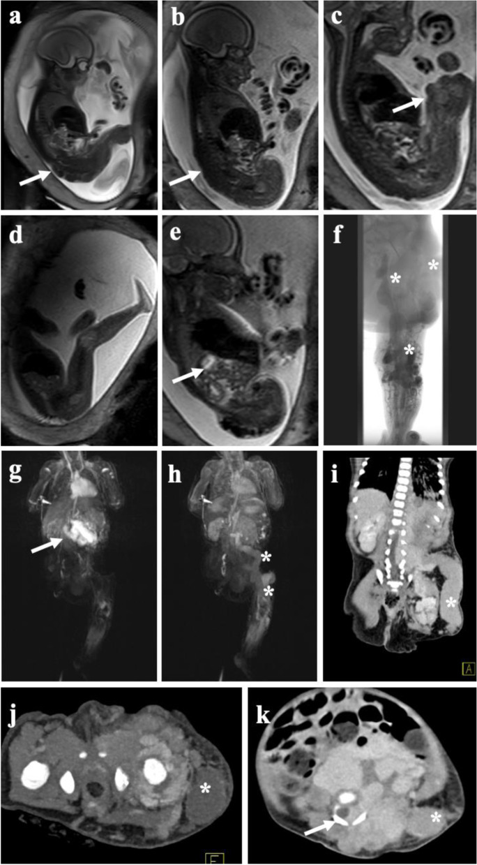
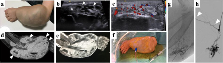
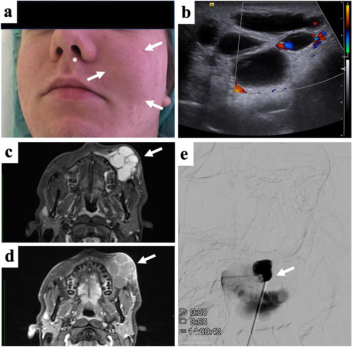
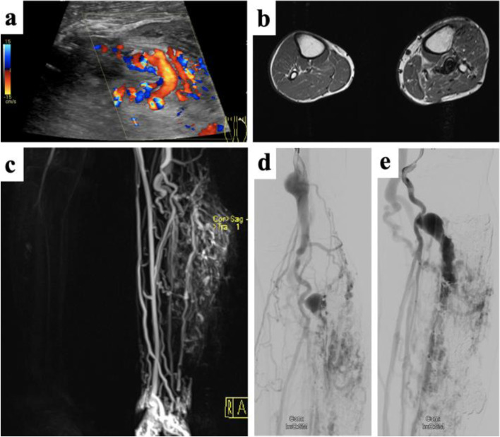
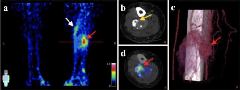
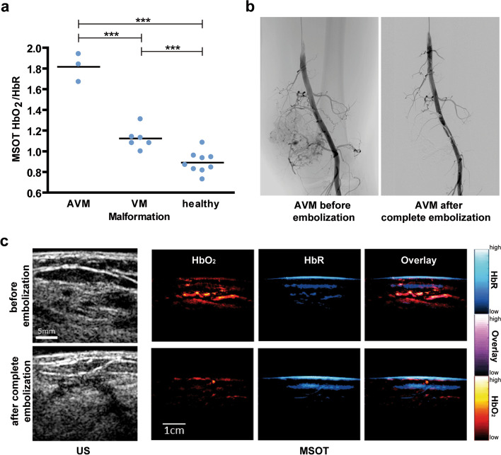
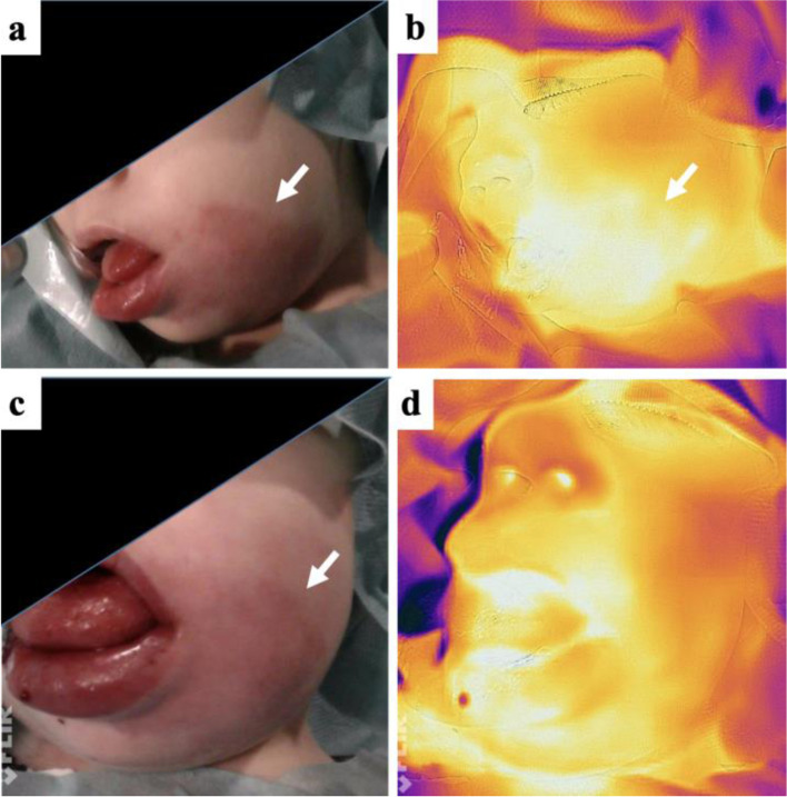
Vascular Malformations belong to the spectrum of orphan diseases and can involve all segments of the vascular tree: arteries, capillaries, and veins, and similarly the lymphatic vasculature. The classification according to the International Society for the Study of Vascular Anomalies (ISSVA) is of major importance to guide proper treatment. Imaging plays a crucial role to classify vascular malformations according to their dominant vessel type, anatomical extension, and flow pattern. Several imaging concepts including color-coded Duplex ultrasound/contrast-enhanced ultrasound (CDUS/CEUS), 4D computed tomography angiography (CTA), magnetic resonance imaging (MRI) including dynamic contrast-enhanced MR-angiography (DCE-MRA), and conventional arterial and venous angiography are established in the current clinical routine. Besides the very heterogenous phenotypes of vascular malformations, molecular and genetic profiling has recently offered an advanced understanding of the pathogenesis and progression of these lesions. As distinct molecular subtypes may be suitable for targeted therapies, capturing certain patterns by means of molecular imaging could enhance non-invasive diagnostics of vascular malformations. This review provides an overview of subtype-specific imaging and established imaging modalities, as well as future perspectives of novel functional and molecular imaging approaches. We highlight recent pioneering imaging studies including thermography, positron emission tomography (PET), and multispectral optoacoustic tomography (MSOT), which have successfully targeted specific biomarkers of vascular malformations.

Keywords: Duplex ultrasound; Magnetic resonance imaging; Molecular imaging; Multispectral optoacoustic tomography; Positron emission tomography; Thermography; Vascular malformations.


Conflict of interest statement

The authors declare that they have no competing interests.

Figures

Fig. 1
Fig. 1
Fetal MRI and post-partum MRI/CT of complex vascular malformation. Fetal MRI using dedicated T2-weighted sequences reveal extensive venous malformation along the entire left leg (a) extending along the pelvis and lower back (b). c Large venous channel (arrow) raised suspicion of a large persistent marginal vein. d Healthy right leg in comparison. e Extension of the malformation in the retroperitoneal space. f Post-natal phlebography depicting large marginal vein together with extensive venous malformation of the lower and upper leg with extension into the pelvis (asterisk). Large blood volumes trapped in the marginal vein induced a localized consumptive coagulopathy in the child requiring immediate anticoagulation upon delivery. g Postnatal MRI revealing early arterial filling of an additional mesenteric arteriovenous malformation in the same patient, being asymptomatic at time of diagnosis. The course of the marginal vein, as depicted on MRI (h) and CT (i) showing large venous drainage into the pelvis (asterisk). j Transverse CT image in the arterial phase demonstrating multiple AV fistula paraspinal and along the left hip joint (arrows) together with the not yet contrasted marginal vein (asterisk). k Venous phase CT depicting the extension of intraabdominal, retroperitoneal, and paraspinal venous cavities with infiltration of the spinal canal (arrow) by the large slow-flow malformation, at time with contrast in the large marginal vein (asterisk). The large venous malformations required multiple rounds of image-guided embolization followed by stepwise surgery of the marginal vein
Fig. 2
Fig. 2
Venous Malformation a four-year-old child of the forearm with bone disfigurement. a Clinical presentation shows large, non-pulsatile painless soft tissue mass with bluish discoloration and visible dysplastic veins. Wrist function was restricted but finger motility was not significantly impaired. The heavy weight of the arm made the boy carry his left arm with the right hand. b B-Mode ultrasound imaging shows cavernous dysplastic veins (white arrowhead) and thrombosed parts (white asterisks) of the lesion. c Color-coded Duplex ultrasound reveals slow blood flow within the dysplastic vessels. d STIR MRI imaging shows typical hyperintense signal with multiple hypointense spots representing phleboliths (white arrowheads). T1-weighted MRI imaging after contrast administration shows enhancement of the large venous cavities of the VM (e). Venous malformations are treated by percutaneous sclerotherapy. The periprocedural image (f) shows confirmation of blood aspiration after percutaneous access to the dysplastic vessels, before using a contrast agent and a sclerosing agent subsequently. g Angiographic depiction of VM after contrast agent administration via percutaneous access displays a fine reticular network of dysplastic vessels and drainage to deep veins (h) not visible on MRI
Fig. 3
Fig. 3
Subcutaneous Lymphatic malformation of the face in an adolescent. a Clinical photograph showing local swelling and moderate discoloration of the cheek (white arrows) and local deviation of the left ala of the nose (asterisk). The patient was suffered from esthetic disfigurement with the mass itself being painless. b Color-coded Duplex ultrasound documents the absence of intralesional flow, only focal vascularization along the septa. c Fat-saturated STIR MRI shows fluid-filled cavities in the subcutaneous tissue of the mixed-type lymphatic malformation (arrow). d Following contrast administration only septal structures show moderate enhancement (arrow). e Percutaneous contrast administration before sclerotherapy reveals partial communication between adjacent lymphatic cysts. Sclerotherapy resulted in subtotal regression of the mass
Fig. 4
Fig. 4
Arteriovenous malformation of the lower limb with associated soft-tissue proliferation. a Color-coded Duplex ultrasound reveals vigorous flow along the AVM nidus. b Cross-sectional MRI reveals multiple flow-voids as sign of arterialized flow patterns together with soft-tissue proliferation adjacent to the AVM. The mass was clinically pulsatile without ulceration at the time of clinical presentation. c Dynamic MR angiography shows rapid filling of the AVM along the left lower limb at time with only initial enhancement of the arteries of the non-affected leg. Pre-embolization angiogram in the late arterial phase (d) and early venous phase (e) depicting dilated aneurysms along the venous outflow tract
Fig. 5
Fig. 5
68Ga-RGB PET/CT in 38-year-old man with right-leg AVM. Maximal-intensity 68Ga-RGD PET projection (a), CTA (b) and 68Ga-RGD PET/CT (d), and 3D reconstruction of 4D-CTA (c) show nidus (SUVmax 3.2; SUVpeak 2.5, red arrows). Axial plane in (b) and (d) is at location of line in (a). Heterogeneous pattern of enhanced uptake is seen in tissue adjacent to nidus at more proximal part of the right leg (white arrow); bone deformation in the fibula caused by compression and infiltration of vessels is also seen (yellow arrow), along with large venous aneurysm (asterisk). Arterial flow with nidus and fistula is seen in (c), along with upcoming venous flow and the large venous aneurysm. This research was originally published in JNM. Lobeek D, Bouwman FCM, Aarntzen EHJG, Molkenboer-Kuenen JDM, Flucke UE, Nguyen HL, Vikkula M, Boon LM, Klein W, Laverman P, Oyen WJG, Boerman OC, Terry SYA, Schultze Kool LJ, Rijpkema M. A Clinical Feasibility Study to Image Angiogenesis in Patients with Arteriovenous Malformations Using 68Ga-RGD PET/CT. J Nucl Med 2020; 61:270–275.© SNMMI
Fig. 6
Fig. 6
Multispectral optoacoustic tomography (MSOT) of vascular malformations. a Novel hybrid MSOT/US technique was shown to enable distinction of arteriovenous (AVM) from venous (VM) malformations as well as from healthy tissue by non-invasive, contrast-free, and radiation-free imaging of the lesions at the patients’ bedside. Further, as exemplarily shown for a patient receiving complete embolization of an AVM (b), MSOT/US depicted therapy response by showing a reduced signal of oxygenated and deoxygenated hemoglobin (c). ***p < 0.001. Figure adapted from Masthoff M, Helfen A, Claussen J, Karlas A, Markwardt NA, Ntziachristos V, Eisenblätter M, Wildgruber M. Use of Multispectral Optoacoustic Tomography to Diagnose Vascular Malformations. JAMA Dermatol. 2018 Dec 1;154(12):1457–1462. doi: 10.1001/jamadermatol.2018.3269. PMID: 30267083; PMCID: PMC6583374© 2018, American Medical Association
Fig. 7
Fig. 7
Infrared thermography of an arteriovenous malformation of the face. a The patient (m, age: 3 years) suffers from an AVM of the left cheek and lower lip. Pink discoloration of the skin is characteristic of the arterial component in this vascular malformation. b Infrared thermography before embolization. Note the locally increased temperature, color-coded as white areas. c Clinical presentation and d infrared thermography immediately after successful embolization. The locally increased temperature is gone. Note a local swelling, induced by the embolization disappearing after a few weeks. AVM is marked with arrow, a more whitish color indicating elevated temperature

References

    1. Mulligan PR, Prajapati HJ, Martin LG, Patel TH. Vascular anomalies: classification, imaging characteristics and implications for interventional radiology treatment approaches. Br J Radiol. 2014;87(1035):20130392. doi: 10.1259/bjr.20130392. - DOI - PMC - PubMed
    1. Greene AK, Liu AS, Mulliken JB, Chalache K, Fishman SJ. Vascular anomalies in 5,621 patients: guidelines for referral. J Pediatr Surg. 2011;46(9):1784–1789. doi: 10.1016/j.jpedsurg.2011.05.006. - DOI - PubMed
    1. Mulliken JB, Glowacki J. Hemangiomas and vascular malformations in infants and children: a classification based on endothelial characteristics. Plast Reconstr Surg. 1982;69(3):412–422. doi: 10.1097/00006534-198203000-00002. - DOI - PubMed
    1. Mulliken JB, Glowacki J. Classification of pediatric vascular lesions. Plast Reconstr Surg. 1982;70(1):120–121. doi: 10.1097/00006534-198207000-00041. - DOI - PubMed
    1. ISSVA Classification of Vascular Anomalies @2018 International Society for the Study of Vascular Anomalies. Available at: issva.org/classification. Accessed 16 April 2021