

Caveolin-1 promotes cancer progression via inhibiting ferroptosis in head and neck squamous cell carcinoma
- PMID: 34874578
- PMCID: PMC9300096
- DOI: 10.1111/jop.13267
Caveolin-1 promotes cancer progression via inhibiting ferroptosis in head and neck squamous cell carcinoma
Abstract
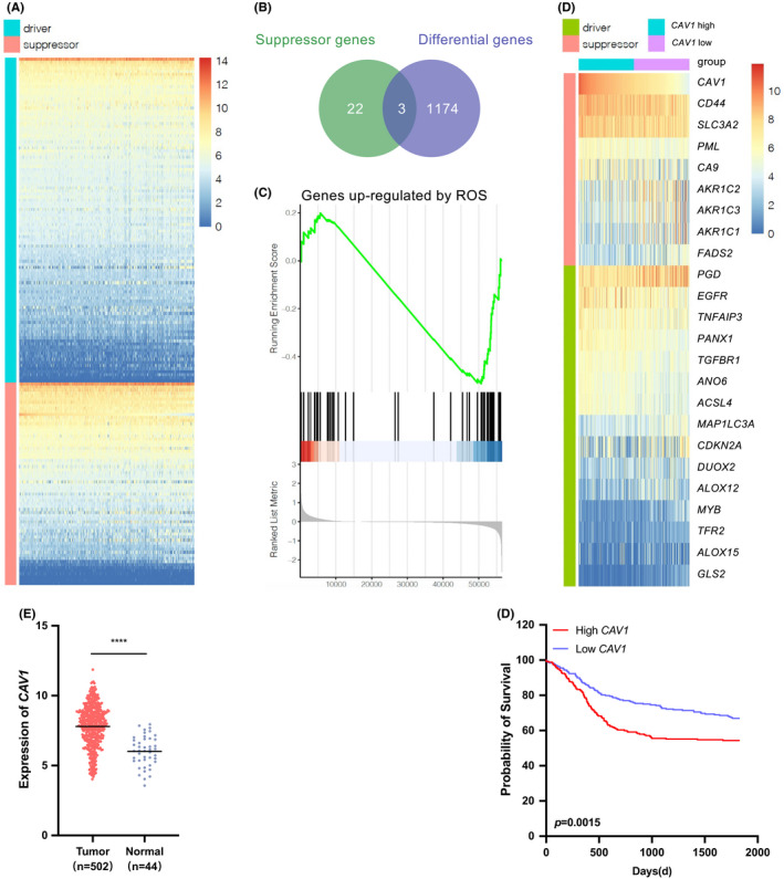
Background: Head and neck squamous cell carcinoma (HNSCC) is an aggressive disease worldwide. Much progress has been made in exploring mechanisms and improving the therapy of HNSCC, but only a few studies have focused on the role of ferroptosis on HNSCC progression. The current study aimed to reveal the underlining mechanisms that caveolin-1 (CAV1)-ROS (reactive oxygen species)-ferroptosis axis affect the process of HNSCC and discover novo therapeutic targets or strategies.
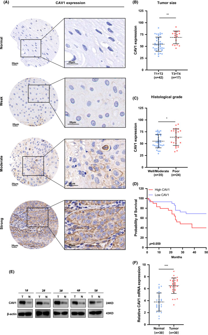
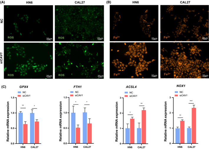
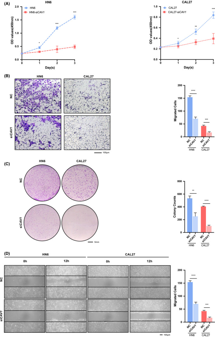
Methods: The role of CAV1 in ferroptosis was analyzed by FerrDb, and its clinical significance was examined by TCGA dataset of HNSCC. The expressions of caveolin-1 (CAV1) in HNSCC tissues were measured by immunohistochemistry, western blot, and real-time PCR assay. Three siRNA sequences were designed to silence CAV1 mRNA in HNSCC cells. Cell proliferation, colony formation, wound-healing, and transwell assays were used to examine the proliferation, migration, and invasion of cancer cells. ROS evaluation and intracellular Fe2+ content assays were performed to examine the levels of ferroptosis.
Results: Through the analysis with published data, CAV1 was found to overexpress in HNSCC than normal tissues, and was one of the vital suppressors of ferroptosis pathway. Our study showed that CAV1 was over expressed in HNSCC tissues and the high level of CAV1 predicted poorer prognosis. Further experiments indicated that CAV1 could inhibit the ferroptosis of cancer cells and promote the proliferation, migration and invasion.
Conclusions: Overexpression of CAV1 in HNSCC inhibited the process of ferroptosis, leading to aggressive phenotypes, as well as worse prognosis. The regulatory pathway of CAV1 and ferroptosis are potential targets for designing diagnostic and combined therapeutic strategies for HNSCC patients.
Keywords: caveolin-1; ferroptosis; head and neck; squamous cell carcinoma.
© 2021 The Authors. Journal of Oral Pathology & Medicine published by John Wiley & Sons Ltd.
Conflict of interest statement
The authors declare no competing interests.
Figures
Similar articles
-
SENP1 inhibits ferroptosis and promotes head and neck squamous cell carcinoma by regulating ACSL4 protein stability via SUMO1.Oncol Rep. 2024 Feb;51(2):34. doi: 10.3892/or.2023.8693. Epub 2024 Jan 8. Oncol Rep. 2024. PMID: 38186303 Free PMC article.
-
Caveolin-1 modulates cisplatin sensitivity in oral squamous cell carcinoma through ferroptosis.Clin Transl Oncol. 2025 May;27(5):2160-2173. doi: 10.1007/s12094-024-03724-w. Epub 2024 Sep 26. Clin Transl Oncol. 2025. PMID: 39322925
-
HSPA5, as a ferroptosis regulator, may serve as a potential therapeutic for head and neck squamous cell carcinoma.Mol Immunol. 2023 Jun;158:79-90. doi: 10.1016/j.molimm.2023.05.001. Epub 2023 May 10. Mol Immunol. 2023. PMID: 37172353
-
Advances in the study of regulators of ferroptosis in head and neck squamous cell carcinoma (Review).Int J Mol Med. 2023 Jun;51(6):45. doi: 10.3892/ijmm.2023.5248. Epub 2023 Apr 13. Int J Mol Med. 2023. PMID: 37052257 Free PMC article. Review.
-
'Toxic Masculinity': What Is Known about the Role of Androgen Receptors in Head and Neck Squamous Cell Carcinoma.Int J Mol Sci. 2023 Feb 13;24(4):3766. doi: 10.3390/ijms24043766. Int J Mol Sci. 2023. PMID: 36835177 Free PMC article. Review.
Cited by
-
Caveolin-1 promotes glioma progression and maintains its mitochondrial inhibition resistance.Discov Oncol. 2023 Aug 29;14(1):161. doi: 10.1007/s12672-023-00765-5. Discov Oncol. 2023. PMID: 37642765 Free PMC article.
-
Identification of a ferroptosis-related gene signature predicting recurrence in stage II/III colorectal cancer based on machine learning algorithms.Front Pharmacol. 2023 Aug 30;14:1260697. doi: 10.3389/fphar.2023.1260697. eCollection 2023. Front Pharmacol. 2023. PMID: 37711170 Free PMC article.
-
MicroRNA-Based Markers of Oral Tongue Squamous Cell Carcinoma and Buccal Squamous Cell Carcinoma: A Systems Biology Approach.Biochem Res Int. 2023 Apr 24;2023:5512894. doi: 10.1155/2023/5512894. eCollection 2023. Biochem Res Int. 2023. PMID: 37143570 Free PMC article.
-
Caveolin-1 promotes glioma proliferation and metastasis by enhancing EMT via mediating PAI-1 activation and its correlation with immune infiltrates.Heliyon. 2024 Jan 14;10(2):e24464. doi: 10.1016/j.heliyon.2024.e24464. eCollection 2024 Jan 30. Heliyon. 2024. PMID: 38298655 Free PMC article.
-
SENP1 inhibits ferroptosis and promotes head and neck squamous cell carcinoma by regulating ACSL4 protein stability via SUMO1.Oncol Rep. 2024 Feb;51(2):34. doi: 10.3892/or.2023.8693. Epub 2024 Jan 8. Oncol Rep. 2024. PMID: 38186303 Free PMC article.
References
MeSH terms
Substances
Grants and funding
LinkOut - more resources
Full Text Sources
Medical
