. 2022 Jan;36(1):e21995.
doi: 10.1096/fj.202101098R.

Uveitis-mediated immune cell invasion through the extracellular matrix of the lens capsule

Affiliations

Uveitis-mediated immune cell invasion through the extracellular matrix of the lens capsule

JodiRae DeDreu et al. FASEB J. 2022 Jan.

Abstract

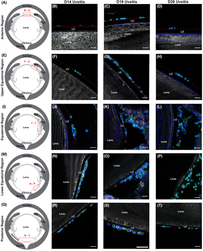
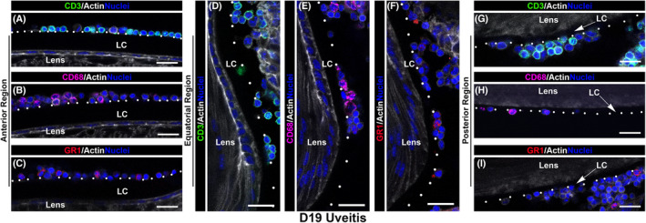
While the eye is considered an immune privileged site, its privilege is abrogated when immune cells are recruited from the surrounding vasculature in response to trauma, infection, aging, and autoimmune diseases like uveitis. Here, we investigate whether in uveitis immune cells become associated with the lens capsule and compromise its privilege in studies of C57BL/6J mice with experimental autoimmune uveitis. These studies show that at D14, the peak of uveitis in these mice, T cells, macrophages, and Ly6G/Ly6C+ immune cells associate with the lens basement membrane capsule, burrow into the capsule matrix, and remain integrated with the capsule as immune resolution is occurring at D26. 3D surface rendering image analytics of confocal z-stacks and scanning electron microscopy imaging of the lens surface show the degradation of the lens capsule as these lens-associated immune cells integrate with and invade the lens capsule, with a subset infiltrating both epithelial and fiber cell regions of lens tissue, abrogating its immune privilege. Those immune cells that remain on the surface often become entwined with a fibrillar net-like structure. Immune cell invasion of the lens capsule in uveitis has not been described previously and may play a role in induction of lens and other eye pathologies associated with autoimmunity.

Keywords: basement membrane; immune cell attachment; immune cell infiltration; immune cell migration; immune privilege; inflammation; lens; matrix; uveitic.


Figures

FIGURE 1
FIGURE 1
Inflammatory cells populate the environments surrounding the lens in EAU. Whole eye cryosections at D19 post‐induction of uveitis were imaged by confocal microscopy following immunolabeling immune cells for CD45 or β2 integrin. Sections were co‐labeled for nuclei and F‐actin and ciliary zonules detected by labeling for MAGP1. Z‐stacks were collected in the regions of the eye indicated in the diagrams including the aqueous humor (A–C), the ciliary zonules (D–F), the vitreous humor just beneath the zonules (G–I), a region of the vitreous mid‐way between the zonules and the posterior‐most aspect of the eye, and the posterior vitreous humor just adjacent to the retina (M–O). Images are shown as both a single 0.5 µm optical plane (B, E, H, K, and N) and the respective 3D surface renderings (C, F, I, L, and O). CD45 was rendered transparent in the inset in (C). The inset in E is a 3D surface structure of the region noted by an asterisk, which is shown zoomed‐in with perlecan rendered transparent in F. CB, ciliary body; CZ, ciliary zonules. Mag bar 20 µm (B, C, E, H, I, K, L, N, and O), 5 µm (E insert, F), and 2 µm (C insert). The data presented represents at least 3 independent studies
FIGURE 2
FIGURE 2
Immune cells become linked to the lens capsule in uveitic eyes. Whole eye cryosections from mice with EAU were co‐labeled at D14, D19, and D26 for CD45 (green), F‐actin (white) and nuclei (blue), and imaged by high resolution confocal microscopy. Z‐stacks were collected in the following regions of the eye, as indicated in the diagrams: the anterior surface of the lens (A–D), the upper region of the lens equator at its border with the lens anterior zone (E–H), the lens equator (I–L), the lower region of the lens equator at its border with the lens posterior surface (M–P), and the posterior surface of the lens (Q–T). Red dotted lines identify the superficial surface of the lens basement membrane capsule (LC). CD45+ immune cells were detected along all surfaces of the lens capsule at D14, D19, and D26 post‐induction of uveitis. CB, ciliary body. Mag bar 20 µm. The data presented represents at least 3 independent studies
FIGURE 3
FIGURE 3
Quantification of the relative distribution of immune cells in different regions of uveitic eyes. Immune cell presence was quantified as indicated in the diagram (A) for the aqueous humor, the anterior, equatorial and posterior surfaces of the lens capsule, and the vitreous and compared at D14 (B), D19 (C), and D26 (D) post‐induction of EAU. *p < .02, **p < .005, ***p < .001
FIGURE 4
FIGURE 4
T cells, macrophages and Ly6G/Ly6C+ immune cells become associated with the lens surface in the eyes of mice with EAU. Whole eye cryosections from mice with EAU at D19 were immunolabeled for the T‐cell antigen CD3 (A, D, and G), the macrophage antigen CD68 (B, E, and H), or the Ly6G/Ly6C antigen (GR‐1) expressed by leukocytes and myeloid‐derived suppressor cells (C, F, and I) and co‐labeled for F‐actin and nuclei. Single optical planes (0.5 µm) from confocal z‐stacks acquired along the lens anterior, equatorial and posterior capsules show that T cells, macrophages and Ly6G/Ly6C+ immune cells associate with the lens capsule in uveitic eyes. White dotted lines identify the superficial surface of the lens basement membrane capsule (LC). Mag bar 20 µm. The data presented represents at least 3 independent studies
FIGURE 5
FIGURE 5
Immune cells induced in the eyes of mice with EAU become integrated with and migrate within the matrix of the anterior lens basement membrane capsule. Confocal z‐stacks of whole eye cryosections of normal (B) and uveitic eyes at D14 (C), D19 (D), and D26 (E), were acquired along the lens anterior capsule, as indicated in the schematic (Aa). Normal eyes were co‐labeled for perlecan and nuclei, uveitic eyes for CD45, perlecan and nuclei. Images are presented either as single 0.5 µm optical planes (Ba; Ca; Da–c; Ea,b) or 3D surface renderings (Bb; Cb,c; Dd–g; Ec–f) of the acquired z‐stacks. The 3D surface renderings showing the integration of immune cells with the lens capsule at D14 (Cb), D19 (Dd) and D26 (Ec) were also created for the perlecan label alone (Cc, De, Ed, respectively) to show the impact of the immune cells on the degradation of the perlecan matrix. Zoomed‐in views at D19 (Df) and D26 (Ee) are also shown with perlecan rendered transparent (Dg and Ef), which reveals the highly migratory morphology of the CD45+ immune cells as they burrow within the anterior lens capsule and the morphology and position of the CD45+ immune cells that have located within the lens capsule. The single optical plane of a uveitic eye at D26 (Ea) shows regions of the perlecan matrix that have been removed by the immune cells (arrowheads), and immune cells that have migrated across the lens capsule and come to reside amongst the cells of the lens anterior epithelium (arrow). SEM images acquired along the anterior surface (schematic, Ab) of lenses isolated from uveitic eyes at D14 (Cd,e), D19 (Dh,i), and D26 (Eg,h) provide a unique view of the morphology of the immune cells associated with the anterior capsule and their movement within the capsule matrix. Mag bars: 50 µm (Eg), 20 µm (Ba,b; Ca,d; Da,b,c,h; Ea,b,h), 10 µm (Cb,c,e; Dd,e; Ec,d), 5 µm (Di), 3 µm (Ee,f), 2 µm (Df,g). The data presented represents at least 3 independent studies
FIGURE 6
FIGURE 6
Immune cells induced in the eyes of mice with EAU rapidly become integrated with and migrate within the lens equatorial capsule. Confocal z‐stacks of whole eye cryosections of normal (B) and uveitic eyes at D14 (C), D19 (D), and D26 (E), were acquired at the equatorial zone of the lens capsule, as indicated in the schematic (Aa). Normal eyes were co‐labeled for perlecan, nuclei, and MAGP1, uveitic eyes for CD45, perlecan and nuclei. Images are presented either as single 0.5 µm optical planes (Ba; Ca; Da,b; Ea) or 3D surface renderings (Bb,c; Cb‐d; Dc‐f; Eb‐e) of the acquired z‐stacks. The 3D surface renderings created to view immune cells association with the lens capsule at D14 (Cb), D19 (Dc,e) and D26 (Eb,d) were also created for the perlecan label alone (Cd; Dd,f; Ec,e, respectively), revealing the degradation and removal of the perlecan matrix by the immune cells as they invade the lens capsule over time. The zoomed‐in view at D14 (Cb) is also shown with perlecan rendered transparent (Cc) to reveal this immune cell burrowing into the perlecan matrix. SEM images are presented of ciliary zonules on the surface of the equatorial capsule of lenses (schematic, Ab) isolated from normal eyes (Bd,e), and of the immune cells associated with the surface of the equatorial lens capsule at D14 (Ce,f), D19 (Dg,h), and D26 (Ef,g) post‐induction of uveitis. These images highlight the rounded morphology of the immune cells associated with the equatorial capsule and the fibrillar structures that link them to the capsule surface. Mag bars: 40 µm (Ce), 30 µm (Cf), 20 µm (Ba–e; Ca; Da–d; Dg; Ea–c; Eg; Eh), 5 µm (Dh), 3 µm (Cb–d), 2 µm (De,f; Ed,e). The data presented represents at least 3 independent studies
FIGURE 7
FIGURE 7
Immune cells induced in the eyes of mice with EAU become integrated with the lens posterior capsule. Confocal z‐stacks of whole eye cryosections of normal (B) and uveitic eyes at D14 (C), D19 (D), and D26 (E), were acquired along the posterior the lens capsule, as indicated in the schematic (Aa). Normal eyes were co‐labeled for perlecan and nuclei, uveitic eyes for CD45, perlecan and nuclei. Images are presented either as single 0.5 µm optical planes (Ba, Ca, Da, Ea) or 3D surface renderings (Bb,c; Cb‐g; Db‐g; Eb‐e) of the acquired z‐stacks. The 3D surface renderings created to view immune cells association with the lens capsule at D14 (Cb,d,f), D19 (Db,d,f) and D26 (Eb,d) were also created for the perlecan label alone (Cc,e,g; Dc,e,g; Ec,e, respectively), revealing the degradation and removal of the perlecan matrix by the immune cells as they integrate with and invade the lens capsule over time. SEM images presented of the immune cells associated with the posterior lens capsule (schematic, Ab) at D14 (Ch,i), D19 (Dh,i), and D26 (Ef,g) post‐induction of uveitis highlight the rounded morphology of the immune cells associated with the posterior capsule and the fibrillar structures that link them to the capsule surface. Mag bars: 50 µm (Dh), 20 µm (Ba; Ca–c,h; Da–c,i; Ea–c,f,g), 10 µm (Bb), 5 µm (Ci), 3 µm (Df,g), 2 µm (Cd–g; Dd,e; Ed,e). The data presented represents at least 3 independent studies
FIGURE 8
FIGURE 8
Infiltration of the lens by immune cells in mice with EAU. Whole eye cryosections from mice with EAU were examined at (A) D14, (B) D19, and (C) D26 post‐induction of uveitis for the presence of immune cells that had infiltrated the lens. Immune cells were detected by immunolabeling for either CD45 or β2 integrin and the sections co‐labeled for nuclei. Sections were also labeled for either F‐actin or perlecan. Confocal z‐stacks were acquired in the region of the lens equatorial epithelium (Ab–d, Bb–e), the lens anterior zone (Bg–i), and the lens cortical fiber zone, a region between the equatorial epithelium and central fiber cells (Bk–n; Cb–d), as indicated in the diagrams. Images shown are either single 0.5 µm optical planes (Ab; Bb,g,h,k), 3D surface structures (Ac,d; Bc–e,i,l–n; Cb–d), or 3D surface structures in which F‐actin is rendered transparent to highlight the presence of the immune cells within lens tissue (Ad; Bd,e,l–n). By D19 after induction of uveitis immune cells have crossed the lens anterior and equatorial capsules and infiltrated lens tissue. LC, lens capsule. Mag bars: 20 µm (Ab, Ac, Bb, Bc, Bg, Bh, Cb), 10 µm (Bi), 5 µm (Ab, inserts), 4 µm (Bn), 3 µm (Ad, Bd, Bl, Bm, Cc, Cd), 2 µm (Be). The data presented represents at least 3 independent studies

Similar articles

Cited by

References

    1. Streilein JW. Ocular immune privilege: the eye takes a dim but practical view of immunity and inflammation. J Leukoc Biol. 2003;74:179‐185. - PubMed
    1. Perez VL, Caspi RR. Immune mechanisms in inflammatory and degenerative eye disease. Trends Immunol. 2015;36: 354‐363. - PMC - PubMed
    1. Rosenbaum JT, Rosenzweig HL, Smith JR, Martin TM, Planck SR. Uveitis secondary to bacterial products. Ophthalmic Res. 2008;40:165‐168. - PubMed
    1. Sharma SM, Jackson D. Uveitis and spondyloarthropathies. Best Pract Res Clin Rheumatol. 2017;31:846‐862. - PubMed
    1. Rosenzweig HL, Jann MM, Glant TT, et al. Activation of nucleotide oligomerization domain 2 exacerbates a murine model of proteoglycan‐induced arthritis. J Leukoc Biol. 2009;85:711‐718. - PMC - PubMed

Publication types