. 2022 Jan 25;145(4):279-294.
doi: 10.1161/CIRCULATIONAHA.121.055801. Epub 2021 Dec 7.

Identification of Drug Transporter Genomic Variants and Inhibitors That Protect Against Doxorubicin-Induced Cardiotoxicity

Affiliations

Identification of Drug Transporter Genomic Variants and Inhibitors That Protect Against Doxorubicin-Induced Cardiotoxicity

Tarek Magdy et al. Circulation. .

Abstract

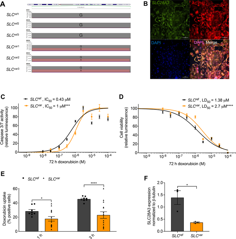
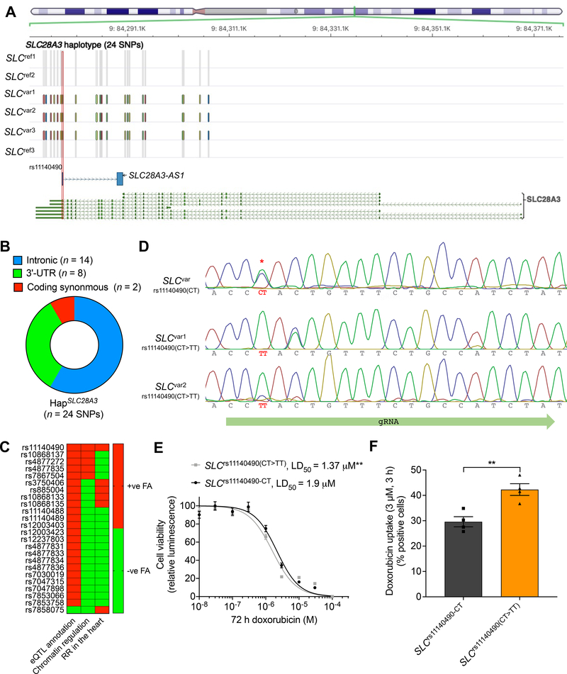
Background: Multiple pharmacogenomic studies have identified the synonymous genomic variant rs7853758 (G > A, L461L) and the intronic variant rs885004 in SLC28A3 (solute carrier family 28 member 3) as statistically associated with a lower incidence of anthracycline-induced cardiotoxicity. However, the true causal variant(s), the cardioprotective mechanism of this locus, the role of SLC28A3 and other solute carrier (SLC) transporters in anthracycline-induced cardiotoxicity, and the suitability of SLC transporters as targets for cardioprotective drugs has not been investigated.

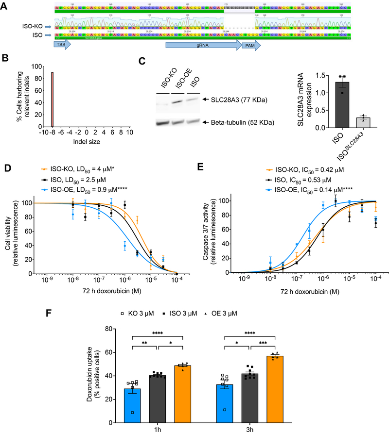
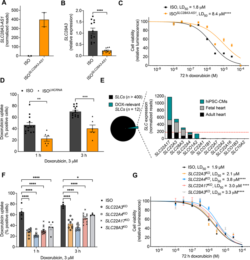
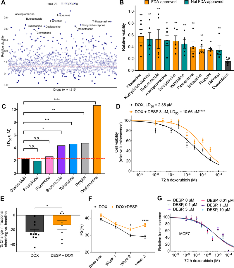
Methods: Six well-phenotyped, doxorubicin-treated pediatric patients from the original association study cohort were recruited again, and human induced pluripotent stem cell-derived cardiomyocytes were generated. Patient-specific doxorubicin-induced cardiotoxicity (DIC) was then characterized using assays of cell viability, activated caspase 3/7, and doxorubicin uptake. The role of SLC28A3 in DIC was then queried using overexpression and knockout of SLC28A3 in isogenic human-induced pluripotent stem cell-derived cardiomyocytes using a CRISPR/Cas9 (Clustered Regularly Interspaced Short Palindromic Repeats/CRISPR-associated protein 9). Fine-mapping of the SLC28A3 locus was then completed after SLC28A3 resequencing and an extended in silico haplotype and functional analysis. Genome editing of the potential causal variant was done using cytosine base editor. SLC28A3-AS1 overexpression was done using a lentiviral plasmid-based transduction and was validated using stranded RNA-sequencing after ribosomal RNA depletion. Drug screening was done using the Prestwick Chemical Library (n = 1200), followed by in vivo validation in mice. The effect of desipramine on doxorubicin cytotoxicity was also investigated in 8 cancer cell lines.

Results: Here, using the most commonly used anthracycline, doxorubicin, we demonstrate that patient-derived cardiomyocytes recapitulate the cardioprotective effect of the SLC28A3 locus and that SLC28A3 expression influences the severity of DIC. Using Nanopore-based fine-mapping and base editing, we identify a novel cardioprotective single nucleotide polymorphism, rs11140490, in the SLC28A3 locus; its effect is exerted via regulation of an antisense long noncoding RNA (SLC28A3-AS1) that overlaps with SLC28A3. Using high-throughput drug screening in patient-derived cardiomyocytes and whole organism validation in mice, we identify the SLC competitive inhibitor desipramine as protective against DIC.

Conclusions: This work demonstrates the power of the human induced pluripotent stem cell model to take a single nucleotide polymorphism from a statistical association through to drug discovery, providing human cell-tested data for clinical trials to attenuate DIC.

Keywords: CRISPR-Cas systems; cardiotoxicity; doxorubicin; human induced pluripotent stem cells; myocytes, cardiac.


Conflict of interest statement

Conflict of Interest Disclosures. None declared

Figures

Figure 1.
Figure 1.. Patient-specific hiPSC–CMs recapitulate the cardioprotective effect of SLC28A3 variant rs7853758.
Comparison of hiPSC–CMs derived from three patients harboring the heterozygous rs7853758 variant and were protected from DIC after DOX treatment (SLCvar1, SLCvar2, SLCvar3; collectively SLCvar), to hiPSC-CMs from three control patients who did not carry this protective SNP and developed DIC upon same DOX treatment (SLCref1, SLCref2, SLCref3 SLCref3SLCref3; collectively SLCref). A, Nanopore sequencing reads at SNP rs7853758 locus confirming its genotypes in all patient–derived hiPSC lines. B, Immunofluorescent staining showing the expression and localization of SLC28A3 throughout the cell in patient–derived hiPSC–CMs. C, Effect of DOX (72 h) on cell viability in SLCvar (n = 126) and SLCref (n = 81) hiPSC–CMs measured by a CellTiter-Glo 2.0 assay. D, Effect of DOX (72 h) on apoptosis measured by activated caspase 3/7 in SLCvar (n = 20) and SLCref (n = 20) hiPSC–CMs. E, Assessment of DOX uptake via measurement of percentage of cells with DOX intrinsic fluorescence using a flow cytometry-based assay in patient–derived hiPSC–CMs (n = 8–13). F, SLC28A3 expression in SLCref (n = 3), and SLCvar (n = 3) hiPSC-CMs using western blot. n = full independent experimental replicates, Error bars, s.e.m, *P ≤ 0.05, **P ≤ 0.01, ***P ≤ 0.001, ****P ≤ 0.0001 by unpaired two-tailed Student’s t-test (E and F). For (C and D) log-logistic non-linear regression model was used to estimate the value of the four parameters, and t-statistic was used to test for significant difference in LD50 between different groups.
Figure 2.
Figure 2.. SLC28A3 expression affects the severity of DIC by regulating DOX uptake into cardiomyocytes.
A, Validation of CRISPR/Cas9–mediated SLC28A3 knockout (KO) in an isogenic hiPSC line detected by Sanger sequencing, showing 8 bp deletion downstream of the transcription start site (TSS). PAM, protospacer adjacent motif. B, Demonstration that 91% of the cell population acquire the introduced deletion. C, Validation of KO and AAVS1-based SLC28A3 overexpression (OE) by western blot and RT-PCR. D, Effect of DOX (72 h) on viability in ISO (n = 45), ISO–OE (n = 14), and ISO–KO (n = 6) hiPSC–CMs. E, Effect of doxorubicin (72 h) on apoptosis measured by activated caspase 3/7 in ISO (n = 8), ISO–OE (n = 10), and ISO–KO (n = 6) hiPSC–CMs. F, Assessment of DOX uptake via measurement of DOX intrinsic fluorescence using flow cytometry-based assay (n = 6–9). n = full independent experimental replicates, Error bars, s.e.m, *P ≤ 0.05, **P ≤ 0.01, ***P ≤ 0.001, ****P ≤ 0.0001 by unpaired two-tailed Student’s t-test (f). For (d and e) log-logistic non–linear regression model was used to estimate the value of the four parameters, and t-statistic was used to test for significant difference in LD50 between different groups.
Figure 3.
Figure 3.. Fine–mapping at the SLC28A3–SLC28A3-AS1 locus identifies rs11140490 as the potential causal cardioprotective variant.
A, Location of the HapSLC28A3 comprising 24 SNPs that are co–inherited only in SLCvar protected patients. SNP rs11140490 marked by red rectangle is located at the splice site of the first exon of an overlapping long non–coding RNA, SLC28A3-AS1 (adapted from Magdy et al.). B, Consequence of co–inherited HapSLC28A3 SNPs (n = 24). C, Overall prioritization of candidate causal SNPs based on functional annotation analyses including, eQTL annotation, chromatin regulatory analyses, and overlapping with regulatory regions (RR) in cardiac tissues. FA, functional annotation (adopted from Magdy et al.). D, Editing of rs11140490 (CT>TT) in two patient-specific hiPSC lines using cytosine base editor (Target-AID-NG), rs11140490 is marked by red asterisk. E, Effect of DOX (72 h) on viability in SLCvar-rs11140490 (CT) (2 lines, n = 31) and SLCvar-rs11140490(CT>TT edited) (2 lines, n = 75) hiPSC–CMs. F, Assessment of DOX uptake via measurement percentage of cells with DOX intrinsic fluorescence using flow cytometry-based assay in SLCvar-rs11140490 (CT) and SLCvar-rs11140490(CT>TT edited) (n = 4). n = full independent experimental replicates, Error bars, s.e.m, *P ≤ 0.05, **P ≤ 0.01, ***P ≤ 0.001, ****P ≤ 0.0001 by unpaired two-tailed Student’s t-test (F). For (E) log-logistic non-linear regression model was used to estimate the value of the four parameters, and t-statistic was used to test for significant difference in LD50 between different groups.
Figure 4.
Figure 4.. The cardioprotective role of SLC28A3-AS1.
A, Relative Expression of SLC28A3-AS1 in ISO hiPSC compared to ISO transduced by viral plasmid encoding SLC28A3-AS1 cDNA to overexpress SLC28A3-AS1 (ISOSLC28A3-AS1) assessed by RNA–Seq (n = 2). B, SLC28A3 relative expression in ISO and ISOSLC28A3-AS1 hiPSC-CMs (n = 5–11) assessed by real-time PCR. C, Effect of SLC28A3-AS1 overexpression on cell viability after DOX (72 h) treatment, ISO (n = 17), ISOSLC28A3-AS1 (n = 12). D, Effect of SLC28A3-AS1 overexpression on DOX uptake 1 h and 3 h post DOX treatment (n = 6–14). E, Relative human cardiomyocyte expression of SLC transporters (n = 12) previously identified as transporting DOX or a DOX metabolite and/or by genetic associations with DOX clinical outcomes. Red dashed line denoted for the expression cutoff for SLC transporter selection. F, Effect of knocking out DOX–relevant SLC transporters on DOX uptake into patient–derived cardiomyocytes [SLC28A3KO, SLC22A4KO, SLC22A3KO, and SLC22A17KO (n = 5–13)]. G, Effect of knocking out potential cardiac-specific SLC transporters on cell viability after DOX treatment [SLC28A3KO (n = 14), SLC22A4KO (n = 58), SLC22A3KO (n = 17), and SLC22A17KO, (n = 10), ISO (n = 128)]. n = full independent experimental replicates, Error bars, s.e.m, *P ≤ 0.05, **P ≤ 0.01, ***P ≤ 0.001, ****P ≤ 0.0001 by unpaired two-tailed Student’s t-test (B, D, and F). For (C and G) log-logistic non-linear regression model was used to estimate the value of the four parameters, and t-statistic was used to test for significant difference in LD50 between different groups.
Figure 5.
Figure 5.. Multi-modality drug screening identifies desipramine as a novel effective cardioprotectant against DIC.
A, Prestwick drug library screening (n = 1219) in relation to DIC (n = 5). All drugs were used at 3 μM. Red dashed line represents cell viability 72 h post DOX (10 μM) treatment; the top ten significant cardioprotective drugs based on cell viability are labeled. B, Bar plot showing top ten significant cardioprotective (based on P value) compared to DOX alone (72 h, 10 μM) treated cells. Non–FDA–approved drugs are represented by teal bars. C, Further validation of top FDA-approved drugs (identified from the Prestwick library screening) against 10 log–doses of doxorubicin. LD50, median lethal dose. D, Effect of co-treatment of desipramine (3 μM) and doxorubicin (72 h) on hiPSC-CM viability [DOX (n = 42), DOX + DESP (n = 35)]. E, Percent change in ventricular fraction shortening (FS) normalized to baseline, after 3 weeks of doxorubicin treatment (3 mg/kg, ip, n = 10) compared co-treatment (n = 8) of desipramine (20 mg/kg/day, Alzet pump) and doxorubicin (3 mg/kg, ip) in mice. F, Ventricular fractional shortening at baseline, 1-, 2-, 3-weeks post treatment. G, Assessment of cell viability of MCF breast cancer cell line after 72 h of DOX and desipramine (DESP) cotreatment (n = 12–20). f = full independent experimental replicates, Error bars, s.e.m, *P < 0.05, **P ≤ 0.01, ***P < 0.001, ****P < 0.0001 by unpaired two-tailed Student’s t-test (A-C and E) and by ANOVA with post-hoc testing (F). For (D and G) log-logistic non-linear regression model was used to estimate the value of the four parameters, and t-statistic was used to test for significant difference in LD50 between different groups.

Comment in

References

    1. Cardinale D, Colombo A, Bacchiani G, Tedeschi I, Meroni CA, Veglia F, Civelli M, Lamantia G, Colombo N, Curigliano G, et al. Early detection of anthracycline cardiotoxicity and improvement with heart failure therapy. Circulation. 2015;131:1981–8. - PubMed
    1. Swain SM, Whaley FS and Ewer MS. Congestive heart failure in patients treated with doxorubicin: A retrospective analysis of three trials. Cancer. 2003;97:2869–2879. - PubMed
    1. Avila MS, Ayub-Ferreira SM, de Barros Wanderley MR Jr., das Dores Cruz F, Goncalves Brandao SM, Rigaud VOC, Higuchi-Dos-Santos MH, Hajjar LA, Kalil Filho R, Hoff PM, et al. Carvedilol for Prevention of Chemotherapy-Related Cardiotoxicity: The CECCY Trial. J Am Coll Cardiol. 2018;71:2281–2290. - PubMed
    1. Magdy T, Burmeister BT and Burridge PW. Validating the pharmacogenomics of chemotherapy-induced cardiotoxicity: What is missing? Pharmacol Ther. 2016. - PMC - PubMed
    1. Aminkeng F, Ross CJD, Rassekh SR, Rieder MJ, Bhavsar AP, Sanatani S, Bernstein D, Hayden MR, Amstutz U and Carleton BC. Pharmacogenomic screening for anthracycline-induced cardiotoxicity in childhood cancer. Br J Clin Pharmacol. 2017;83:1143–1145. - PMC - PubMed

Publication types

LinkOut - more resources