. 2022 Jan 25;7(2):e154573.
doi: 10.1172/jci.insight.154573.

Alveolar epithelial glycocalyx degradation mediates surfactant dysfunction and contributes to acute respiratory distress syndrome

Affiliations

Alveolar epithelial glycocalyx degradation mediates surfactant dysfunction and contributes to acute respiratory distress syndrome

Alicia N Rizzo et al. JCI Insight. .

Abstract

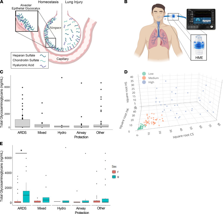
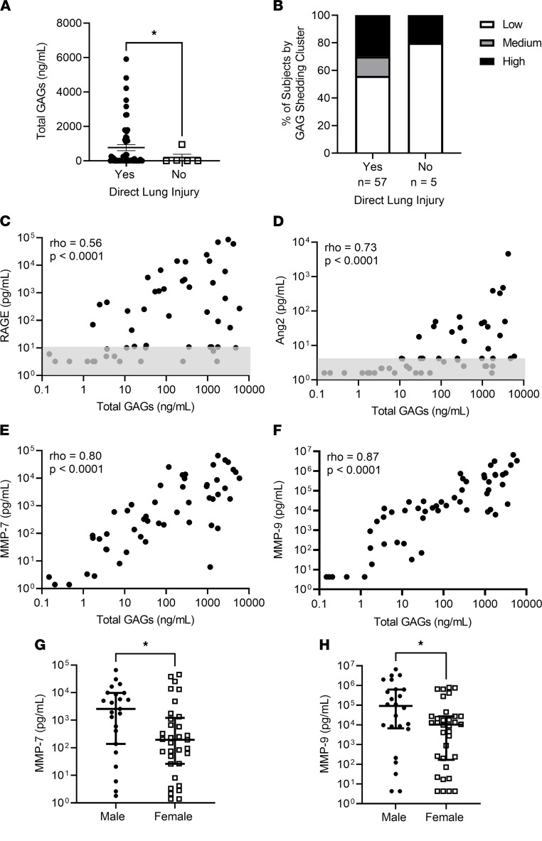
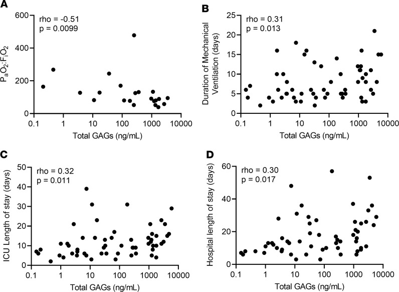
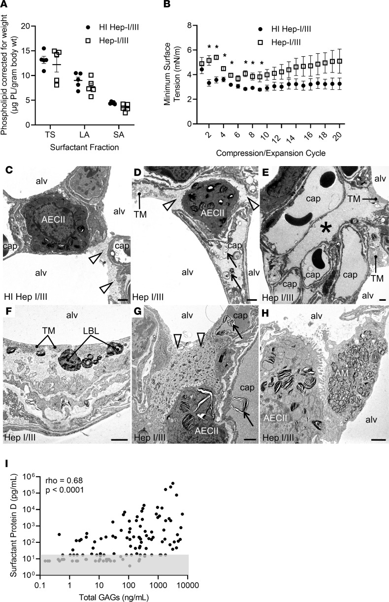
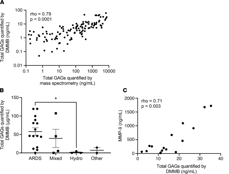
Acute respiratory distress syndrome (ARDS) is a common cause of respiratory failure yet has few pharmacologic therapies, reflecting the mechanistic heterogeneity of lung injury. We hypothesized that damage to the alveolar epithelial glycocalyx, a layer of glycosaminoglycans interposed between the epithelium and surfactant, contributes to lung injury in patients with ARDS. Using mass spectrometry of airspace fluid noninvasively collected from mechanically ventilated patients, we found that airspace glycosaminoglycan shedding (an index of glycocalyx degradation) occurred predominantly in patients with direct lung injury and was associated with duration of mechanical ventilation. Male patients had increased shedding, which correlated with airspace concentrations of matrix metalloproteinases. Selective epithelial glycocalyx degradation in mice was sufficient to induce surfactant dysfunction, a key characteristic of ARDS, leading to microatelectasis and decreased lung compliance. Rapid colorimetric quantification of airspace glycosaminoglycans was feasible and could provide point-of-care prognostic information to clinicians and/or be used for predictive enrichment in clinical trials.

Keywords: Glycobiology; Proteoglycans; Pulmonary surfactants; Pulmonology.


Conflict of interest statement

Conflict of interest: The authors have declared that no conflict of interest exists.

Figures

Figure 1
Figure 1. GAG shedding is heterogeneous in ARDS.
(A) Graphical representation (not to scale) of the alveolar epithelial glycocalyx, a layer of GAGs — heparan sulfate (HS), chondroitin sulfate (CS), and hyaluronic acid (HA) — that lines the apical alveolar epithelial surface. In murine models of acute lung injury, the glycocalyx is damaged, leading to shedding of GAGs into the airspace fluid. (B) Graphical representation of a patient on a ventilator circuit containing a heat and moisture exchanger (HME) filter, from which airspace fluid can be noninvasively sampled in mechanically ventilated patients. (C) Assessment of airspace fluid GAG shedding in mechanically ventilated patients was performed by mass spectrometry analysis of HMEF. (D) Partitioning around k-medoids clustering using the variables HS, CS, and HA was performed to group patients into high, medium, and low shedding clusters based on the degree of GAG shedding. (E) Assessment of the effect of patients’ sex on airspace GAG levels in patients with respiratory failure. n = 153 participants with respiratory failure. *P < 0.05 by Wilcoxon’s rank sum test. Data are represented as median and IQR with individual data points for those outside of 1.5 × IQR. A and B were created with BioRender (biorender.com).
Figure 2
Figure 2. Alveolar epithelial glycocalyx degradation occurs in patients with direct lung injury and is associated with upregulation of matrix metalloproteinases.
(A) Assessment of total GAG shedding by clinically determined risk factor for lung injury. Direct lung injury indicates patients who were diagnosed with either pneumonia or aspiration at the time of intubation. n = 62 patients with ARDS. P = 0.045 by Kruskal-Wallis test. Data are represented as mean ± SEM. (B) Assessment of the percentage of patients in each GAG shedding cluster for patients with and without direct lung injury. (CF) Assessment of the relationship between GAG shedding and the epithelial cellular injury marker receptor for advanced glycation end products (RAGE) (n = 56) (C), the endothelial cellular injury marker angiopoietin-2 (Ang2) (n = 56) (D), and the matrix metalloproteinases MMP-7 (n = 56) (E) and MMP-9 (n = 58) (F). Gray boxes (C and D) represent values at or below the lower limit of detection (variability of this limit reflects different sample dilutions). Spearman’s ρ and P values are as indicated on each graph. (G and H) Assessment of the airspace MMP-7 (G) and MMP-9 (H) levels based on sex. P values were computed by Wilcoxon’s rank sum test. *P < 0.05. Data are represented as mean ± SEM.
Figure 3
Figure 3. GAG shedding predicts ARDS severity and duration.
(A) Assessment of the relationship between GAG shedding and PaO2:FiO2. n = 25 patients with ARDS in whom arterial blood gas data were available. (BD) Assessment of the relationship between GAG shedding and duration of mechanical ventilation (B), ICU length of stay (C), and hospital length of stay (D) in patients with ARDS. n = 64 patients with ARDS in whom full data regarding duration of critical illness were available. Spearman ρ and P values were computed as indicated on each graph.
Figure 4
Figure 4. Alveolar epithelial glycocalyx degradation impairs lung compliance by causing microatelectasis.
(AC) After treatment with heat-inactivated heparinases I/III (HI Hep-I/III; 15 U, intratracheal), mouse lungs demonstrated colocalization of HS (HS 10E4, green) and the alveolar epithelium (Lycopersicon esculentum agglutinin lectin, red). Mice treated with active heparinases I/III (Hep-I/III, 15 U, intratracheal) demonstrated a denuded epithelial layer without HS staining at 12 hours, which persisted at 72 hours. As lungs were not perfusion-fixed, the endothelial glycocalyx is not visible. Scale bars: 100 μm. (D and E) Assessment of the lung mechanics of mice treated with Hep-I/III and HI Hep-I/III at t = 24 hours. n = 10 mice per group. Unpaired Student’s t test was used for comparisons between 2 groups. *P < 0.05, ***P < 0.0005. (FK) Lung histology of Hep-I/III– and HI Hep-I/III–treated mice (24 hours). Dilated alveolar ducts (arrowhead) and microatelectasis (arrow) were noted at increased frequency in Hep-I/III–treated mice. (LO) Quantification of the histologic changes present in Hep-I/III–treated mice as assessed by unbiased stereologic assessment of the lung architecture. There was no evidence of fibrosis in either group. n = 4–5 mice per group. Unpaired t test was used for comparisons between 2 groups. *P < 0.05, **P < 0.005, ****P < 0.0001. Data are represented as mean ± SEM.
Figure 5
Figure 5. Alveolar epithelial glycocalyx degradation impairs surfactant function.
(A) The effect of Hep-I/III treatment on the quantity of total surfactant (TS) and its subfractions, the surface-active large aggregates (LA) and the inactive small aggregates (SA). (B) The minimum surface tension of the LA surfactant subfraction from Hep-I/III–treated mice compared with HI Hep-I/III–treated controls. n = 5 mice per group. *P < 0.05 by 2-tailed unpaired t test. Data are represented as mean ± SEM. (CH) In both HI Hep-I/III–treated (C) and Hep-I/III–treated (DH) animals, transmission electron microscopy revealed a thin layer of electron-dense alveolar liquid film (open arrowheads) containing lipid fragments on top of the alveolar epithelium. This film had an increased thickness in Hep-I/III–treated mice (D). Higher amounts were particularly found in alveolar corners and interalveolar pores of Kohn or covering the secretory surface of type II alveolar epithelial cells (AECII; C, D, G, and H). Alveolar liquid–filled infoldings of the alveolar epithelium (asterisk) were present to a larger extent in the Hep-I/III–treated mice (E). In Hep-I/III–treated mice, intra-alveolar surfactant subtypes, in particular freshly secreted lamellar body–like forms (LBL) in close proximity to tubular myelin (TM) at the air-liquid interface, were more abundant (F). The electron density of the liquid film and the quantity of contained small lipid fragments were increased in the Hep-I/III group (G). Lamellar lipid accumulations were also found in some alveolar macrophages (AM; H) and capillaries (arrows; D and G). alv, alveolar lumen; cap, capillary lumen. Scale bars: 1 μm. (I) The relationship between GAG shedding and surfactant protein D in HMEF. Gray box represents values at or below the lower limit of detection (variability of this limit reflects different sample dilutions). n = 115 participants with respiratory failure. Spearman ρ and P values were calculated as indicated.
Figure 6
Figure 6. Point-of-care detection of alveolar epithelial glycocalyx degradation approximates mass spectrometry and is feasible in the ICU.
(A) Assessment of the relationship between GAG shedding as quantified by state-of-the-art HPLC-MS and GAG shedding as quantified by colorimetric DMMB assay. n = 132 participants in whom sufficient fluid was available to run the DMMB assay. Spearman ρ and P values are as indicated on the graph. (B) Assessment of the relationship between GAG shedding by DMMB assay and cause of respiratory failure in a second cohort of patients in whom filters were collected only at routine filter changes, rather than through a standardized research study protocol. n = 24 participants with respiratory failure. *P < 0.05 by Wilcoxon’s rank sum test. Data are presented with mean ± SEM. (C) Assessment of the relationship between GAG shedding by DMMB assay and MMP-9 expression in the cohort of patients with filters collected only as part of routine care. n = 16 participants with respiratory failure. Spearman ρ and P values are as indicated on the graph.

Similar articles

Cited by

References

    1. Bellani G, et al. Epidemiology, patterns of care, and mortality for patients with acute respiratory distress syndrome in intensive care units in 50 countries. JAMA. 2016;315(8):788–800. doi: 10.1001/jama.2016.0291. - DOI - PubMed
    1. Matthay MA, et al. Clinical trials in acute respiratory distress syndrome: challenges and opportunities. Lancet Respir Med. 2017;5(6):524–534. doi: 10.1016/S2213-2600(17)30188-1. - DOI - PubMed
    1. Calfee CS, et al. Subphenotypes in acute respiratory distress syndrome: latent class analysis of data from two randomised controlled trials. Lancet Respir Med. 2014;2(8):611–620. doi: 10.1016/S2213-2600(14)70097-9. - DOI - PMC - PubMed
    1. Meyer NJ, Calfee CS. Novel translational approaches to the search for precision therapies for acute respiratory distress syndrome. Lancet Respir Med. 2017;5(6):512–523. doi: 10.1016/S2213-2600(17)30187-X. - DOI - PMC - PubMed
    1. Shaver CM, Bastarache JA. Clinical and biological heterogeneity in acute respiratory distress syndrome: direct versus indirect lung injury. Clin Chest Med. 2014;35(4):639–653. doi: 10.1016/j.ccm.2014.08.004. - DOI - PMC - PubMed

Publication types

MeSH terms

Substances

Supplementary concepts