

doi: 10.1093/infdis/jiab587.
SARS-CoV-2 Interference of Influenza Virus Replication in Syrian Hamsters
Affiliations
- PMID: 34875072
- PMCID: PMC8689717
- DOI: 10.1093/infdis/jiab587
Item in Clipboard
SARS-CoV-2 Interference of Influenza Virus Replication in Syrian Hamsters
J Infect Dis.
.
Abstract
In hamsters, SARS-CoV-2 infection at the same time as or before H3N2 influenza virus infection resulted in significantly reduced influenza virus titers in the lungs and nasal turbinates. This interference may be correlated with SARS-CoV-2-induced expression of MX1.
Keywords: MX1; Coinfection; Influenza; SARS-CoV-2; antiviral.
© The Author(s) 2021. Published by Oxford University Press for the Infectious Diseases Society of America. All rights reserved. For permissions, e-mail: journals.permissions@oup.com.
Figures
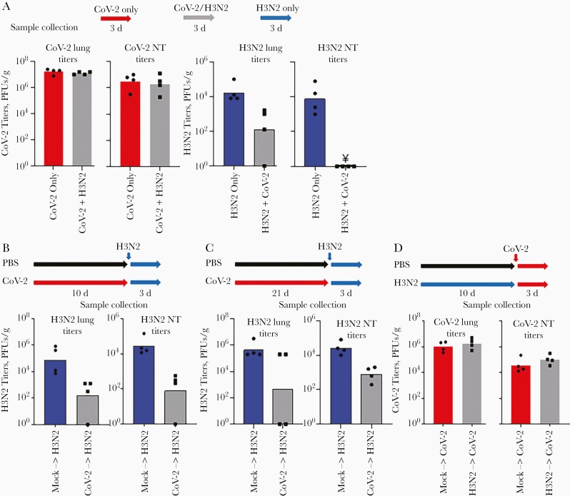
Simultaneous and serial infection studies. A, Groups of hamsters were infected with severe acute respiratory syndrome coronavirus 2 (SARS-CoV-2) only, influenza virus (H3N2), or simultaneously with influenza virus and SARS-CoV-2 (CoV-2). Viral titers in the lungs and nasal turbinates (NTs) were determined 3 days after infection. Note: ¥ represents titers below the limit of detection (10 plaque-forming units [PFUs]/g. B, C, For serial infection studies, groups of hamsters were first inoculated with phosphate-buffered saline (PBS; mock) or infected with SARS-CoV-2 and then infected with influenza virus after 10 (B) or 21 (C) days. D, In addition, groups of hamsters were first inoculated with PBS (mock) or infected with influenza virus and then infected with SARS-CoV-210 days later. Viral titers in the lungs and NTs were determined 3 days after influenza infection.

Immunohistochemical staining of myxovirus resistance protein 1 (Mx1) in lung tissues. A, B, Representative images of Mx1 staining from fixed lung tissues collected 10 days after inoculation with phosphate-buffered saline (PBS) only (A) or infection with severe acute respiratory syndrome coronavirus 2 (SARS-CoV-2) only (B). C, D, In a serial infection study, animals were first inoculated with PBS (C) or SARS-CoV-2 (D) and then infected with influenza virus 10 days later. Mx1 staining was performed on fixed lung tissues collected 3 days after influenza virus infection. (Scale bars represent 100 µm.).
References
-
- Liang L, He C, Lei M, et al. Pathology of guinea pigs experimentally infected with a novel reovirus and coronavirus isolated from SARS patients. DNA Cell Biol 2005; 24:485–90. - PubMed
Publication types
MeSH terms
Substances
Grants and funding
LinkOut - more resources
Full Text Sources
Medical
Miscellaneous
