Review
. 2021 Dec;11(12):210110.
doi: 10.1098/rsob.210110. Epub 2021 Dec 8.

Fibro-adipogenic progenitors in skeletal muscle homeostasis, regeneration and diseases

Affiliations
Review

Fibro-adipogenic progenitors in skeletal muscle homeostasis, regeneration and diseases

Thomas Molina et al. Open Biol. 2021 Dec.

Abstract

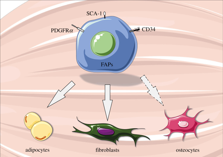
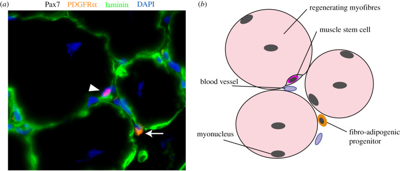
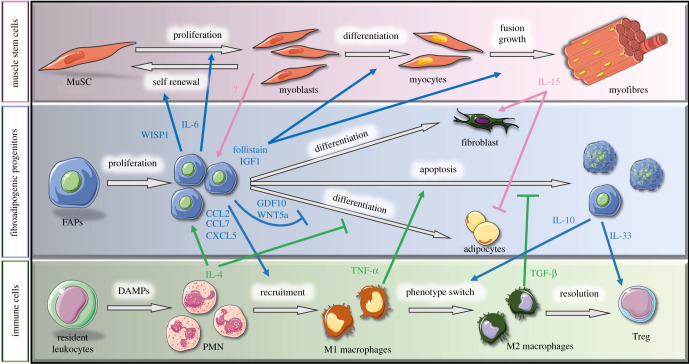
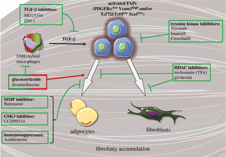
Skeletal muscle possesses a remarkable regenerative capacity that relies on the activity of muscle stem cells, also known as satellite cells. The presence of non-myogenic cells also plays a key role in the coordination of skeletal muscle regeneration. Particularly, fibro-adipogenic progenitors (FAPs) emerged as master regulators of muscle stem cell function and skeletal muscle regeneration. This population of muscle resident mesenchymal stromal cells has been initially characterized based on its bi-potent ability to differentiate into fibroblasts or adipocytes. New technologies such as single-cell RNAseq revealed the cellular heterogeneity of FAPs and their complex regulatory network during muscle regeneration. In acute injury, FAPs rapidly enter the cell cycle and secrete trophic factors that support the myogenic activity of muscle stem cells. Conversely, deregulation of FAP cell activity is associated with the accumulation of fibrofatty tissue in pathological conditions such as muscular dystrophies and ageing. Considering their central role in skeletal muscle pathophysiology, the regulatory mechanisms of FAPs and their cellular and molecular crosstalk with muscle stem cells are highly investigated in the field. In this review, we summarize the current knowledge on FAP cell characteristics, heterogeneity and the cellular crosstalk during skeletal muscle homeostasis and regeneration. We further describe their role in muscular disorders, as well as different therapeutic strategies targeting these cells to restore muscle regeneration.

Keywords: fibro-adipogenic progenitors; fibrosis; mesenchymal stromal cells; muscle stem cells; muscular disorders; myogenesis.


Figures

Figure 1.
Figure 1.
Schematic of fibro-adipogenic progenitors (FAPs). FAPs are muscle resident multipotent mesenchymal stem cells that can differentiate into adipocytes, fibroblasts or osteocytes (under specific conditions). They express key surface markers such as platelet-derived growth factor receptor α (PDGFRα), stem cell antigen-1 (Sca-1) and cluster of differentiation 34 (CD34). This figure was created with the Servier Medical Art service (https://smart.servier.com/), which is licensed under a Creative Commons Attribution 3.0 Unported License (https://creativecommons.org/licenses/by/3.0/).
Figure 2.
Figure 2.
Fibro-adipogenic progenitors (FAPs) in regenerating skeletal muscle. (a) Immunofluorescence of Pax7 (red), PDGFRα (orange), laminin (green) and DAPI (nuclei, blue) on regenerating skeletal muscle section (14 days post-cardiotoxin injury). Muscle stem cell (Pax7+ cell in red; identified with white arrowhead) is visible in its niche under the basal membrane (laminin; green). FAP (PDGFRα+ cell in orange) is identified with a white arrow in the interstitial space. (b) Schematic of the location of muscle stem cell and FAP in skeletal muscle.
Figure 3.
Figure 3.
Cellular interactions during muscle regeneration. Schematic of the known interactions between muscle stem cells (MuSC, pink background), fibro-adipogenic progenitors (FAPs, blue background) and immune cells (green background) throughout muscle regeneration. The blue arrows represent molecules secreted by FAPs that act on other cell types, whereas pink and green arrows indicate paracrine factors secreted by muscle stem cells and immune cells, respectively, that affect FAPs. IL, interleukin; WISP1, Wnt1-inducible-signalling pathway protein 1; IGF1, insulin growth factor-1; GDF10, growth differentiation factor 10; TNF-α, tumour necrosis factor alpha; TGF-β, transforming growth factor-β; DAMPs, damage-associated molecular pattern; CCL, C–C motif chemokine ligand; CXCL5, C–X–C motif chemokine ligand 5, PMN, polymorphonuclear cells. This figure was created with the Servier Medical Art service (https://smart.servier.com/), which is licensed under a Creative Commons Attribution 3.0 Unported License (https://creativecommons.org/licenses/by/3.0/).
Figure 4.
Figure 4.
Molecular approaches targeting fibro-adipogenic progenitors in muscular dystrophies. Schematic of different drugs tested to inhibit fibro-adipogenic progenitor (FAP) activity, proliferation and/or their differentiation into adipocytes or fibroblasts. Arrows and T-shaped lines indicate stimulatory and inhibitory effects, respectively. DMD, Duchenne muscular dystrophy; HDAC, histone deacetylase; Vcam1, vascular cell adhesion molecule 1; PDGFRα, platelet-derived growth factor receptor α; TCF7L2/TCF4, transcription factor 4; Sca1, stem cells antigen-1; TGF-β, transforming growth factor-β; MMP, matrix metalloproteinase; GSK3, glycogen synthase kinase-3. This figure was created with the Servier Medical Art service (https://smart.servier.com/), which is licensed under a Creative Commons Attribution 3.0 Unported License (https://creativecommons.org/licenses/by/3.0/).

References

    1. Proctor DN, O'Brien PC, Atkinson EJ, Nair KS. 1999. Comparison of techniques to estimate total body skeletal muscle mass in people of different age groups. Am. J. Physiol. 277, E489-E495. (10.1152/ajpendo.1999.277.3.E489) - DOI - PubMed
    1. Mauro A. 1961. Satellite cell of skeletal muscle fibers. J. Biophys. Biochem. Cytol. 9, 493-495. (10.1083/jcb.9.2.493) - DOI - PMC - PubMed
    1. Dumont NA, Bentzinger CF, Sincennes MC, Rudnicki MA. 2015. Satellite cells and skeletal muscle regeneration. Compr. Physiol. 5, 1027-1059. (10.1002/cphy.c140068) - DOI - PubMed
    1. Dumont NA, Wang YX, Rudnicki MA. 2015. Intrinsic and extrinsic mechanisms regulating satellite cell function. Development 142, 1572-1581. (10.1242/dev.114223) - DOI - PMC - PubMed
    1. Bentzinger CF, Wang YX, Dumont NA, Rudnicki MA. 2013. Cellular dynamics in the muscle satellite cell niche. EMBO Rep. 14, 1062-1072. (10.1038/embor.2013.182) - DOI - PMC - PubMed

Publication types

LinkOut - more resources