Review
. 2022 Jan:178:189-201.
doi: 10.1016/j.freeradbiomed.2021.12.002. Epub 2021 Dec 4.

Breaking barriers: Neurodegenerative repercussions of radiotherapy induced damage on the blood-brain and blood-tumor barrier

Affiliations
Review

Breaking barriers: Neurodegenerative repercussions of radiotherapy induced damage on the blood-brain and blood-tumor barrier

Barrett D Allen et al. Free Radic Biol Med. 2022 Jan.

Abstract

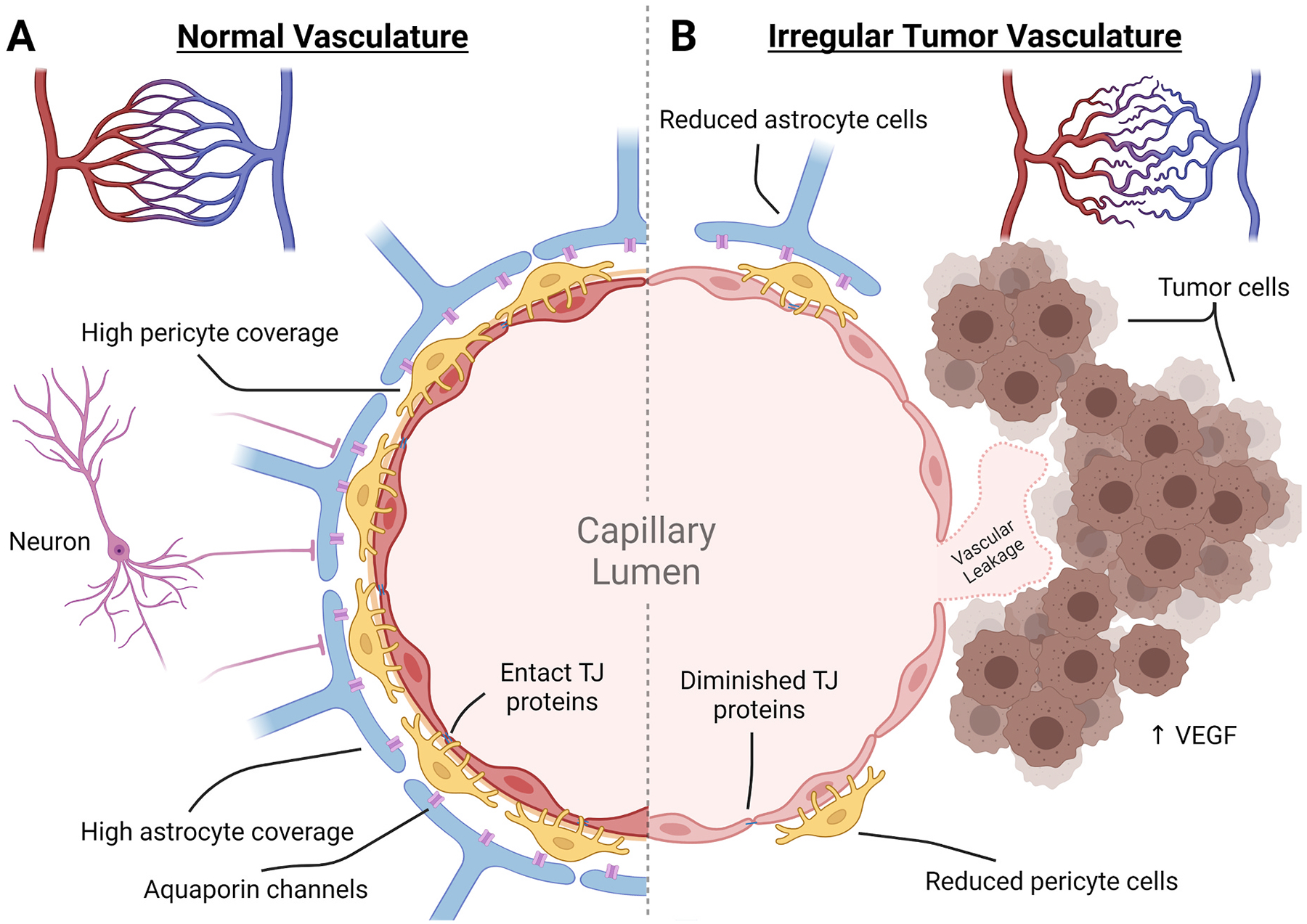
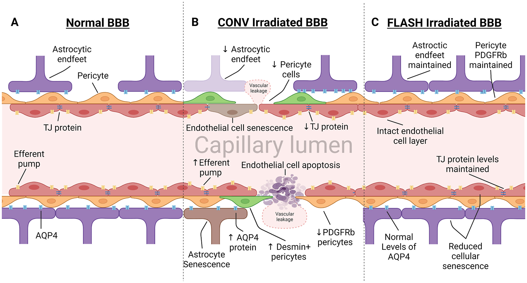
Exposure to radiation during the treatment of CNS tumors leads to detrimental damage of the blood brain barrier (BBB) in normal tissue. Effects are characterized by leakage of the vasculature which exposes the brain to a host of neurotoxic agents potentially leading to white matter necrosis, parenchymal calcification, and an increased chance of stroke. Vasculature of the blood tumor barrier (BTB) is irregular leading to poorly perfused and hypoxic tissue throughout the tumor that becomes resistant to radiation. While current clinical applications of cranial radiotherapy use dose fractionation to reduce normal tissue damage, these treatments still cause significant alterations to the cells that make up the neurovascular unit of the BBB and BTB. Damage to the vasculature manifests as reduction in tight junction proteins, alterations to membrane transporters, impaired cell signaling, apoptosis, and cellular senescence. While radiotherapy treatments are detrimental to normal tissue, adapting combined strategies with radiation targeted to damage the BTB could aid in drug delivery. Understanding differences between the BBB and the BTB may provide valuable insight allowing clinicians to improve treatment outcomes. Leveraging this information should allow advances in the development of therapeutic modalities that will protect the normal tissue while simultaneously improving CNS tumor treatments.

Keywords: Blood brain barrier; Blood tumor barrier; Neurovascular unit; Radiation.


Figures

Fig. 1.
Fig. 1.. Comparison of the neurovascular unit of the blood brain barrier and blood tumor barrier.
Normal vasculature of the brain develops from pial arteries to arterioles to capillaries where transfer of nutrients and oxygen can take place. In the normal tissue, capillaries are spread out somewhat uniformly to ensure equal distribution. In the BTB, there is a reduced presence of capillary endings and an irregular overlay creating areas of hypoxic tumor tissue. (A) In the normal tissue capillaries, the neurovascular unit controls the influx of materials by forming a non-fenestrated endothelial cell layer that is tightly bound together with TJ proteins. These are enveloped by pericyte cells and astrocytic endfeet that aid in maintaining the endothelial cell transport proteins, TJ proteins, induction of angiogenesis, and influx of ions and fluids. This is controlled by local input as well as neuronal demand that can interact directly or through astrocytic channels. (B) Within the BTB, integrity is compromised. Tumor cells attach to the outer vascular wall and induce vascular growth through VEGF expression. The new capillary bed is irregular and leaves areas of the tumor hypoxic. The new vasculature becomes the foundation of the BTB and has been found to have reductions in astrocytic endfeet coverage, pericyte cells, TJ proteins, and neuron attachment. The resulting capillary wall of the BTB is susceptible to neurotoxic leakage.
Fig. 2.
Fig. 2.
Irradiation of the blood brain barrier leads to damage of the neurovascular unit. Vasculature of the BBB is susceptible to radiotherapy treatment, inducing modifications to protein expression and apoptosis and senescence of cells that alter homeostasis. (A) Normal BBB expresses high levels of TJ proteins, sealing gaps between endothelial cells, preventing paracellular crossing. Expression of efferent pumps on the lumen side of endothelial cells actively work to remove foreign materials and chemotherapeutic drugs from the brain. Pericytes embedded within the basement membrane provide support and regulation over dilation and TJ expression. Astrocytic endfeet express aquaporin protein (AQP4), regulating fluid transfer across the membrane. (B) Radiotherapy induces leakage of the BBB through apoptosis and senescence in cells of the NVU. Additionally, declines in TJ protein expression lead to vascular leakage through paracellular channels. Pericyte subpopulations respond to BBB damage by reducing expression of PDGFRβ and replacing it with desmin. Astrocytes produce irregular expression of AQP4, contributing directly to dysregulated fluid transfer into the CNS, causing edema. (C) Ultra-high dose rate FLASH irradiation protects the vasculature through reduced induction of cellular apoptosis and conservation of TJ proteins. Due to the normal tissue sparing effect observed in previous FLASH irradiation studies, we hypothesize that protection of the BBB is due to reduced toxicity to endothelial cells, pericytes, and astrocytic endfeet.

References

    1. Patel MM, Patel BM, Crossing the blood–brain barrier: recent advances in drug delivery to the brain, CNS Drugs 31 (2017) 109–133, 10.1007/s40263-016-0405-9. - DOI - PubMed
    1. Sanchez-Covarrubias L, Slosky LM, Thompson BJ, Davis TP, Ronaldson PT, Transporters at CNS barrier sites: obstacles or opportunities for drug delivery? Curr. Pharmaceut. Des 20 (2014) 1422. /pmc/articles/PMC3913737/. (Accessed 8 July 2021). - PMC - PubMed
    1. Quail DF, Joyce JA, The microenvironmental landscape of brain tumors,, Cancer Cell 31 (2017) 326–341, 10.1016/j.ccell.2017.02.009. - DOI - PMC - PubMed
    1. Jain RK, Di Tomaso E, Duda DG, Loeffler JS, Sorensen AG, Batchelor TT, Angiogenesis in brain tumours, Nat. Rev. Neurosci 8 (2007) 610–622, 10.1038/nrn2175. - DOI - PubMed
    1. Arvanitis CD, Ferraro GB, Jain RK, The blood–brain barrier and blood–tumour barrier in brain tumours and metastases,, Nat. Rev. Cancer 20 (2020) 26–41, 10.1038/s41568-019-0205-x. - DOI - PMC - PubMed

Publication types