. 2021 Dec 7;21(1):656.
doi: 10.1186/s12935-021-02374-0.

MiR-155-5p suppresses SOX1 to promote proliferation of cholangiocarcinoma via RAF/MEK/ERK pathway

Affiliations

MiR-155-5p suppresses SOX1 to promote proliferation of cholangiocarcinoma via RAF/MEK/ERK pathway

Da Wang et al. Cancer Cell Int. .

Abstract

Background: Accumulating evidence has demonstrated the close relation of SOX1 with tumorigenesis and tumor progression. Upregulation of SOX1 was recently shown to suppress growth of human cancers. However, the expression and role of SOX1 in cholangiocarcinoma (CCA) is not well characterized.

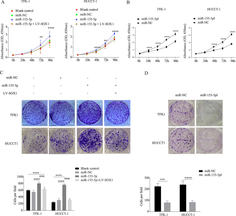
Methods: Expression levels of SOX1 in CCA tissues and normal bile duct tissues were examined using public GEO database. Western blot and immunohistochemistry were used to confirm the expression levels. Cell proliferation assay (CCK-8) and colony formation assay were performed to assess proliferation of CCA cells. A mouse model of subcutaneous transplantable tumors was used to evaluated proliferation of CCA in vivo. The putative regulating factor of SOX1 were determined using Targetscan and dual-luciferase reporter assay.

Results: SOX1 was downregulated in CCA tissues. Overexpression of SOX1 significantly inhibited cell proliferation in vitro and suppressed tumor growth in vivo. miR-155-5p directly targeted the 3'-untranslated region (3'UTR) of SOX1 and inhibited expression of SOX1, resulting in the activation of RAF, MEK and ERK phosphorylation, and thus CCA proliferation. However, restoration of SOX1 expression in miR-155-5p overexpressing cell lines decreased the phosphorylation level of RAF, MEK and ERK, as well as the proliferation of CCA cells.

Conclusion: MiR-155-5p decreased the expression of SOX1 by binding to its 3'UTR, which activated the RAF/MEK/ERK signaling pathway and promoted CCA progression.

Keywords: Cholangiocarcinoma; ERK; RAF; SOX1; miRNA.


Conflict of interest statement

The authors declare that they have no competing interests.

Figures

Fig. 1
Fig. 1
SOX1 is down-regulated in CCA tissues. A Box plots of SOX1 expression in cholangiocarcinoma (CCA) tissues compared to normal bile duct tissues. Data sourced from GEO databases GSE32225 (A) and GSE76297 (B). ***p  < 0.001, ****p  < 0.0001. B Representative IHC images showing SOX1 expression in normal bile duct tissue and CCA tissue specimens. Upper panel shows the overall view of the entire section. Scale bars are shown. The SOX1 expression were examined by semiquantitatively analyzed. **p  < 0.01. C Results of Western blotting showing the expression levels of SOX1 protein in nine CCA tissue specimens and four normal bile duct tissue specimens. N normal bile duct tissues; C CCA tissues
Fig. 2
Fig. 2
SOX1 inhibits CCA cell proliferation in vitro and suppresses tumor growth in vivo. A TFK-1 and HUCCT-1 cells were transfected with NC-SOX1, LV-SOX1 and SOX1-KD for 72 h. SOX1 protein level was assessed by Western blotting. B Representative images of colony formation assay (left panel) and analysis of colony numbers (right panel). *p  < 0.05, **p  < 0.01, ****p  < 0.0001. C Cell proliferation was assessed by CCK-8 assay. ***p  < 0.001, ****p  < 0.0001. D Above panel: Xenograft tumors at day 18 after implantation of NC-SOX1 or LV-SOX1 cells into the right flank of nude mice. Below panel: comparison of tumor volumes between NC-SOX1 and LV-SOX1 xenograft mice. **p  < 0.01, ***p  < 0.001. E Protein of xenograft tumors was extract and PCNA, BCL2, SOX1 was assessed by Western blotting. F Flow cytometry detected apoptotic cells after cells were transfected with LV-SOX1 and NC-SOX1
Fig. 3
Fig. 3
MicroRNA-155-5p directly targets 3′UTR of SOX1 and inhibits expression of SOX1, and is overexpression in CCA tissues. A Targetscan was used to screen candidate miRNAs regulating SOX1. B SOX1 protein levels in CCA cells with overexpression or inhibition of candidate miRNAs. C The relative mRNA level of SOX1 and miR-155-5p were determined respectively in TFK-1 and HUCCT-1 cells transfected with miR-NC, miR-155-5p and miR-155-5pI by RT-PCR. *p  < 0.05, **p  < 0.01, *** p  < 0.001. D The relative levels of miR-155-5p expression in four normal bile duct tissues and nine CCA tissues was analysis by RT-PCR. ****p  < 0.0001. E Box plots of miR-155-5p expression in CCA tissues compared to normal bile duct tissues. Data sourced from GEO databases GSE32957. ****p  < 0.0001. F Schematic illustration of the potential biding motifs for miR-155-5p in the wild-type (WT) 3′-UTR of SOX1 and their mutant-type (MT). G The plasmid map of psiCHECK2 which contained a SOX1 3′UTR-WT predicted binding site. H Relative activity of luciferase reporters with SOX1 3′UTR-WT and SOX1 3′UTR-MT after co-transfection with miR-155-5p mimics in HEK-293 T cells. ***p  < 0.001
Fig. 4
Fig. 4
Mir-155-5p inhibits SOX1 leading to activation of the Raf/MEK/ERK pathway. A Cells were transfected with lentiviral negative control vector (NC-SOX1) or lentiviral SOX1 (LV-SOX1) for 72 h. Protein expressions of SOX1, HES1, PROX1, p-AKT, p-JNK, and p-P38 were examined by Western blot. B Above panel: protein levels of ERK and p-ERK in HUCCT-1 and TFK-1 cells transfected with miR-negative control (miR-NC), miR-155-5p-mimic (miR-155-5p), and miR-155-5p-inhibitor (miR-155-5pI). Below panel: TFK-1 and HUCCT-1 cells was treated with different concentrations of miR-155-5pI. The protein level of ERK and p-ERK were detected by Western blot. C Protein levels of central members of MAPK/ERK signaling (RAF, p-RAF, MEK, p-MEK, ERK and p-ERK) and downstream of ERK (PCNA) in TFK-1 and HUCCT-1 cells were detected by Western blot. D TFK-1 and HUCCT-1 cells was transfected with miR-negative control (miR-NC) and miR-15-5p inhibitor (miR-155-5pI), then protein levels of central members of MAPK/ERK signaling (RAF, p-RAF, MEK, p-MEK, ERK and p-ERK) and PCNA were detected by Western blot. E TFK-1 and HUCCT-1 cells was transfected with lenvisual carrying SOX1-RNAi (SOX1-KD), then protein levels of central members of MAPK/ERK signaling (RAF, p-RAF, MEK, p-MEK, ERK and p-ERK) and PCNA were detected by Western blot. F Cells were infected with NC-SOX1, LV-SOX1, LV-SOX1  +  NC-ERK and LV-SOX1  +  LV-ERK, and then detect changes in ERK and PCNA protein levels by western blot. G Protein levels of central members of MAPK/ERK signaling (RAF, p-RAF, MEK, p-MEK, ERK and p-ERK) was detected in xenograft tumor samples which had been transfected with NC-SOX1 and LV-SOX1
Fig. 5
Fig. 5
The miR-155-5p/SOX1 axis regulates the proliferation of CCA cells. A, B CCK-8 assay was performed to compare cell proliferation in blank control, miR-NC, miR-155-5p, miR-155-5p  +  SOX1 and miR-155-5pI in TFK-1 and HUCCT-1 cells. All experiments were performed in triplicate and data are presented as mean  ±  standard deviation. **p  < 0.01, ****p  < 0.0001. C, D Representative images of colony formation assays of TFK-1 and HUCCT-1 cells in groups of blank control, miR-NC, miR-155-5p, miR-155-5p  +  SOX1 and miR-155-5pI. ***p  < 0.001, ****p  < 0.0001
Fig. 6
Fig. 6
A Schematic illustration of the mechanism by which Mir-155-5p promotes progression of CCA. Mir-155-5p represses SOX1 expression by binding to the 3′UTR region of SOX1, which activates the Raf/MEK/ERK pathway, thus promoting CCA progression. However, low expression level of miR-155-5p would not lead to inhibition of SOX1; in this setting, SOX1 would suppress the Raf/MEK/ERK pathway and inhibit CCA progression

Similar articles

Cited by

References

    1. Xie Y, Wang Y, Li J, Hang Y, Jaramillo L, Wehrkamp CJ, et al. Cholangiocarcinoma therapy with nanoparticles that combine downregulation of MicroRNA-210 with inhibition of cancer cell invasiveness. Theranostics. 2018;8(16):4305–4320. doi: 10.7150/thno.26506. - DOI - PMC - PubMed
    1. Rizvi S, Khan SA, Hallemeier CL, Kelley RK, Gores GJ. Cholangiocarcinoma—evolving concepts and therapeutic strategies. Nat Rev Clin Oncol. 2018;15(2):95–111. doi: 10.1038/nrclinonc.2017.157. - DOI - PMC - PubMed
    1. Radtke A, Konigsrainer A. Surgical therapy of cholangiocarcinoma. Visc Med. 2016;32(6):422–426. doi: 10.1159/000452921. - DOI - PMC - PubMed
    1. Wang P, Song X, Utpatel K, Shang R, Yang YM, Xu M, et al. MEK inhibition suppresses K-Ras wild-type cholangiocarcinoma in vitro and in vivo via inhibiting cell proliferation and modulating tumor microenvironment. Cell Death Dis. 2019;10(2):120. doi: 10.1038/s41419-019-1389-4. - DOI - PMC - PubMed
    1. Doherty B, Nambudiri VE, Palmer WC. Update on the diagnosis and treatment of cholangiocarcinoma. Curr Gastroenterol Rep. 2017;19(1):2. doi: 10.1007/s11894-017-0542-4. - DOI - PubMed