

Basal lamina changes in neurodegenerative disorders
- PMID: 34876200
- PMCID: PMC8650282
- DOI: 10.1186/s13024-021-00502-y
Basal lamina changes in neurodegenerative disorders
Abstract
Background: Neurodegenerative disorders are a group of age-associated diseases characterized by progressive degeneration of the structure and function of the CNS. Two key pathological features of these disorders are blood-brain barrier (BBB) breakdown and protein aggregation.
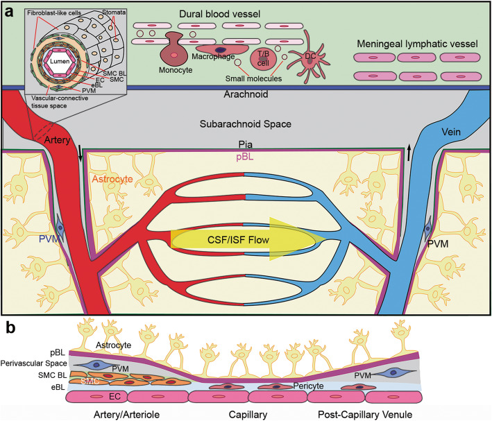
Main body: The BBB is composed of various cell types and a non-cellular component---the basal lamina (BL). Although how different cells affect the BBB is well studied, the roles of the BL in BBB maintenance and function remain largely unknown. In addition, located in the perivascular space, the BL is also speculated to regulate protein clearance via the meningeal lymphatic/glymphatic system. Recent studies from our laboratory and others have shown that the BL actively regulates BBB integrity and meningeal lymphatic/glymphatic function in both physiological and pathological conditions, suggesting that it may play an important role in the pathogenesis and/or progression of neurodegenerative disorders. In this review, we focus on changes of the BL and its major components during aging and in neurodegenerative disorders, including Alzheimer's disease (AD), Parkinson's disease (PD), and amyotrophic lateral sclerosis (ALS). First, we introduce the vascular and lymphatic systems in the CNS. Next, we discuss the BL and its major components under homeostatic conditions, and summarize their changes during aging and in AD, PD, and ALS in both rodents and humans. The functional significance of these alterations and potential therapeutic targets are also reviewed. Finally, key challenges in the field and future directions are discussed.
Conclusions: Understanding BL changes and the functional significance of these changes in neurodegenerative disorders will fill the gap of knowledge in the field. Our goal is to provide a clear and concise review of the complex relationship between the BL and neurodegenerative disorders to stimulate new hypotheses and further research in this field.
Keywords: Basal lamina; Blood-Brain Barrier; Laminin; Neurodegenerative disorders; Perlecan.
© 2021. The Author(s).
Conflict of interest statement
The authors declared no competing interests.
Figures


References
Publication types
MeSH terms
Grants and funding
LinkOut - more resources
Full Text Sources
Medical
Miscellaneous
