. 2021 Dec 8;158(1):47.
doi: 10.1186/s41065-021-00213-w.

TGF-β1/SMOC2/AKT and ERK axis regulates proliferation, migration, and fibroblast to myofibroblast transformation in lung fibroblast, contributing with the asthma progression

Affiliations

TGF-β1/SMOC2/AKT and ERK axis regulates proliferation, migration, and fibroblast to myofibroblast transformation in lung fibroblast, contributing with the asthma progression

Yuebin Wang et al. Hereditas. .

Abstract

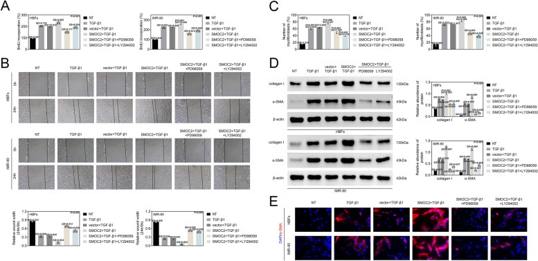
Background: Asthma is a common chronic respiratory disease that influences 300 million people all over the world. However, the pathogenesis of asthma has not been fully elucidated. It has been reported that transforming growth factor-β (TGF-β) can activate myofibroblasts. Moreover, the fibroblast to myofibroblast transformation (FMT) can be triggered by TGF-β, which is a major mediator of subepithelial fibrosis. Secreted modular calcium-binding protein 2 (SMOC2) is a member of cysteine (SPARC) family and is involved in the progression of multiple diseases. However, its role in asthma remains poorly understood. RT-qPCR evaluated the expression of SMOC2. Bromodeoxyuridine assay and wound-healing assay detected the proliferation and migration of lung fibroblasts, respectively. IF staining was performed to assess the expression of α-smooth muscle actin (α-SMA). Western blot analysis detected the levels of proteins. Flow cytometry was utilized for determination of the number of myofibroblasts.

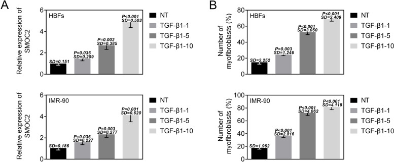
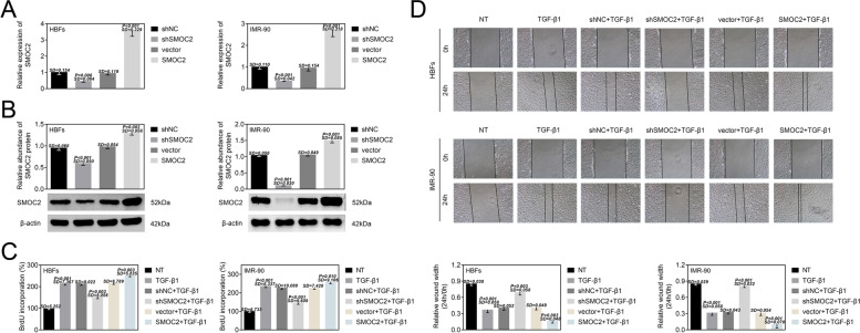
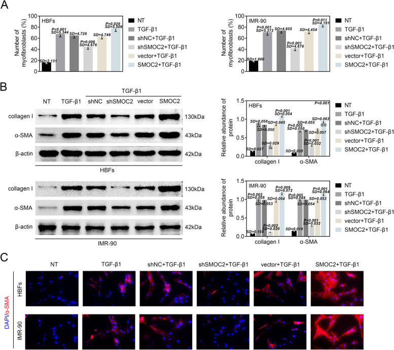
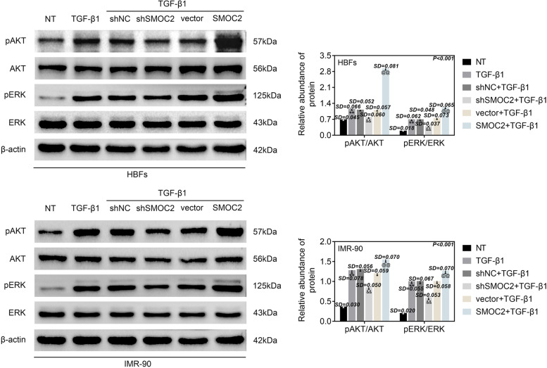
Results: We found the expression of SMOC2 was upregulated by the treatment of TGF-β1 in lung fibroblasts. In addition, SMOC2 promoted the proliferation and migration of lung fibroblasts. More importantly, SMOC2 accelerated FMT of lung fibroblasts. Furthermore, SMOC2 was verified to control the activation of AKT and ERK. Rescue assays showed that the inhibition of AKT and ERK pathway reversed the promoting effect of SMOC2 overexpression on proliferation, migration and FMT in lung fibroblasts.

Conclusions: This work demonstrated that SMOC2 modulated TGF-β1-induced proliferation, migration and FMT in lung fibroblasts and may promote asthma, which potentially provided a novel therapeutic target for the management of asthma.

Keywords: Asthma; Lung fibroblasts; Myofibroblast transformation; SMOC2; TGF-β1.


Conflict of interest statement

The authors declare that they have no competing interests, and all authors should confirm its accuracy.

Figures

Fig. 1
Fig. 1
TGF-β1 induced the upregulation of SMOC2 expression in lung fibroblasts. (A) RT-qPCR was implemented to detect the expression of SMOC2 in HBFs and IMR-90 cells following different concentration of TGF-β1 treatment (0, 1, 5, 10 ng/mL). (B) The numbers of myofibroblasts were detected by flow cytometry analysis in HBFs and IMR-90 cells following different concentration of TGF-β1 treatment (0, 1, 5, 10 ng/mL). *p < 0.05 compared with NT group, **p < 0.01 compared with NT group
Fig. 2
Fig. 2
SMOC2 modulated the proliferation and migration of lung fibroblasts. (A) RT-qPCR was carried out to disclose the expression of SMOC2 in HBFs and IMR-90 cells after transfection of shNC, shSMOC2, vector or SMOC2. **p < 0.01 compared with NT group; &&p < 0.01 compared with vector group. (B) The protein level of SMOC2 was assessed via western blot analysis in HBFs and IMR-90 cells after transfection of shNC, shSMOC2, vector or SMOC2. **p < 0.01 compared with NT group; &&p < 0.01 compared with vector group. (C) BrdU assay was performe to assess the proliferation of HBFs and IMR-90 cells after TGF-β1 treatment or combined with transfection of shNC, shSMOC2, vector or SMOC2. **p < 0.01 compared with NT group; &&p < 0.01 compared with shNC+TGF-β1 group; @@p < 0.01 compared with vector+TGF-β1 group. (D) The migration ability was detected by wound-healing assay in HBFs and IMR-90 cells after TGF-β1 treatment or combined with transfection of shNC, shSMOC2, vector or SMOC2. **p < 0.01 compared with NT group; &&p < 0.01 compared with shNC+TGF-β1 group; @@p < 0.01 compared with vector+TGF-β1 group
Fig. 3
Fig. 3
SMOC2 regulated FMT of lung fibroblasts. HBFs and IMR-90 cells were transfected with shNC, shSMOC2, vector or SMOC2, and then treated with TGF-β1. (A) Flow cytometry analysis was performed to demonstrate the number of myofibroblasts in HBFs and IMR-90 cells. **p < 0.01 compared with NT group; &&p < 0.01 compared with shNC+TGF-β1 group; @p < 0.05 compared with vector+TGF-β1 group. (B) The protein levels of collagen I and α-SMA in HBFs and IMR-90 cells were evaluated via western blot analysis. **p < 0.01 compared with NT group; &&p < 0.01 compared with shNC+TGF-β1 group; @@p < 0.01 compared with vector+TGF-β1 group. (C) IF staining was perfoemed to measure the protein content and distribution of α-SMA in HBFs and IMR-90 cells
Fig. 4
Fig. 4
SMOC2 controlled the activation of AKT and ERK. HBFs and IMR-90 cells were transfected with shNC, shSMOC2, vector or SMOC2, and then treated with TGF-β1. Western blot analysis was implemented to evaluate the protein levels of p-AKT and p-ERK in HBFs and IMR-90 cells. **p < 0.01 compared with NT group; &&p < 0.01 compared with shNC+TGF-β1 group; @@p < 0.01 compared with vector+TGF-β1 group
Fig. 5
Fig. 5
Inhibition of AKT and ERK pathway reversed the promoting effect of SMOC2 overexpression on FMT. HBFs and IMR-90 cells were transfected with vector or SMOC2, and then treated with TGF-β1 or combined with PD98059 or LY294002. BrdU (A) and wound healing (B) assays were performed to assess cell proliferation and migration in HBFs and IMR-90 cells. **p < 0.01 compared with NT group; &&p < 0.01 compared with vector+TGF-β1 group; @@p < 0.01 compared with SMOC2 + TGF-β1 group. (C) The number of myofibroblasts in HBFs and IMR-90 cells was detected by flow cytometry analysis. **p < 0.01 compared with NT group; &&p < 0.01 compared with vector+TGF-β1 group; @@p < 0.01 compared with SMOC2 + TGF-β1 group. (D) The protein levels of collagen I and α-SMA in HBFs and IMR-90 cells were measured by western blot analysis. **p < 0.01 compared with NT group; &&p < 0.01 compared with vector+TGF-β1 group; @@p < 0.01 compared with SMOC2 + TGF-β1 group. (E) IF staining was performed to examine the protein content and distribution of α-SMA in HBFs and IMR-90 cells

Similar articles

Cited by

References

    1. Masoli M, Fabian D, Holt S, Beasley R. The global burden of asthma: executive summary of the GINA dissemination committee report. Allergy. 2004;59:469–478. doi: 10.1111/j.1398-9995.2004.00526.x. - DOI - PubMed
    1. To T, Stanojevic S, Moores G, Gershon AS, Bateman ED, Cruz AA, et al. Global asthma prevalence in adults: findings from the cross-sectional world health survey. BMC Public Health. 2012;12:204. doi: 10.1186/1471-2458-12-204. - DOI - PMC - PubMed
    1. Djukanovic R, Lai CK, Wilson JW, Britten KM, Wilson SJ, Roche WR, et al. Bronchial mucosal manifestations of atopy: a comparison of markers of inflammation between atopic asthmatics, atopic nonasthmatics and healthy controls. Eur Respir J. 1992;5:538–544. - PubMed
    1. Roche W, Williams J, Beasley R, Holgate S. Subepithelial fibrosis in the bronchi of asthmatics. Lancet. 1989;333:520–524. doi: 10.1016/S0140-6736(89)90067-6. - DOI - PubMed
    1. Beasley R, Roche WR, Roberts JA, Holgate ST. Cellular events in the bronchi in mild asthma and after bronchial provocation. Am Rev Respir Dis. 1989;139:806–817. doi: 10.1164/ajrccm/139.3.806. - DOI - PubMed

Substances