. 2021 Dec;23(12):1255-1270.
doi: 10.1038/s41556-021-00800-z. Epub 2021 Dec 7.

Reciprocal regulation of chaperone-mediated autophagy and the circadian clock

Affiliations

Reciprocal regulation of chaperone-mediated autophagy and the circadian clock

Yves R Juste et al. Nat Cell Biol. 2021 Dec.

Abstract

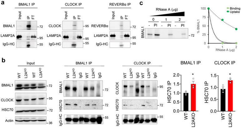
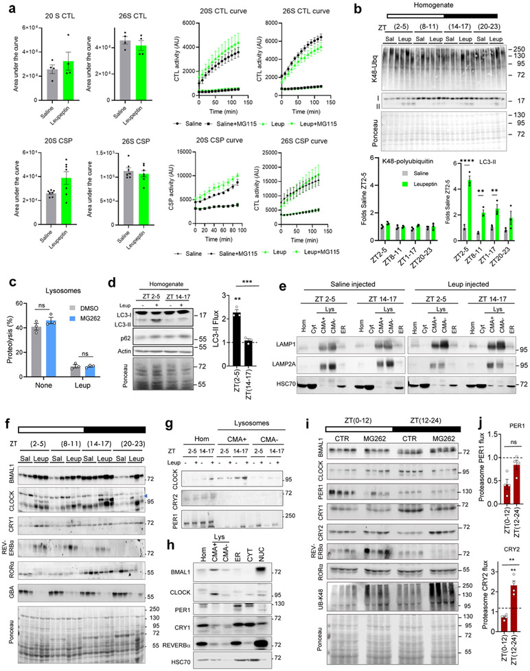
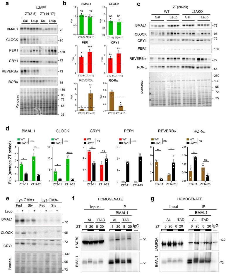
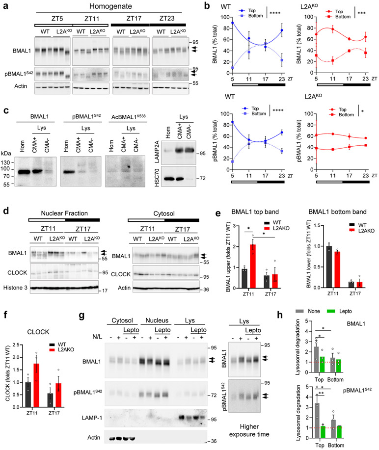
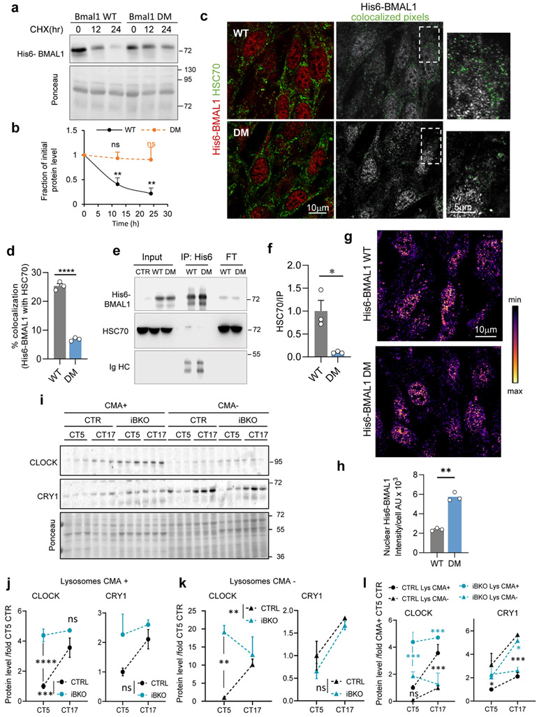
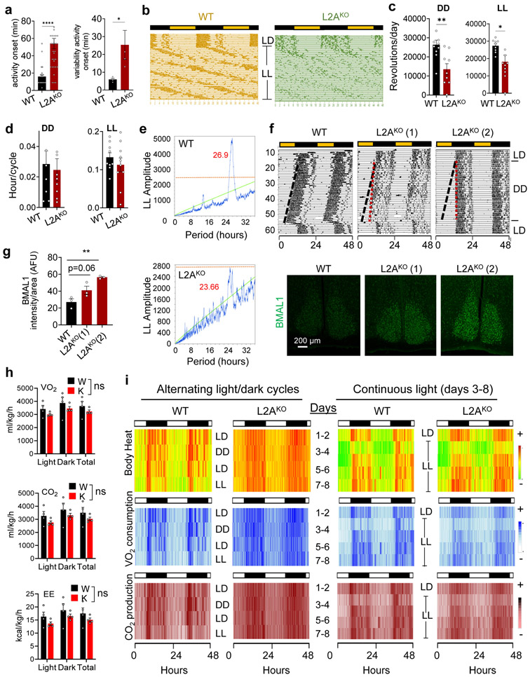
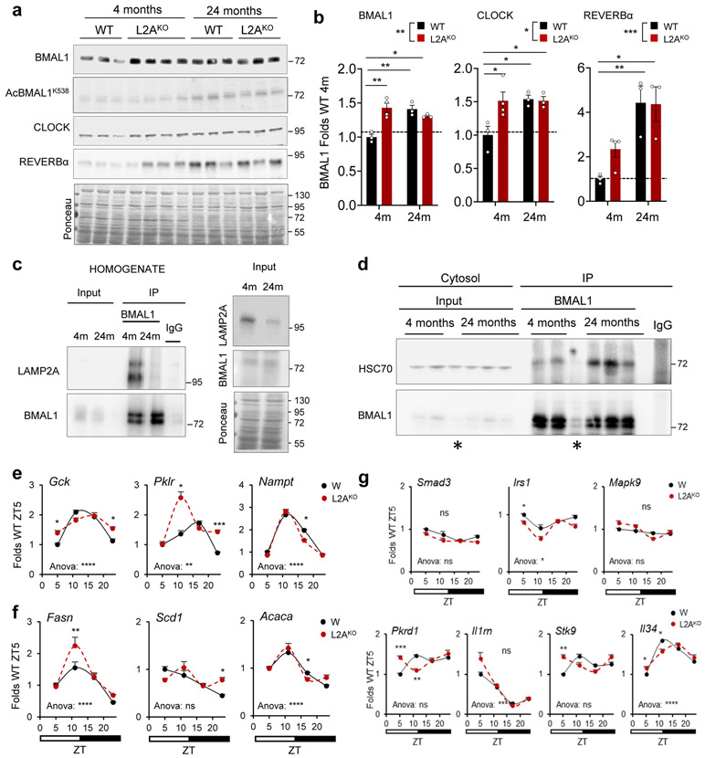
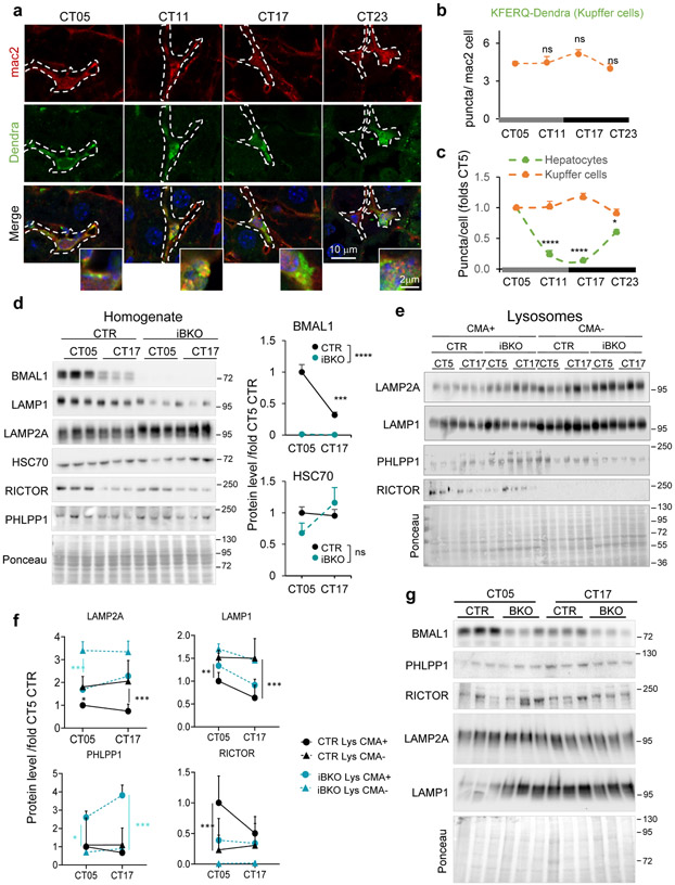
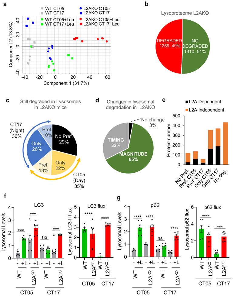
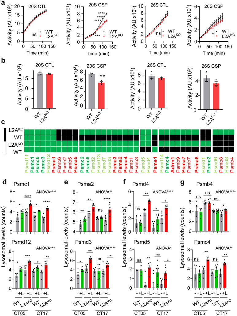
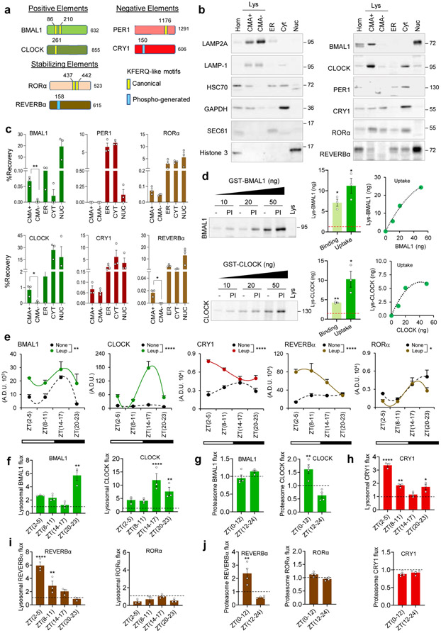
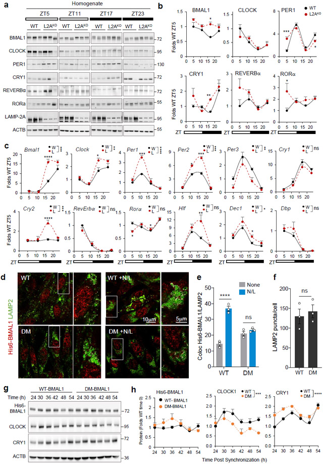
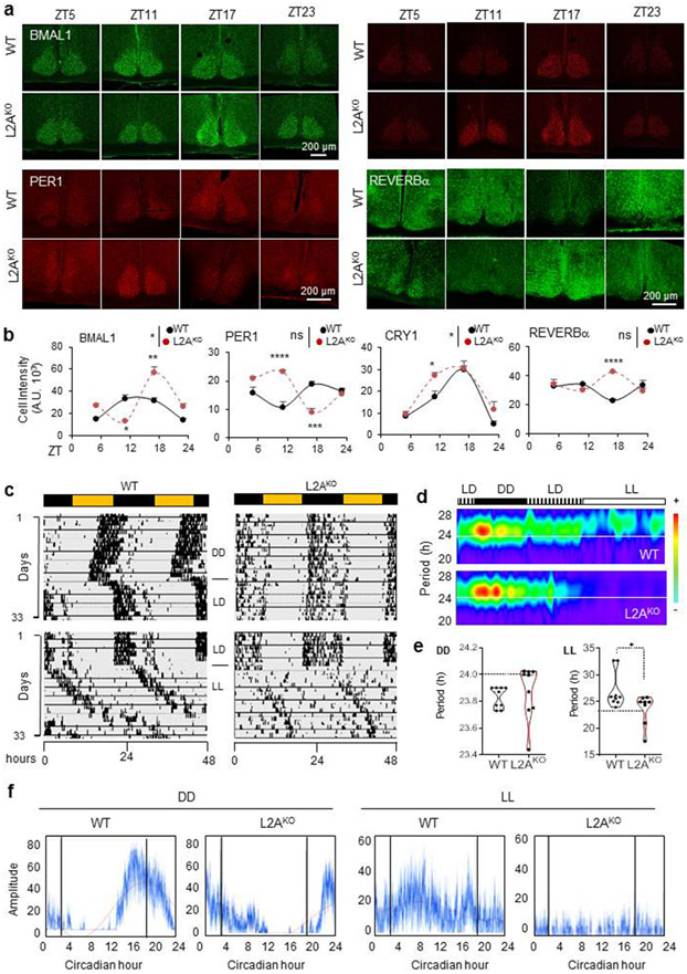
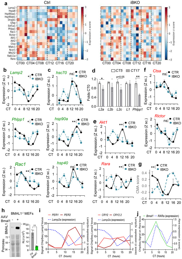
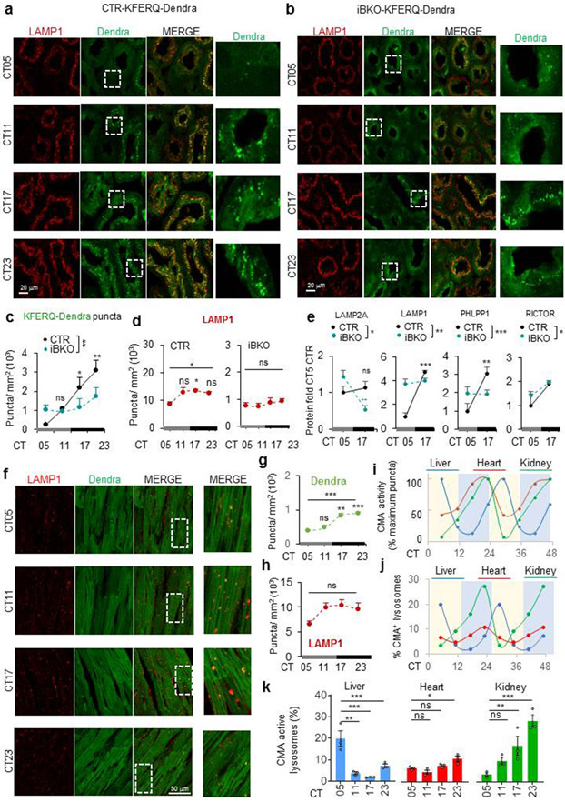
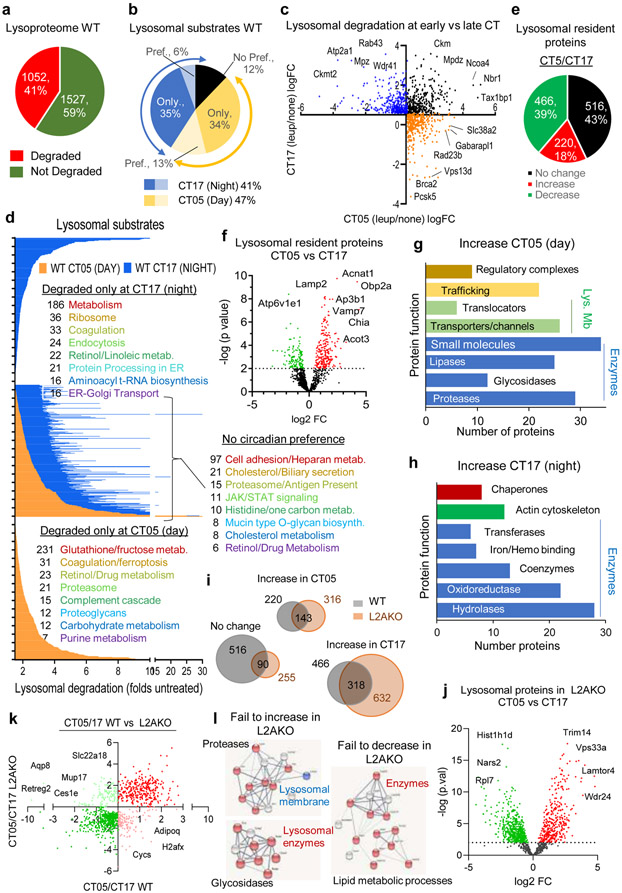
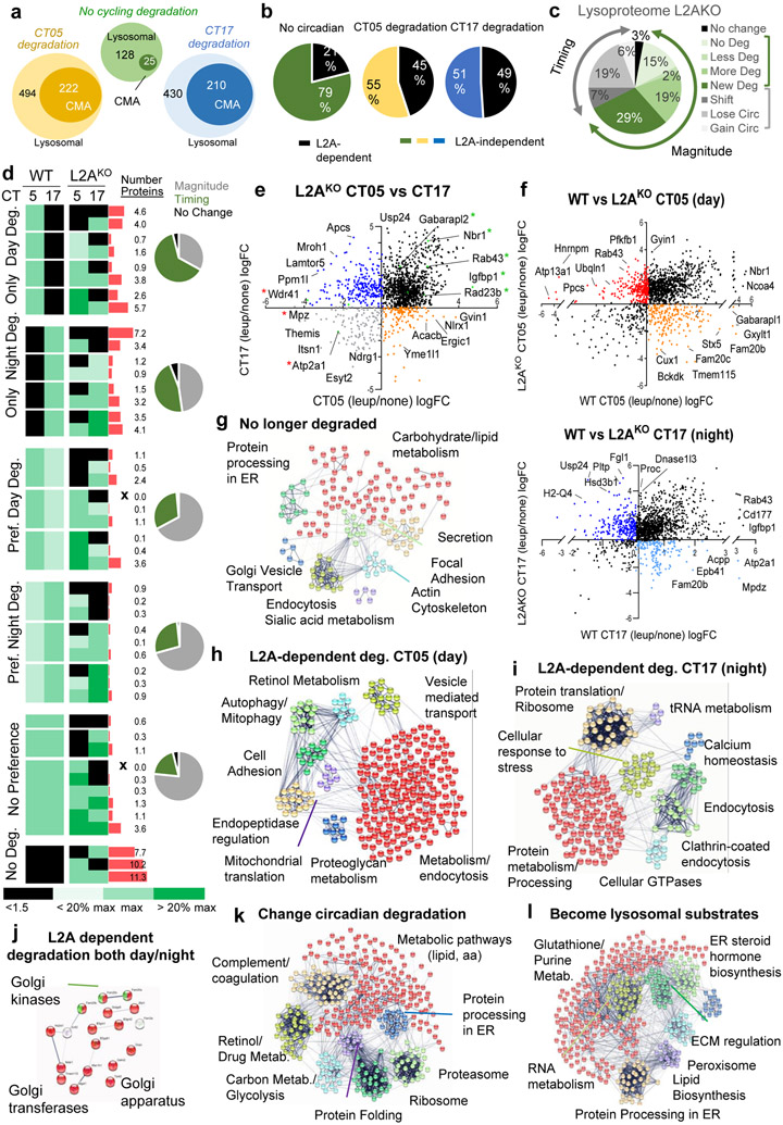
Circadian rhythms align physiological functions with the light-dark cycle through oscillatory changes in the abundance of proteins in the clock transcriptional programme. Timely removal of these proteins by different proteolytic systems is essential to circadian strength and adaptability. Here we show a functional interplay between the circadian clock and chaperone-mediated autophagy (CMA), whereby CMA contributes to the rhythmic removal of clock machinery proteins (selective chronophagy) and to the circadian remodelling of a subset of the cellular proteome. Disruption of this autophagic pathway in vivo leads to temporal shifts and amplitude changes of the clock-dependent transcriptional waves and fragmented circadian patterns, resembling those in sleep disorders and ageing. Conversely, loss of the circadian clock abolishes the rhythmicity of CMA, leading to pronounced changes in the CMA-dependent cellular proteome. Disruption of this circadian clock/CMA axis may be responsible for both pathways malfunctioning in ageing and for the subsequently pronounced proteostasis defect.


Figures

Extended Data Fig. 1
Extended Data Fig. 1. Circadian proteins display properties of bona fide CMA substrates.
a. Representative immunoblot for LAMP2A from rat liver lysosomes immunoprecipitated for BMAL1 (left), CLOCK (middle), or REVERBα (right). The heavy chain (HC) of IgG used in the immunoprecipitation is shown. Input is 5% amount used for IP. b. Representative immunoblots for HSC70 from wild-type (WT) or LAMP2A knockout (L2AKO) cell lysates immunoprecipitated for BMAL1 or CLOCK. Heavy chain (HC) for IgG used in the immunoprecipitation is shown. Input is 5% concentration used for IP. Right, quantification of HSC70 normalized by the amount of BMAL1 or CLOCK immunoprecipitated. An increase in the amount of CMA substrates bound at a given time to HSC70, similar to the one observed here for BMAL1 and CLOCK, has previously been described upon blockage of CMA. n=3 independent experiments. Individual values (b) and mean+s.e.m are shown. Unpaired two-tailed t test was used, and differences were significant for *p<0.05. c. Representative immunoblot of competition of BMAL1 lysosomal uptake by ribonuclease A (RNase A). Left: representative immunoblot. Right: effect of increasing concentrations of RNase A on binding and uptake of 20ng of BMAL1. Numerical source data, statistics and exact p values and unprocessed blots are available as source data.
Extended Data Fig. 2
Extended Data Fig. 2. Circadian lysosomal and proteasomal protein degradation in liver
a. Chymotrypsin-like (CTL) and caspase-like (CSP) activities of the 20S and 26S proteasome in livers from mice injected with saline (Sal) or leupeptin (Leup; 40mg/kg b.w.) 3h before tissue collection. Area under the curve after discounting residual MG115-resistent activity (left) and time-course kinetics (right) are shown. n=4 (CTL), 7 (CSP) mice. b. Top: Immunoblot from livers from mice treated as in a collected at the indicated Zeitgeber times (ZT). Ponceau staining is shown as loading control. Bottom: Levels of K48-polyubiquitinated proteins (left) and LC3-II (right) relative to ZT2-5 saline injected. n=3 mice per ZT and treatment. c. Proteolytic activity of liver lysosomes from mice injected with MG262 in 60% dimethyl sulfoxide (DMSO) or only DMSO (control; CTR) and incubated with a pool of radiolabeled proteins with or without leupeptin. n=3 mice per treatment. d. Left: Representative immunoblot of homogenate of mice injected (+) or not (−) with leupeptin (leup) as in a. Right: LC3-II flux (fold increase upon leupeptin injection). n=3 mice per ZT. e-g. Immunoblot of liver homogenates (Hom), lysosomes (Lys) active (+) or not (−) for CMA, ER, and cytosol (Cyt) (e), CMA+ lysosomes (f) and CMA+ and CMA (−) lysosomes (g) isolated from mice treated as in a. Loading controls for f are in e. h. Immunoblot of rat liver fractions at ZT17 to compare with ZT5 (Fig. 1b). i. Immunoblot of liver homogenates from mice treated as in c. UB-K48 is used as a control for efficacy of MG262 injection. j. Proteasome degradation of PER1 and CRY2 (>1.5 fold increase upon MG262 injection) at each time. n=4 mice per ZT. Dotted line: no degradation. Individual values (a-d,j) and mean+s.e.m are shown. Unpaired t-test (a left) and Two-way ANOVA followed by Sidak (a right, b and c) or Bonferroni’s (c, d, j) multiple comparisons post-hoc tests were used Differences were significant for *p<0.05, **p<0.01, ***p<0.001 and ****p<0.0001. ns=not significant. Numerical source data, statistics and exact p values and unprocessed blots are available as source data.
Extended Data Fig. 3
Extended Data Fig. 3. Lysosomal degradation of clock proteins is dependent on CMA but independent of the nutritional status.
a. Immunoblot of liver homogenates from LAMP2A knockout mice (L2AKO) intraperitoneally injected (i.p.) with saline (Sal) or a single dose of leupeptin 3h before tissue collection at the indicated circadian times (ZT). b. Lysosomal flux for the indicated circadian proteins calculated as the increase in >1.5 folds above the dotted line (value 1) upon leupeptin injection. n=3 mice per ZT. c. Immunoblot of livers from WT and L2AKO mice treated as in a and collected at ZT(20-23), time of maximal BMAL1 and CLOCK lysosomal degradation, and run in the same membrane for comparative purposes. d. Lysosomal flux for the indicated proteins in WT and L2AKO mice livers from blots in Extended Data Fig. 2f and 3a,c. n=3 mice per ZT and genotype. A lane with the same sample in all gels was used for normalization across membranes. Note that PER1 was not detected in WT lysosomes (Extended Data Fig. 2g). e. Immunoblot for the indicated proteins in CMA-active lysosomes (Lys CMA+) and CMA-inactive lysosomes (Lys CMA−) from mice fed ad libitum or starved for 24h (Stv) and injected or not with leupeptin. A representative immunoblot is shown (the experiment was replicated 3 times). f,g. Immunoblot for HSC70 (f) and LAMP2A (g) of BMAL1 immunoprecipitated (IP) from liver homogenates of mice fed ad libitum (AL) or maintained in an isocaloric twice-a-day feeding (iTAD) during the light time for 6 months collected at ZT8 and ZT20. The experiment was repeated 3 times. Individual values and mean+s.e.m are shown. Two-way ANOVA followed by Bonferroni's multiple comparisons post-hoc test was used to determine differences in protein flux at each time in L2AKO mice (b) and differences between genotypes (d). Differences in flux between both ZT are shown in the legend and between genotypes on top of the bars. Absolute protein levels are included in the raw data file. *p<0.05, **p<0.01, ***p<0.001 and ****p<0.0001. ns = not significant. Numerical source data, statistics and exact p values and unprocessed blots are available as source data.
Extended Data Fig. 4
Extended Data Fig. 4. Properties of CMA targeted BMAL1.
a. Immunoblot for BMAL1 phosphorylated on serine 42 (pBMAL1S42) in wild-type (WT, W) and LAMP2A knockout mice (L2AKO, L) livers at the indicated Zeitgeber times (ZT). Total BMAL1 and actin immunoblots from Fig. 2a are shown here for comparison. Arrows: BMAL1 bands of different electrophoretic mobility. All membranes contained a common sample for normalization across membranes. b. Levels of the two bands of total (top) and pBMAL1S42 (bottom) in WT (left) and L2AKO mice (right) from immunoblots as in a. Values are percentage of total BMAL1 contributed by each band. n=3 mice per ZT. c. Immunoblot for total BMAL1, pBMAL1S42 and BMAL1 acetylated on lysine 538 (AcBMAL1K538) in mouse liver homogenates (Hom) and lysosomes (Lys) active (+) or not (−) for CMA at the time of maximal BMAL1 lysosomal degradation. LAMP2A and HSC70 from the same fractions are shown on the right. d. Immunoblot of nuclear (left) and cytosolic fractions (right) from WT and L2AKO mice livers collected at the indicated Zeitgeber times (ZT), n=3 mice per ZT and genotype. Actin is shown as cytosolic loading control and Histone 3 as marker of the nuclear fraction. e, f. Levels of the top (left) and bottom (right) bands of BMAL1 (e) and of CLOCK (f) shown in d. Values are folds of ZT11 WT (arbitrary value of 1). n=3 mice per ZT and genotype in e and f. g. Immunoblot for total BMAL1 and pBMAL1S42 in fractions from synchronized NIH3T3 cells cultured or not with leptomycin to block nuclear export and (+) ammonium chloride and leupeptin (N/L) to block lysosomal degradation. Right: higher exposure of lysosome lanes. h. Lysosomal degradation (fold increase upon N/L) of the top and bottom BMAL1 (top) and pBMAL1S42 bands (bottom) in cells in g. Red dotted line: no degradation. n=3 mice per treatment. Individual values (e,f,h) and mean+s.e.m are shown. Two-way ANOVA followed by Bonferroni's multiple comparisons post-hoc test was used. *p<0.05, **p<0.01, ***p<0.001 and ****p<0.0001. Numerical source data, statistics and exact p values and unprocessed blots are available as source data.
Extended Data Fig. 5
Extended Data Fig. 5. Expression of BMAL1 protein mutated in the CMA-targeting motifs.
a. Immunoblot for His6 tag of NIH3T3 cells stably expressing His6-tagged BMAL1 wild-type (WT) or mutated in both CMA-targeting motifs (DM) at the indicated times after addition of cycloheximide (CHX) to stop protein synthesis. Ponceau is shown as loading control. b. Quantification of BMAL1 in experiments as in a. Values are expressed as fraction of the initial BMAL1 remaining at each time. n=3 independent experiments. c. Immunofluorescence for HSC70 (green) and His6 tag (red) in NIH3T3 cells stably expressing WT and DM BMAL1. Merged channels and colocalization mask are shown. Insets: boxed areas at higher magnification. d. Fraction of His6-BMAL1 colocalizing with HSC70 (Mander’s Coefficient - M1), n=3 independent experiments. e. Co-immunoprecipitation (IP) of HSC70 with BMAL1 in the same cells. Input and flow through (FT) are also shown. f. Amount of HSC70 co-immunoprecipitated with BMAL1 expressed as folds that in WT (arbitrary value of 1), n=3 independent experiments. g. Immunofluorescence for His6 tag in cells as in c. Image shows pseudocolor by gradient intensity. h. Intensity of nuclear BMAL1 in images as in g. n=3 independent experiments (55 cells were quantified per experiment and the mean value of intensity in each experiment was used for statistics). i-l. Immunoblot for CLOCK and CRY1 in lysosomes active (+) or not (−) for CMA isolated from livers of WT or BMAL1 knockout mice (iBKO) at the indicated CT times (i). Quantification of the levels of both proteins in CMA+ (j) and CMA− (k) lysosomes and in the four fractions (l) together for comparative purposes from fractions as the ones in I. Values are expressed as fold those in fractions from CT5 WT. n=3 mice per CT and genotype (in j, k and l). Individual values (d,f,h) and mean+s.e.m are shown. Two-way ANOVA followed by Bonferroni’s (b, j, k) or Tukey’s (l) post-hoc test for multiple variable comparisons and two-sided unpaired T-test (d, f, h) were used. *p<0.05, **p<0.01, ***p<0.001 and ****p<0.0001. ns = not significant. Numerical source data, statistics and exact p values and unprocessed blots are available as source data.
Extended Data Fig. 6
Extended Data Fig. 6. Free running behavior and metabolic characteristics of CMA-deficient mice.
a. Average activity onset relative to light offset (left) and variability of activity onset (right) of wild-type (WT) and LAMP-2A knock out mice (L2AKO) under 12:12 light:dark (LD) cycle. n=32 recordings in 4 mice per genotype (left) and n=4 mice per genotype (right). b. Representative actograms of WT and L2AKO mice during continuous light exposure (LL) n=9 mice per genotype. c, d. Average wheel running revolutions per day (c) and strength of rhythmicity (d) of WT and L2AKO mice in continuous dark (DD) or light (LL), n=10 (WT) and 9 (L2AKO) mice in c and d. e. Representative chi-square periodogram of WT and L2AKO mice in LL. The mean period is in red. n=9 mice per genotype. Significance level (above green line) with chi-squared threshold of 0.001. Orange dotted line: peak period of L2AKO mice (weaker periodicity than WT). f. Double-plotted actograms of WT and L2AKO mice with moderate (1) or severe (2) disruption of circadian properties at the indicated light conditions. Discontinuous lines: corrective shift in response to continuous darkness in WT (black) or L2AKO mice (red). Bottom: BMAL1 immunostaining of SCN at ZT17 in the mice of the actograms shown on top. g. Average intensity per area of BMAL1 staining in SCN of WT and L2AKO mice separated by the severity of their actogram changes, n=3 mice per group. h. Average value during the LD cycles or throughout a 24h period (total) of O2 consumption, CO2 production and energy expenditure (EE) in WT or L2AKO mice. n=4 mice per genotype. i. Median body heat production, VO2 consumption and CO2 production plots from WT and L2AKO mice, n=4 mice per genotype, subjected to alternating light/dark cycles each 2 days (left) or continuous light for 8 days (right). Data is single plotted in two-day intervals. Individual values and mean+s.e.m are shown. Unpaired two tailed t test (a, c and d), one-way (g) and two-way (h) ANOVA followed by Bonferroni’s multiple comparisons post-hoc test were used. *p<0.05, **p<0.01, ***p<0.001. ns: not significant. Numerical source data, statistics and exact p values are available as source data.
Extended Data Fig. 7
Extended Data Fig. 7. Changes in clock components and in the circadian transcriptome with age in mice with active or inactive CMA activity.
a. Immunoblot for the indicated proteins in livers from 4m and 24m old wild-type (WT) and LAMP2A knockout (L2AKO) mice. n=3 (WT) and 4 (L2AKO) mice per age. Ponceau staining is shown as loading control. b. Levels of the indicated proteins in the same mice obtained by densitometric quantification of immunoblots as in a. Values are expressed as fold levels in 4m old WT (dotted line) n=3 (WT) and 4 (L2AKO) mice per age. Animals were analyzed at ZT2 to catch the end of the nocturnal degradation of BMAL1 and CLOCK and the beginning of the diurnal degradation of REVERBα. c, d. Co-immunoprecipitation of LAMP2A (c) and HSC70 (d) with BMAL1 in livers of 4m and 24m old mice. * sample lost during processing. Experiments were repeated 3 times. e-g. Temporal changes in the mRNA of representative genes involved in carbohydrate (e), lipid metabolism (f) and in cytokine and interleukin signaling (g) shown to undergo reprograming of their circadian expression in old mice in the liver of young WT (W) or L2AKO (L) mice at the indicated ZT times. n=3 mice per ZT and genotype. Individual values (b) and mean+s.e.m are shown. Two-way ANOVA test followed by Tukey's multiple comparisons post-hoc test was used in b, and by multiple two-sided unpaired t-test in e-g. In b, significant differences between genotypes are marked in the legend and differences with 4m WT in the graph. In e-g, significant differences between genotypes are marked in the graph and ANOVA indicates p value for time differences. *p<0.05, **p<0.01, ***p<0.001 and ****p<0.0001. ns: not significant. Numerical source data, statistics and exact p values and unprocessed blots are available as source data.
Extended Data Fig. 8
Extended Data Fig. 8. Cell-type and tissue-specific changes in hepatic CMA activity during the light/dark cycle in different liver cells.
a. Immunofluorescence for Dendra and mac2 (to label hepatic Kupffer cells) in liver sections from KFERQ-Dendra mice at the indicated circadian times (CT). Nuclei are highlighted with Hoechst. Individual and merged channels are shown. Insets: higher magnification images. Dotted white line: Kupffer cells profile. b. Quantification of Dendra+ puncta per mac2+ cell section. n=3 mice per CT (50 cells were quantified per mouse and the mean value of puncta per cell in each mouse was used for statistics). c. Quantification of temporal changes in Dendra+ puncta number in hepatocytes and Kupffer cells relative to CT05 values (arbitrary value of 1). n=3 mice per CT (65 cells of each type were quantified per mouse and the mean value of puncta per cell in each mouse was used for statistics). d. Immunoblot of homogenate from control (CTR) and BMAL1 knockout (iBKO) mice livers at two circadian times (CT). Ponceau staining is shown as loading control. Right: Quantification of BMAL1 and HSC70, n=3 mice per CT and genotype. e,f. Immunoblot of lysosomes active (+) or inactive (−) for CMA isolated from livers of CTR and iBKO mice (e). Ponceau staining is shown as loading control. Quantification of immunoblots as the ones shown in e (f). n = 6 (CTR) and 3 (iBKO) mice per CT. g. Immunoblot for the indicated proteins in homogenate from kidneys of control (CTR) and BMAL1 knockout (iBKO) mice at two circadian times (CT). Three mice per condition are shown. Ponceau staining is shown as loading control. All values are mean+s.e.m. One-way ANOVA test followed by Tukey HSD post-hoc tests (b) or two-way ANOVA test followed by Bonferroni’s (c) or Tukey’s (d,f) (post-hoc tests for multiple variable comparisons were used. Significant differences between genotypes are shown in the legend and between times in the graph in d. Significant differences between CMA+ and CMA− lysosomes for each genotype are marked in the graph in f and between lysosomes, times and genotype are summarized in the raw data file. *p<0.05, **p<0.01, ***p<0.001 and ****p<0.0001. ns = not significant. Numerical source data, statistics and exact p values and unprocessed blots are available as source data.
Extended Data Fig. 9
Extended Data Fig. 9. Changes in the cyclic degradation of macroautophagy components upon CMA blockage.
Comparative differential proteomics of lysosomes from livers of wild-type (WT) or LAMP2A knockout mice (L2AKO) injected or not with leupeptin (Leu) and isolated at circadian time CT05 (day) or CT17 (night). All values are from n=3 mice per CT, genotype and treatment group. a. Principal component analysis showing the multivariate variation among the different genotype, time, and treatment groups. b. Percentage and number of proteins undergoing or not lysosomal degradation (increase upon leupeptin treatment). c. Preferences in degradation time of the subset of proteins degraded in lysosomes from L2AKO mice. d. Percentage of proteins in lysosomes from L2AKO mice displaying changes in their rate (magnitude) or time (timing) of degradation. e. Number of proteins in lysosomes according to their preference in degradation time and their dependence on the presence of LAMP2A in lysosomes. f,g. Lysosomal degradation of macroautophagy proteins LC3 (f) and p62 (g) calculated from the proteomic data. Graphs show quantification of levels of each of the proteins in lysosomes isolated from mice untreated (−) or injected with leupeptin (+L) (left) and lysosomal flux for both proteins calculated as differences in their lysosomal abundance between untreated or leupeptin injected mice (right). n=3 mice per CT, genotype and treatment with technical replicates for each one. Individual values per mouse (f,g) and mean+s.e.m are shown. Two-way ANOVA test followed by Bonferroni’s (f, g) post-hoc test (for multiple variable comparisons) was used. Differences were significant for ***p<0.001 and ****p<0.0001. ns = not significant. Numerical source data, statistics and exact p values are available as source data.
Extended Data Fig. 10
Extended Data Fig. 10. Changes in proteasome components upon CMA blockage.
a, b. Chymotrypsin-like (CTL) and caspase-like (CSP) activities of the 20S and 26S proteasome in livers from WT or L2AKO mice. Time-course kinetics (a) and area under the curve (b) are shown. n=3 mice per genotype in a and b. c-g. Lysosomal degradation of proteasome subunits in the same mice. c shows heatmaps of degradation (green) or not (black) at the two time points and d-g shows changes in lysosomal levels of the indicated proteasome proteins as representative examples of the 4 identified patterns: subunits with comparable degradation and cycling in WT and L2AKO mice (d), subunits preferentially degraded during the day (e) or during the night (f) in WT mice that become continuously degraded in L2AKO mice, and subunits normally not degraded in lysosomes that become now lysosomal substrates (g). n=3 mice per CT, genotype and treatment with technical replicates for each one. Individual values per mouse and mean+s.e.m are shown. Two-way ANOVA test followed by Bonferroni’s (a) or Sidak’s (d-g) post-hoc tests (for multiple variable comparisons), and two-sided unpaired T-test (b) were used. Differences were significant for *p<0.05, **p<0.01, ***p<0.001 and ****p<0.0001. ns = not significant. Numerical source data, statistics and exact p values are available as source data.
Fig. 1.
Fig. 1.. Components of the molecular clock are degraded in lysosomes via CMA.
a, Canonical (yellow) and phosphorylation generated (blue) CMA-targeting motifs in mammalian clock proteins. b, Immunoblot for circadian clock proteins in rat liver homogenates (Hom), lysosomes (Lys) active (+) or not (−) for CMA, ER, cytosol (Cyt) and nuclear fractions (Nuc). Left: markers for lysosomes (LAMP2A and LAMP1), CMA+ lysosomes (HSC70), cytosol (GAPDH), ER (SEC61) and nucleus (Histone 3). c, Percentage of the positive (green), negative (red) and stabilizing (brown) clock elements recovered in each fraction relative to their total cellular content. n=3 rats. d, Immunoblot of BMAL1 (top) and CLOCK (bottom) associated with lysosomes pre-incubated or not with protease inhibitors (PI) and then incubated with recombinant BMAL1 and CLOCK. Lysosomal binding and uptake (middle) and uptake upon incubation with increasing protein concentrations (right). n=3 independent experiments. e-j, Temporal changes of clock elements in lysosomes from mice injected or not with leupeptin (e, f, h, i) or in homogenates of mice injected or not with MG262 (g, h, j) calculated from blots in Extended Data Fig. 2f, i, respectively. n=3 mice per CT and treatment in e-j. Individual values (c,d,f-j) and mean±s.e.m are shown. One-way ANOVA was used in c to determine differences in recovery among fractions and unpaired two-tailed t test (shown here) to determine differences between CMA+ and CMA− lysosomes. One sample t and Wilcoxon test with hypothetical value of 1 for binding and uptake was used in d. Two-way ANOVA followed by Bonferroni's multiple comparisons post-hoc test was used in e-j. Significant differences between untreated and leupeptin treated samples are shown in e (graph legend) and the specific times at which flux was significantly different are marked in f-j. Statistical analysis was performed in all panels but only significant differences are labeled in the graphs as significant for *p<0.05, **p<0.01 and ****p<0.0001. Numerical source data, statistics and exact p values and unprocessed blots are available as source data.
Fig. 2.
Fig. 2.. Blockage of CMA disrupts the molecular clock.
a, Immunoblot of wild-type (WT, W) and LAMP2A knockout mice (L2AKO, L) livers collected at the indicated Zeitgeber times (ZT). Actin is shown as loading control, and LAMP2A to confirm KO. (Note: the anti-L2A antibody recognizes a non-specific protein in liver, ). ZT5 and ZT17 samples and ZT11 and ZT23 samples were run in the same membrane and all membranes contained a common sample for normalization across membranes. b, Temporal changes of clock proteins in a expressed as fold levels in WT at ZT5. n=3 mice per ZT and genotype. c, Temporal changes in clock components mRNA levels in the same animal groups as a. Values were normalized to TBP as housekeeping gene and are expressed as fold levels in WT at ZT5. n=3 mice per ZT and genotype. d, Immunofluorescence for LAMP2 (green) and His6 tag (red) in NIH3T3 cells stably expressing wild-type (WT) BMAL1 histidine tagged (His6) or the same protein mutated in its two CMA-targeting motifs (DM). Where indicated 20mM NH4Cl, 100μM leupeptin (N/L) was added to prevent lysosomal proteolysis. Insets: boxed area with colocalized pixels (white) at higher magnification. e, f, Fraction of His6-BMAL1 colocalizing with LAMP2 (Mander’s Coefficient - M1) (e) and average number of LAMP2 fluorescent puncta (f) in cells as in d. n=3 independent experiments (30 cells were quantified per experiment and the mean value of puncta per cell per experiment was used for statistics). g, Immunoblot for clock elements in cells expressing WT or DM-BMAL1 collected at the indicated times after dexamethasone-synchronization. Actin is shown as loading control. h, Levels of the indicated clock elements in experiments as in g. n=4 independent experiments. Individual values (e,f) and mean±s.e.m are shown. Unpaired two tailed t test was used in f and two-way ANOVA followed by Bonferroni’s multiple comparison post-hoc tests in b, c, e and h. Significant differences by genotype or mutation are indicated in the legends in c and g, respectively. Differences were significant for *p<0.05, **p<0.01, ***p<0.001 and ****p<0.0001. ns = not significant. Numerical source data, statistics and exact p values and unprocessed blots are available as source data.
Fig. 3.
Fig. 3.. CMA-defective mice display disturbances in circadian patterns.
a, Immunostaining for BMAL1, PER1, CRY1 and REVERBα in the suprachiasmatic nucleus region (SCN) of brains of wild-type (WT) or LAMP2A knockout mice (L2AKO) at the indicated Zeitgeber times (ZT). b, Quantification of the average fluorescent intensity per cell in the SCN area in images as the ones shown in a. n=3 mice per ZT and genotype. c, Double-plotted actograms of WT and L2AKO mice subjected to the light manipulations indicated in the middle for the number of days indicated on the left. LD, 12:12 light:dark; DD, continuous dark; LL, continuous light. d, Representative Morlet Wavelet of WT and L2AKO mice under the same conditions as in c. e, Average free running circadian period in the DD (left) and LL (right) cycles. n=9 WT mice per condition and 9 (for DD) and 8 (for LL) L2AKO mice. f, Representative activity profile of WT and L2AKO mice for an average 10 cycle in DD (left) or LL (right). The red line is a sine-fit to the waveform indicating the rhythm of these mice. Individual values (e) and mean±s.e.m are shown. Unpaired two-tailed t test was used in e and two-way ANOVA test followed by Bonferroni’s multiple comparisons test post-hoc test was used for b. Significant differences by genotype across the ZT are shown in the legends. Differences were significant for *p<0.05, **p<0.01 and ****p<0.0001. ns = not significant. Numerical source data, statistics and exact p values are available as source data.
Fig. 4.
Fig. 4.. CMA displays BMAL1-dependent circadian activity in liver.
a, Immunofluorescence for Dendra in liver sections from KFERQ-Dendra mice control (CTR) or knockout for BMAL1 (iBKO) at the indicated circadian times (CT). Nuclei are highlighted with DAPI. Right shows higher magnification inserts. b, Quantification of the number of Dendra+ puncta per mm2 of liver section in images as the ones shown in a. n=4 mice per CT and genotype. c, Immunofluorescence for Dendra (green) and LAMP1 (red) in liver sections from KFERQ-Dendra mice at the indicated circadian times (CT). Nuclei are highlighted with Hoechst staining. Right: quantification of the number of LAMP1+ puncta per cell section and LAMP1+ and Dendra+ puncta expressed as fold in CT5 (arbitrary value of 1). n=3 mice per CT and genotype. d, Dendra mRNA levels normalized to TBP as housekeeping gene in the same livers at the indicated times. Values are expressed relative to values at CT05 that was given a value of 1. n=4 mice per CT and genotype. e, Levels of the indicated proteins in CMA active (+) and inactive (−) lysosomes isolated from control mice at CT05 and CT17 calculated by densitometry of blots as the ones shown in Extended Data Fig. 8e. n=6 mice per CT. f, g, Levels of the indicated proteins in total liver homogenates (f) and in CMA-active lysosomes (g) isolated from livers of CTR and iBKO mice at CT05 or CT17 calculated by densitometry of blots as the ones shown in Extended Data Fig. 8d and e, respectively. Values are shown relative to values in CTR mice at CT05. n=6 mice per CT and genotype (in f and g). All values are mean+s.e.m. Two-way ANOVA test followed by Tukey HSD post-hoc tests (c left, e-g) or by Bonferroni’s (b,c right and d) or Tukey’s (e-g) multiple comparisons post-hoc tests were used. Significant differences between lysosomal type or genotypes are shown in the legends and between times in the graphs. *p<0.05, **p<0.01, ***p<0.001 and ****p<0.0001. ns = not significant. Numerical source data, statistics and exact p values are available as source data.
Fig. 5.
Fig. 5.. BMAL1 regulates circadian CMA activity in liver at the transcriptional level.
a, Heatmap of the transcriptional activity of CMA-related genes from RNA-seq data of livers from control mice (CTR) and mice knockout for BMAL1 (iBKO) kept under constant darkness. b-f, Normalized expression of known CMA effectors (b and c) and negative regulators (e and f), from the mice in a. n=4 mice per CT and genotype (in b,c,e,f and g). mRNA levels for the indicated spliced variants of Lamp2 in livers of mice at the circadian times of maximal and minimal CMA activity (d). Transcription changes for Lamp1 as an example of other lysosomal membrane proteins and PHLPP1 as a positive control of cycling genes are shown. Values are expressed relative to those in CT5 (arbitrary value of 1). n=3 mice per CT. g, CMA activation score in the same animals as in a, calculated from the transcriptional expression of the components of the CMA network. n=3 mice per CT and genotype. h, Lamp2a promoter expression in Bmal1-deficient MEFs co-transfected with a Lamp2a promoter luciferase reporter plasmid and a control plasmid or a plasmid overexpressing Bmal1 (+). Immunoblot for BMAL1 in the transfected cells (left) and luciferase activity measured 16h post-transfection (right). Values are expressed as relative luciferase units (RLU). n=3 independent experiments. i, Binding of the negative circadian elements PER1, 2 (left) and CRY2 (two binding sites) to the lamp2 locus in mouse liver. Signals for ChipSeq experiments as reported in at different time points. Expression of Lamp2a at the same times is shown as blue discontinuous line. j, Binding of Bmal1 to the RARα locus in mouse liver. Signals for ChipSeq experiments as reported in at different time points. Expression of RARα at the same times is shown as blue discontinuous line, n=3 mice per CT in i and j. Individual values (d,h) and mean±s.e.m are shown. Two-way ANOVA test to determine significance of the interaction between time and genotype (b,c,e,f,g) and unpaired two tailed t test (d, h) were used. *p<0.05, **p<0.01. ns = not significant. Numerical source data, statistics and exact p values and unprocessed blots are available as source data.
Fig. 6.
Fig. 6.. Circadian cycling of CMA activity is tissue specific.
a-f, Immunofluorescence for Dendra and LAMP1 in kidney (a, b) and heart (f) sections from KFERQ-Dendra mice control (CTR) (a,f) or knockout for BMAL1 (iBKO) (b) at the indicated circadian times (CT). Individual and merged channels are shown. Right shows higher magnification inserts of the boxed area. Quantification of the number of Dendra+ (c, g) and of LAMP1+ puncta (d, h) per mm2 area in the tissues shown. n=3 mice per CT and genotype (in c and d) and per CT (in g and h). e, Levels of the indicated proteins in kidney homogenates of CTR and iBKO mice at CT05 or CT17 calculated by densitometry of blots as the ones shown in Extended Data Fig. 8g. n=6 mice per CT and genotpe. i, Changes in CMA activity throughout the circadian cycle in the indicated tissues relative to their maximum period of activity (that was given a value of 100). Data originates from images as the ones shown in Fig. 4a and Fig. 6a,f. n=3 mice per CT. j, k, Percentage of CMA-active lysosomes calculated as the fraction of LAMP1+ puncta also Dendra+ in liver, heart and kidney at the indicated times calculated from images as the ones shown in Fig. 4a and Fig. 6a,f. n=3 mice per CT. Data for each tissue (k) and overlapping of changes in CMA activity throughout the circadian cycles in the three tissues (j) are shown. Individual values (h) and mean±s.e.m are shown. One-way ANOVA test followed by Tukey’s (d,g,h) or Dunnett’s (k) multiple comparisons post-hoc and two-way ANOVA test followed by Tukey’s (c) or Sidak’s multiple comparisons post-hoc tests (e) were used. Significant differences among all conditions are shown above the line and of each time point with CT05 are shown in the graph. *p<0.05, **p<0.01 and ***p<0.001. ns = not significant. Numerical source data, statistics and exact p values are available as source data.
Fig. 7.
Fig. 7.. Cyclic changes in the lysosomal proteome and impact of CMA blockage on lysosomal-resident proteins.
Comparative proteomics of liver lysosomes from leupeptin injected or not mice isolated at CT05 (day) or CT17 (night). n=3 mice per CT and treatment (in a-h) and 3 mice per CT and genotype (in i-l). a,b, Percentage and number of proteins undergoing degradation (increase >1.5 folds after leupeptin treatment) (a) or showing preferential (Pref.) degradation or not during the day or night (b). c, Log2 fold change (LogFC) in lysosomal degradation rates in CT05 vs CT17. No preference for time of degradation (black) and degraded only during the day (orange) or night (blue). d, Degradation rate (as fold levels in lysosomes from untreated mice) of proteins in lysosomes isolated at CT05 (orange) or CT17 (blue). Enriched functional groups and number of proteins per group from proteins degraded at the indicated times is shown on the right. e-h, Lysosomal proteins that do not undergo degradation. Percentage and number of proteins detected at higher (red) or lower (green) levels during the day (e) and log2FC between CT05 and CT17 against their negative log10 transformed p values (f). Proteins with p>0.01 increasing (red) or decreasing (green) levels during the day. Enriched functional terms from lysosomal proteins with increased levels during the day (g) or the night (h). i-l, Comparison of lysosomal proteins that do not undergo degradation in WT and L2AKO mouse liver lysosomes isolated at CT05 (day) or CT17 (night). Venn diagram with number of proteins (i) and volcano plot of log2FC between CT05 and CT17 in L2AKO mice lysosomes (j). LogFC in levels of lysosome-resident proteins between CT05 and CT17 in WT compared to L2AKO mice (k). Proteins that increase during the day (red) or night (green) or fail to increase during the day (pink) or night (light green) in L2AKO mice. STRING analysis of functional terms of proteins with changing timing in their lysosomal abundance in L2AKO mice compared to WT mice (l). Unpaired two-tailed t test was used in f and j. All GO terms are statistically enriched with p<0.001. Numerical source data, statistics and exact p values are available as source data.
Fig. 8.
Fig. 8.. CMA contributes to circadian remodeling of the proteome.
Comparative proteomics of liver lysosomes from leupeptin injected or not wild-type (WT) or LAMP2A knockout mice (L2AKO) isolated at circadian time CT05 (day) or CT17 (night). n=3 mice per CT, genotype and treatment group throughout the figure. a, Number of proteins undergoing lysosomal degradation (increase >1.5 folds after leupeptin treatment) or degradation through CMA (no longer increase upon leupeptin treatment in L2AKO mice) with preferential degradation or not at CT05 or CT17. b, Percentage of proteins degraded in a L2A-dependent (CMA) or -independent manner. c, Percentage of lysosomal proteins changing in magnitude or timing of degradation in L2AKO mice compared to WT. d, Heatmap of the differences in lysosomal degradation rates and timing between WT and L2AKO mice. Black: absence of degradation. Green color gradient: maximal degradation time and changes in >20% from maximal degradation. Red bars: percentage of total proteins in each group. X: no proteins in a group. Pie charts: percentage of proteins with differences in magnitude or time of degradation in each group in L2AKO mice. e, Log2 fold change (Log2FC) in rates of lysosomal degradation for individual proteins during the day and night cycle in L2AKO mice. Black: no cycle preference. Orange and blue: degraded only during the day or night, respectively. Grey: no longer degraded. Labels show examples of proteins in each group. f, Rates of lysosomal degradation (in LogFC) during the day (top) or night (bottom) in WT against L2AKO mice. Black: Proteins with preserved degradation time. Color: proteins displaying shifts in degradation time. g-l, STRING analysis for proteins no longer degraded in lysosomes in L2AKO mice (g), proteins displaying L2A-dependent degradation during the day (h) or during the night (i), proteins showing LAMP2A-dependent degradation both at CT05 and CT17 (j), proteins that change the time of their degradation upon LAMP2A ablation (k) and proteins degraded only in lysosomes from L2AKO mice but not from WT mice (l). All GO terms are statistically enriched with p<0.001. Numerical source data, statistics and exact p values are available as source data.

Comment in

References

    1. Dunlap JC Molecular bases for circadian clocks. Cell 96, 271–290 (1999). - PubMed
    1. Bass J & Takahashi JS Circadian integration of metabolism and energetics. Science 330, 1349–1354 (2010). - PMC - PubMed
    1. Mohawk JA, Green CB & Takahashi JS Central and peripheral circadian clocks in mammals. Annu Rev Neurosci 35, 445–462 (2012). - PMC - PubMed
    1. Lowrey PL & Takahashi JS Genetics of circadian rhythms in Mammalian model organisms. Adv Genet 74, 175–230 (2011). - PMC - PubMed
    1. King DP et al. Positional cloning of the mouse circadian clock gene. Cell 89, 641–653 (1997). - PMC - PubMed

Publication types

LinkOut - more resources