. 2021 Dec 7;5(1):100.
doi: 10.1038/s41698-021-00239-3.

Early identification of disease progression in ALK-rearranged lung cancer using circulating tumor DNA analysis

Affiliations

Early identification of disease progression in ALK-rearranged lung cancer using circulating tumor DNA analysis

Arlou Kristina Angeles et al. NPJ Precis Oncol. .

Abstract

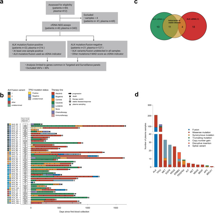
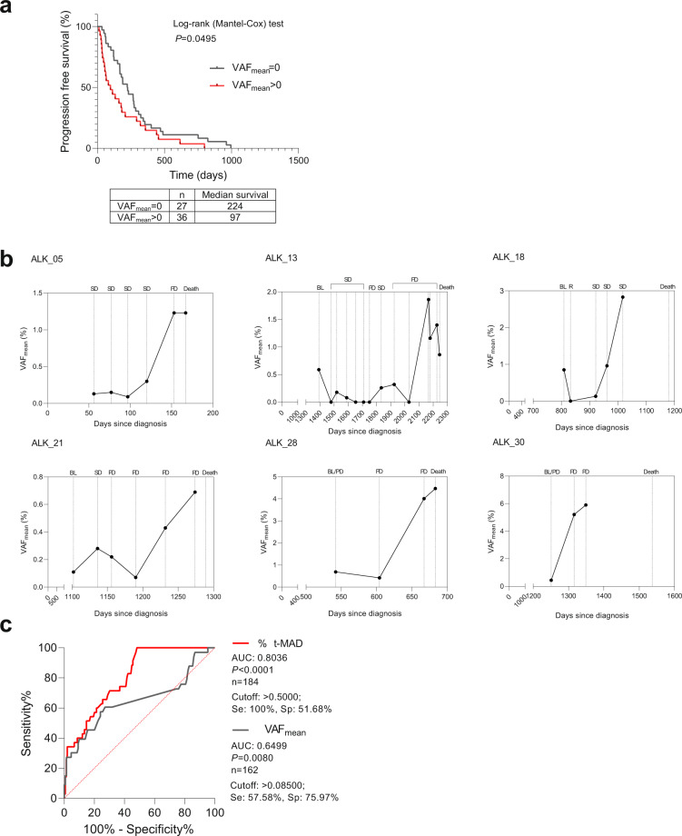
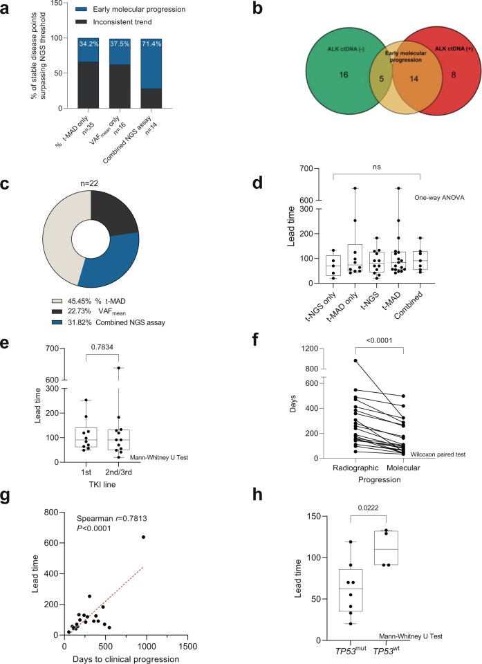
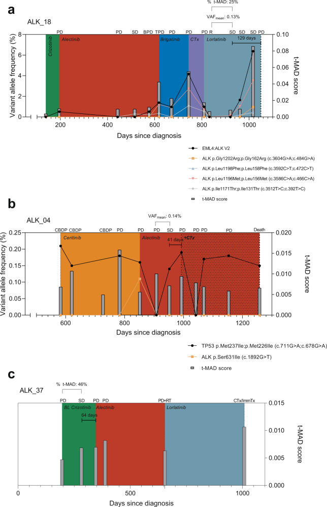
Targeted kinase inhibitors improve the prognosis of lung cancer patients with ALK alterations (ALK+). However, due to the emergence of acquired resistance and varied clinical trajectories, early detection of disease progression is warranted to guide patient management and therapy decisions. We utilized 343 longitudinal plasma DNA samples from 43 ALK+ NSCLC patients receiving ALK-directed therapies to determine molecular progression based on matched panel-based targeted next-generation sequencing (tNGS), and shallow whole-genome sequencing (sWGS). ALK-related alterations were detected in 22 out of 43 (51%) patients. Among 343 longitudinal plasma samples analyzed, 174 (51%) were ctDNA-positive. ALK variant and fusion kinetics generally reflected the disease course. Evidence for early molecular progression was observed in 19 patients (44%). Detection of ctDNA at therapy baseline indicated shorter times to progression compared to cases without mutations at baseline. In patients who succumbed to the disease, ctDNA levels were highly elevated towards the end of life. Our results demonstrate the potential utility of these NGS assays in the clinical management of ALK+ NSCLC.


Conflict of interest statement

P.C. reports research funding from AstraZeneca, Novartis, Roche, and Takeda as well as advisory board and/or lecture fees from Boehringer Ingelheim, Chugai, Lilly, Pfizer, and Takeda, outside the submitted work. M.A.S. reports personal fees from the German Canter for Lung Research (DZL), outside the submitted work. S.D. reports speaker’s honoraria from Roche and research funding from Takeda, outside the submitted work. M.R. reports personal fees from Amgen, AstraZeneca, BMS, Boehringer Ingelheim, Lilly, Merck, MSD, Novartis, Pfizer, Roche, and Samsung, outside the submitted work. M.T. reports advisory board honoraria from Novartis, Lilly, BMS, MSD, Roche, Celgene, Takeda, AbbVie, Boehringer, speaker’s honoraria from Lilly, MSD, Takeda, research funding from AstraZeneca, BMS, Celgene, Novartis, Roche and travel grants from BMS, MSD, Novartis, Boehringer, outside the submitted work. A.S. reports advisory board honoraria from BMS, Bayer, AstraZeneca, Thermo Fisher, Novartis, Seattle Genomics speaker’s honoraria from BMS, Bayer, Illumina, AstraZeneca, Novartis, Thermo Fisher, MSD, Roche, as well as research funding from Chugai, Bayer and BMS, outside the submitted work. H.S. reports grants from Roche Sequencing Solutions, during the conduct of the study, and personal fees from Roche, outside the submitted work. The remaining authors declare no competing interests.

Figures

Fig. 1
Fig. 1. Overview of the ALK+ NSCLC cohort.
a Untargeted sWGS and targeted NGS assays were used to assess ctDNA in 43 ALK+ NSCLC patients corresponding to 343 longitudinal plasma samples. Targeted NGS analysis only included genes common to both the Avenio Targeted and Surveillance panels. In total, 51% (22 of 43) of patients had an ALK alteration in at least one liquid biopsy. b Timeline of plasma collection and therapy administration in the patient cohort. ALK fusion variants and TP53 status from tissue analysis at diagnosis are indicated by colored boxes. c Distribution of patients based on detected ALK alterations—including mutations and rearrangements. The group without detectable ALK alterations (ALK−) is enriched for patients with intracranial progression or stable disease. d Molecular alterations identified in 174 ctDNA (+) plasma samples based on tNGS.
Fig. 2
Fig. 2. Prognostic utility of ctDNA NGS assays.
a Mean variant allele frequency at baseline indicates therapy durability. Progression-free survival plot showing significantly longer therapy durability in instances when ctDNA is undetected (VAFmean = 0) compared to detection (VAFmean > 0) at treatment initiation. b VAF kinetics in representative patients showing elevated VAFmean towards end of life. SD stable disease, PD progressive disease, BL therapy baseline, R response. c Comparison of receiver operating characteristic curves (ROC) using ΔVAFmean (gray) or %Δt-MAD (red) in discriminating progressive and non-progressive disease states.
Fig. 3
Fig. 3. Evaluation of the utility of ΔVAFmean and %Δt-MAD in identifying early molecular progression.
a Number of stable disease points that surpassed the numerical cutoffs per individual NGS assay (%Δt-MAD or ΔVAFmean) and in combination. Gray stacks indicate the percentage of points with inconclusive NGS assay values leading to clinical progression. Blue stacks represent points that showed sustained %Δt-MAD and/or ΔVAFmean increase leading to clinical progression, indicating early molecular progression. b Patients identified with early molecular progression were enriched in the ALK ctDNA (+) group. c Breakdown of 22 patients with early molecular progression based on NGS assays. d Comparison of called lead times based on NGS assays. e Comparison of called lead times based on therapy lines. f Comparison of cases with called lead time showing significant difference in days to radiographic progression versus molecular progression. g Length of lead time per therapy line was significantly correlated with duration of progression-free response to the respective treatment. h Lead times in cases with wild-type TP53 (TP53wt) were significantly longer compared to cases with mutated TP53 (TP53wt) detected by tNGS. The box plot show the the median, upper quartile, and lower quartile values. The whiskers indicate minimum and maximum values.
Fig. 4
Fig. 4. Representative cases illustrating early molecular progression as indicated by elevated ΔVAFmean and %Δt-MAD.
a Patient ALK_18 presented a case—during lorlatinib therapy—where both ΔVAFmean and %Δt-MAD were informative of the lead time. Early molecular progression was apparent 129 days prior to clinical progression, as indicated by ΔVAFmean of 0.13% and %Δt-MAD of 25%. Both values surpass the NGS metric thresholds identified in this study. b Patient ALK_04 depicts a case wherein ΔVAFmean indicated early molecular progression during alectinib therapy. A ΔVAFmean of 0.14% was calculated 41 days prior to clinical progression. c Patient ALK_37 emphasizes the relevance of untargeted NGS in ctDNA monitoring particularly in cases where genetic alterations remained undetected by tNGS. Here, %Δt-MAD of 46% was already apparent at a stable disease time point, 64 days prior to clinical progression. PD progressive disease, SD stable disease, BPD brain PD, TPD thoracic PD, R response, CBDP treatment continuation beyond disease progression, RT radiotherapy, CTx chemotherapy, ImmTx immunotherapy.

References

    1. Skoulidis F, Heymach JV. Co-occurring genomic alterations in non-small-cell lung cancer biology and therapy. Nat. Rev. Cancer. 2019;19:495–509. - PMC - PubMed
    1. Solomon BJ, et al. First-line crizotinib versus chemotherapy in ALK-positive lung cancer. N. Engl. J. Med. 2014;371:2167–2177. - PubMed
    1. Kim DW, et al. Activity and safety of ceritinib in patients with ALK-rearranged non-small-cell lung cancer (ASCEND-1): updated results from the multicentre, open-label, phase 1 trial. Lancet Oncol. 2016;17:452–463. - PMC - PubMed
    1. Shaw AT, et al. Alectinib in ALK-positive, crizotinib-resistant, non-small-cell lung cancer: a single-group, multicentre, phase 2 trial. Lancet Oncol. 2016;17:234–242. - PMC - PubMed
    1. Kim DW, et al. Brigatinib in patients with crizotinib-refractory anaplastic lymphoma kinase-positive non-small-cell lung cancer: a randomized, multicenter phase II trial. J. Clin. Oncol. 2017;35:2490–2498. - PubMed