Review
. 2022 Mar;19(3):384-408.
doi: 10.1038/s41423-021-00791-9. Epub 2021 Dec 7.

Metabolism of tissue macrophages in homeostasis and pathology

Affiliations
Review

Metabolism of tissue macrophages in homeostasis and pathology

Stefanie K Wculek et al. Cell Mol Immunol. 2022 Mar.

Abstract

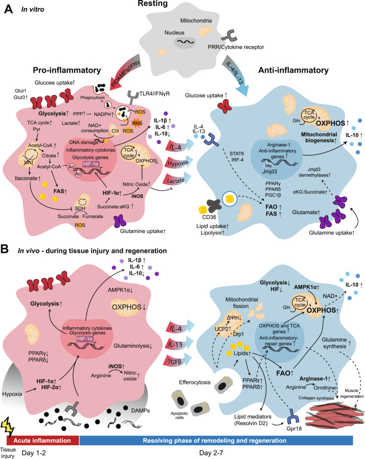
Cellular metabolism orchestrates the intricate use of tissue fuels for catabolism and anabolism to generate cellular energy and structural components. The emerging field of immunometabolism highlights the importance of cellular metabolism for the maintenance and activities of immune cells. Macrophages are embryo- or adult bone marrow-derived leukocytes that are key for healthy tissue homeostasis but can also contribute to pathologies such as metabolic syndrome, atherosclerosis, fibrosis or cancer. Macrophage metabolism has largely been studied in vitro. However, different organs contain diverse macrophage populations that specialize in distinct and often tissue-specific functions. This context specificity creates diverging metabolic challenges for tissue macrophage populations to fulfill their homeostatic roles in their particular microenvironment and conditions their response in pathological conditions. Here, we outline current knowledge on the metabolic requirements and adaptations of macrophages located in tissues during homeostasis and selected diseases.

Keywords: Tissue macrophages; homeostasis; metabolism; pathology; tissue regeneration.


Conflict of interest statement

The authors declare no competing interests.

Figures

Fig. 1
Fig. 1
Metabolic rearrangement in macrophage polarization to proinflammatory or alternatively activated macrophages in vitro and in vivo. A In vitro, resting macrophages can be activated by various pathogen- or danger-associated molecular patterns (PAMPs or DAMPs), and cytokines polarize to classically activated proinflammatory M1 macrophages or alternatively activated anti-inflammatory M2 macrophages. B Upon in vivo tissue injury, damaged cell debris is released into the extracellular microenvironment, and an inflammatory response is mounted. Next, upon clearance of cell debris and DAMPs, the response changes to promote resolution of inflammation. Both in vitro (A left) and in vivo (B left), proinflammatory polarization has been associated with enhanced glycolytic metabolism; however, the majority of the related information has been elucidated in vitro. Both in vitro (A right) and in vivo (B right), increases in OXPHOS, FAO and glutaminolysis are associated with alternatively activated macrophages. Ac, Acetylation; CIII, complex III; Drp1, dynamin-related protein 1; Gln, Glutamine; Gpr18, G protein-coupled receptor 18; Me, Methylation; PRR, pattern recognition receptor; Pyr, Pyruvate; UCP2, mitochondrial uncoupling protein 2; ΔΨm, mitochondrial membrane potential. Solid lines: direct relationships; dashed lines: indirect relationships. Black circles: DAMPs; blue circles: anti-inflammatory cytokines; purple circles: proinflammatory cytokines; orange stars: ROS and RNS; black irregular ovals: phagocytosed particles
Fig. 2
Fig. 2
Lipid handling by tissue macrophages. A The metabolism of alveolar macrophages present in lung tissue is specialized for lipid catabolism and trafficking for effective clearance of pulmonary surfactant. B Excess calorie intake causes adipose tissue hypertrophy, hypoxia and adipocyte death. In response, adipose tissue macrophages become bioenergetically activated, scavenge resulting lipids and elevate their lipid metabolism. Ultimately, they become lipid-laden and proinflammatory and contribute to systemic metabolic syndrome and insulin resistance. C In atherosclerotic lesions, macrophages are exposed to a variety of lipids (i.e., oxLDL, LDL, oxPAPC, long-chain fatty acids, and cholesterol crystals) that either promote or attenuate the proatherogenic environment. Excessive free cholesterol and fatty acids, which are generated in endolysosomes upon lipid uptake, alter the metabolism of macrophages, leading to the production of proinflammatory cytokines. Conversely, effective cholesterol efflux restores macrophage functions, promoting atherosclerosis resolution. CII, complex II; FA, fatty acid; Glc, glucose; Gln, glutamine; LOX1, oxidized low-density lipoprotein receptor 1; NLRP3, NOD-, LRR- and pyrin domain-containing protein 3; Pyr, pyruvate; SRA1, steroid receptor RNA activator 1. Solid lines: direct relationships; dashed lines: indirect relationships. Purple circles: proinflammatory cytokines; gray circles: growth factors; brown and orange circles: bound cholesterol/LDL/oxLDL or oxPARC; yellow and ochre circles: bound cholesterol/HDL; red circles: free cholesterol; pink circles: fatty acids; orange stars: ROS
Fig. 3
Fig. 3
Microenvironmental influence on tissue macrophage metabolism. A Splenic red pulp macrophages scavenge defective erythrocytes for iron recycling. B Large peritoneal macrophages adapt their bioenergetics after detection of different microenvironmental factors, such as yeast, oxLDL or IL-4, to facilitate the respiratory burst. C Osteoclasts shift their cellular metabolism when exposed to bone, promoting bone resorptive activity. CI-III, complex I-III; FA, fatty acid; Gln, glutamine; IRP1, iron-responsive element-binding protein; PKC, protein kinase C; SO, superoxide; SZ, sealing zone. Solid lines: direct relationships; dashed lines: indirect relationships. Purple circles: cytokines; brown circles: bound cholesterol/LDL/oxLDL; red and orange stars: ROS, SO and H2O2
Fig. 4
Fig. 4
Tumor-associated macrophage and cancer cell cross-talk. Cancer cells and tumor-associated macrophages engage in metabolic cross-talk, which has been shown primarily to support tumor cell growth and survival. This includes increased arginase expression by macrophages promoting arginine metabolism (1); tryptophan metabolism in cancer cells via IDO (2); glutamine metabolism in cancer cells to promote their proliferation and in macrophages to drive anti-inflammatory gene expression (3); glucose metabolism-mediated increases in extracellular lactate, which modulates macrophage function (4); the induction of and response to a hypoxic microenvironment (5); cholesterol export from macrophages (via ABC transporters) to cancer cells (via LDLR) (6); heme metabolism and iron efflux from macrophages (7); and mitochondrial ROS production (8). More details of these pathways can be found in the main text. CO, carbon monoxide. Solid lines: direct relationships; dashed lines: indirect relationships. Brown circles: bound cholesterol/LDL/oxLDL; dark green circles: lactate; light green circles: hyaluronic acid; orange stars: ROS

Similar articles

Cited by

References

    1. Nobs SP, Kopf M. Tissue-resident macrophages: guardians of organ homeostasis. Trends Immunol. 2021;42:495–507. - PubMed
    1. Okabe Y, Medzhitov R. Tissue-specific signals control reversible program of localization and functional polarization of macrophages. Cell. 2014;157:832–44. - PMC - PubMed
    1. Lavin Y, Winter D, Blecher-Gonen R, David E, Keren-Shaul H, Merad M, et al. Tissue-resident macrophage enhancer landscapes are shaped by the local microenvironment. Cell. 2014;159:1312–26. - PMC - PubMed
    1. Gautier EL, Shay T, Miller J, Greter M, Jakubzick C, Ivanov S, et al. Gene-expression profiles and transcriptional regulatory pathways that underlie the identity and diversity of mouse tissue macrophages. Nat Immunol. 2012;13:1118–28. - PMC - PubMed
    1. Mass E. Delineating the origins, developmental programs and homeostatic functions of tissue-resident macrophages. Int Immunol. 2018;30:493–501. - PubMed

Publication types