. 2021 Nov 28:2021:2612480.
doi: 10.1155/2021/2612480. eCollection 2021.

Cytological Effects of Serum Isolated from Polytraumatized Patients on Human Bone Marrow-Derived Mesenchymal Stem Cells

Affiliations

Cytological Effects of Serum Isolated from Polytraumatized Patients on Human Bone Marrow-Derived Mesenchymal Stem Cells

Yazhou Long et al. Stem Cells Int. .

Abstract

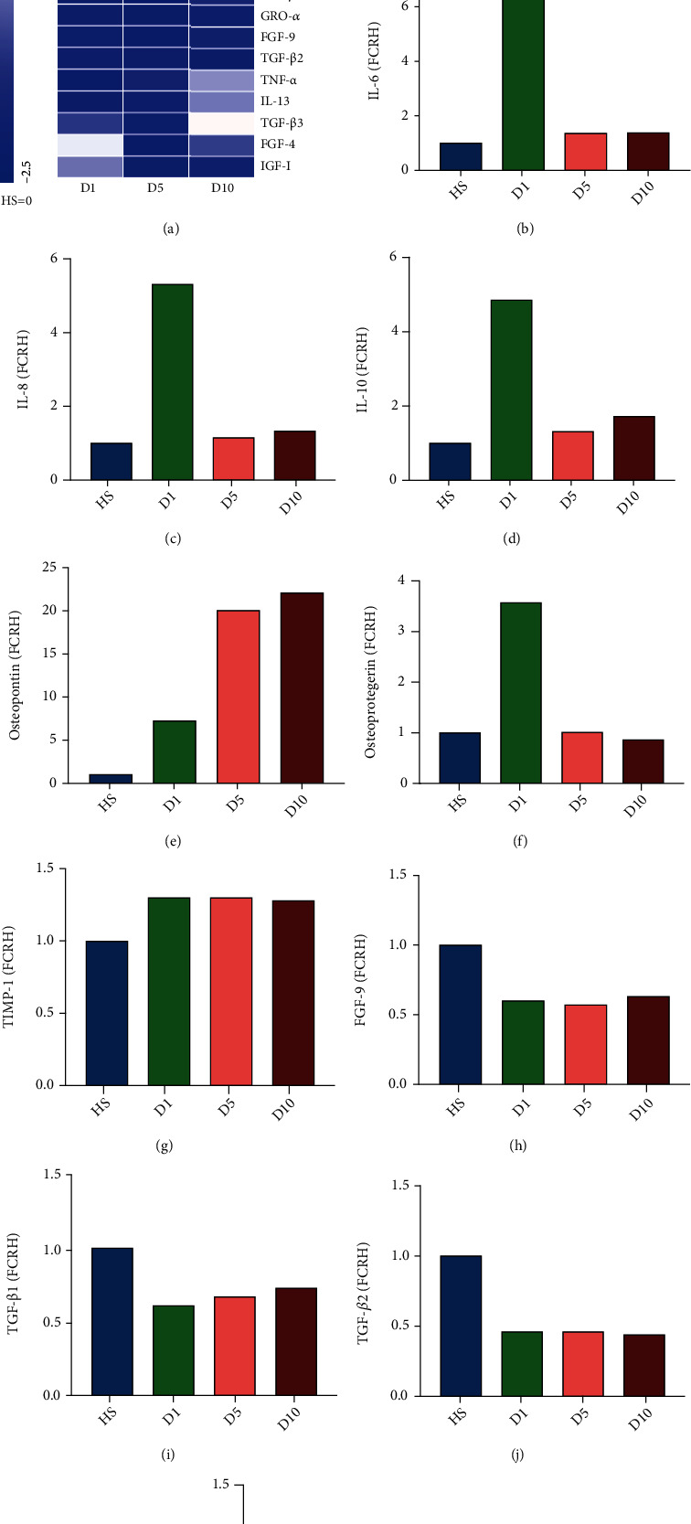
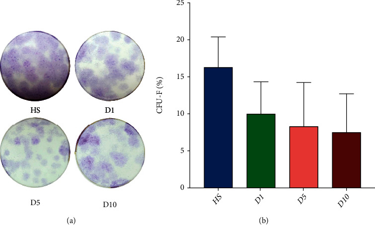
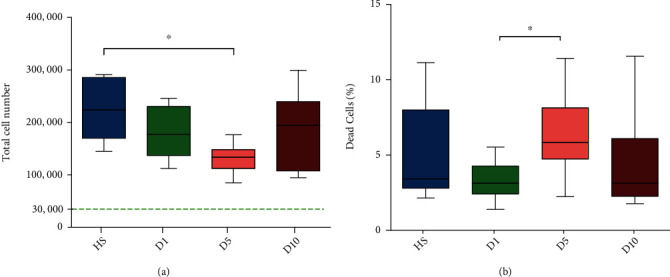
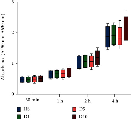
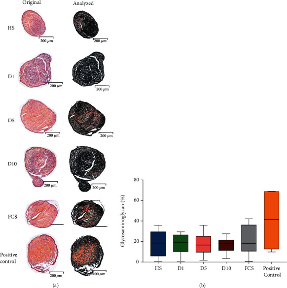
Due to their immunomodulatory and regenerative capacity, human bone marrow-derived mesenchymal stem cells (hBMSCs) are promising in the treatment of patients suffering from polytrauma. However, few studies look at the effects of sera from polytraumatized patients on hBMSCs. The aim of this study was to explore changes in hBMSC properties in response to serum from polytrauma patients taken at different time points after the trauma incident. For this, sera from 84 patients with polytrauma (collected between 2010 and 2020 in our department) were used. In order to test the differential influence on hBMSC, sera from the 1st (D1), 5th (D5), and 10th day (D10) after polytrauma were pooled, respectively. As a control, sera from three healthy donors (HS), matched with respect to age and gender to the polytrauma group, were collected. Furthermore, hBMSCs from four healthy donors were used in the experiments. The pooled sera of HS, D1, D5, and D10 were analyzed by multicytokine array for pro-/anti-inflammatory cytokines. Furthermore, the influence of the different sera on hBMSCs with respect to cell proliferation, colony forming unit-fibroblast (CFU-F) assay, cell viability, cytotoxicity, cell migration, and osteogenic and chondrogenic differentiation was analyzed. The results showed that D5 serum significantly reduced hBMSC cell proliferation capacity compared with HS and increased the proportion of dead cells compared with D1. However, the frequency of CFU-F was not reduced in polytrauma groups compared with HS, as well as the other parameters. The serological effect of polytrauma on hBMSCs was related to the time after trauma. It is disadvantageous to use BMSCs in polytraumatized patients at least until the fifth day after polytrauma as obvious cytological changes could be found at that time point. However, it is promising to use hBMSCs to treat polytrauma after five days, combined with the concept of "Damage Control Orthopedics" (DCO).


Conflict of interest statement

The authors declare to have no potential conflicts of interest.

Figures

Figure 1
Figure 1
Results of the multicytokine array. (a) Heat map depicting the changes in cytokine levels in pooled serum from patients at day 1 (D1), day 5 (D5), and day 10 (D10) after polytrauma relative to cytokine levels in serum from healthy controls. The data are normalized by Z-score (HS = 0) and analyzed by fold change relative to healthy serum (FCRH). A Z-score of 0 is represented by white color, a Z-score of 2.5 by red color, and a Z-score of -2.5 by blue color. Z-scores in between these values are depicted by less saturated colors of the same color palette. Cluster analysis was performed, and the cytokines were ordered accordingly. Some cytokines have an acute peak on the first day after trauma, and some cytokines peak on the 5th or 10th day after trauma, but compared to HS, more than half of the cytokine levels decreased after trauma. The proinflammatory cytokines (b) interleukin- (IL-) 6 and (c) IL-8 as well as the anti-inflammatory cytokine (d) IL-10 were approximately four times higher at D1 in comparison to HS and two or three times higher when compared to D5 and D10. (e) Osteopontin was increased in the serum from all time points postpolytrauma as compared to serum from healthy donors. The increase in D1, D5, and D10 relative to HS was from six to twenty times. (f) Osteoprotegerin was higher only one day after polytrauma in comparison to HS with a fold change for about three times. (g) Tissue inhibitor of metalloproteinase (TIMP-1) was increased at all time points after polytrauma in comparison to serum from healthy controls. (h) Fibroblast growth factor 9 (FGF-9) was decreased in the polytrauma sera as compared to the HS.(i–k) Transforming growth factor-beta 1 (TGF-β1), TGF-β2, and TGF-β3 were all decreased in polytrauma serum at D1 and D5 as compared to healthy serum (n = 1, pooled samples out of 84 donors).
Figure 2
Figure 2
Colony forming unit-fibroblast assay (CFU-F assay). (a) Representative images of CFU-F assays from HS, D1, D5, and D10. (b) Results of the CFU-F assays obtained by seeding 250 cells/well. Calculation was performed using the following formula: CFU‐F% = colonies/250 × 100%. The colony forming ability of polytrauma groups (D1, D5, and D10) had no significant difference when compared to the HS group (n = 4).
Figure 3
Figure 3
Evaluation of the proliferation capacity and viability of stem cells after cultivation in medium containing different sera (HS, D1, D5, or D10) for 72 hours. (a) Proliferation of hBMSCs treated with HS, D1, D5, or D10 serum. The green, dotted line shows the number of seeded cells (30,000 cells). Addition of serum from D5 to the culture medium resulted in a significantly lower proliferation of hBMSCs after 72 hours compared to HS. (b) Amount of dead cells after treatment with HS, D1, D5, or D10 serum. The D5 group demonstrated a significantly increased ratio of dead cells in comparison to D1. p ≤ 0.05 (n = 4; N = 3).
Figure 4
Figure 4
WST assay. Results were observed after cultivation of cells for 24 hours in medium containing different sera (HS, D1, D5, or D10 serum). No significant differences were observed between any of the groups at any of the time points analyzed (30 minutes, 1 hour, 2 hours, and 4 hours) (n = 4; N = 3).
Figure 5
Figure 5
Scratch and transwell migration assay indicating the hBMSC repopulation ability. (a) Representative pictures of the migration and repopulation ability of hBMSCs over 24 hours treated with HS, D1, D5, and D10 sera in a scratch assay. The black area represents the scratched area. As time goes by, the injured area is gradually reduced and dispersed along with the migration of cells. (b) After 4, 8, and 24 hours, no significant increase in migration of the D1, D5, and D10 groups was found compared to the HS group (n = 4; N = 2). (c) In transwell migration assay, no significant differences in the ratio of migration were observed between the HS, D1, D5, and D10 groups 12 hours after seeding (n = 4; N = 3).
Figure 6
Figure 6
Osteogenic differentiation potential. (a) Alizarin red staining showed calcium deposits as red staining after culturing hBMSCs in osteogenic differentiation medium containing 10% serum from HS, D1, D5, D10, or FCS for 14 days. (a1) Macroscopic image of alizarin red staining. (a2) Cells prior to alizarin red staining at 40x magnification. (a3) Images showing the hBMSCs at a 40x magnification after staining with alizarin red. (a4) Analysis of alizarin red staining of calcium deposits using a tool based on OpenCV library (version 4.1.0). Areas without staining were color isolated as black areas. The pixel ratio of the area of interest (nonblack area) in comparison to the black area was obtained. In the groups containing human serum (HS, D1, D5, and D10), a larger area of the 6-well was positive for alizarin red staining than in the FCS group (n = 4). (b) Quantitative analysis of the amount of alizarin red staining quantified using a software tool to compare the relative areas positive for staining. D1 showed significantly more area of alizarin red staining when compared to the FCS group. p ≤ 0.05 (n = 4). (c) Analysis of alizarin red staining indicative of osteogenic differentiation with higher concentrations of calcium deposition after 14 days of culture. HS, D1, and D10 showed significantly higher absorbance as compared to the FCS group. p ≤ 0.05 (n = 4).
Figure 7
Figure 7
Chondrogenic differentiation potential. (a) Chondrogenic differentiation after 21 days of culture in chondrogenic induction medium containing either HS, D1, D5, D10 serum, FCS, or serum-free (positive control). Images were taken at a magnification of 40x. Glycosaminoglycan content is depicted in orange. Demarcation of areas not stained positively for glycosaminoglycans in black using visual analysis software (based on OpenCV library version 4.1.0). Standard parameters were used in the analysis of each image. (b) Quantitative analysis of the glycosaminoglycan content of the chondrogenic pellet showed no significant differences between different groups (p > 0.05, n = 4, N = 2).

Similar articles

Cited by

References

    1. Sakran J. V., Greer S. E., Werlin E., McCunn M. Care of the injured worldwide: trauma still the neglected disease of modern society. Scandinavian Journal of Trauma, Resuscitation and Emergency Medicine . 2012;20(1):p. 64. doi: 10.1186/1757-7241-20-64. - DOI - PMC - PubMed
    1. Unfallchirurgie D. G. F. Definition of polytrauma. 2020, https://www.dgu-online.de/patienten/haeufige-diagnosen/schwerverletzte/p....
    1. Frenzel S., Krenn P., Heinz T., Negrin L. L. Does the applied polytrauma definition notably influence outcome and patient population? - a retrospective analysis. Scandinavian Journal of Trauma, Resuscitation and Emergency Medicine . 2017;25(1):87–87. doi: 10.1186/s13049-017-0400-2. - DOI - PMC - PubMed
    1. Baker S. P., O'Neill B. The injury severity score: an update. The Journal of Trauma . 1976;16(11):882–885. - PubMed
    1. Baker S. P., Oʼneill B., Haddon W., JR., Long W. B. The injury severity SCORE. The Journal of Trauma . 1974;14(3):187–196. doi: 10.1097/00005373-197403000-00001. - DOI - PubMed