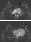
Palpable multifocal and multicentric invasive lobular breast carcinoma in a young female
- PMID: 34876947
- PMCID: PMC8628221
- DOI: 10.1016/j.radcr.2021.10.056
Palpable multifocal and multicentric invasive lobular breast carcinoma in a young female
Abstract
Breast cancer is the most common carcinoma plaguing women in the United States. Invasive lobular carcinoma is the second most prevalent type of breast carcinoma with an incidence rate of 5% and 15% with high propensity for multifocal manifestation of disease. Multifocal disease is defined by two or more malignant foci within a single quadrant. Invasive lobular carcinoma is strongly associated with early menarche, late menopause, late age at first birth, and is typically seen in women ages 50 and older. Invasive lobular carcinoma can be difficult to detect clinically because lesions typically fail to form palpable masses, and it can be challenging to diagnose mammographically due to subtle imaging features of the lesions. Here we present a rare case of a palpable, unilateral, multifocal and multicentric lobular breast carcinoma in a young, previously healthy 41-year-old woman with no family history of breast cancer.
Keywords: Breast carcinoma; Invasive lobular carcinoma; Magnetic resonance imaging; Mammography; Multifocal lobular carcinoma of the breast; Ultrasound.
Published by Elsevier Inc. on behalf of University of Washington.
Figures


Similar articles
-
[Diagnostic imaging of lobular carcinoma of the breast: mammographic, ultrasonographic and MR findings].Radiol Med. 2000 Dec;100(6):436-43. Radiol Med. 2000. PMID: 11307504 Italian.
-
Ultrasound findings in pure invasive lobular carcinoma of the breast: comparison with matched cases of invasive ductal carcinoma of the breast.Breast. 1999 Aug;8(4):188-90. doi: 10.1054/brst.1999.0042. Breast. 1999. PMID: 14731438
-
Non-enhancing malignant lesions of the breast: A case report and review of literature.Heliyon. 2023 Mar 17;9(3):e14498. doi: 10.1016/j.heliyon.2023.e14498. eCollection 2023 Mar. Heliyon. 2023. PMID: 36967981 Free PMC article.
-
Increased incidence of lobular breast cancer in women treated with hormone replacement therapy: implications for diagnosis, surgical and medical treatment.Endocr Relat Cancer. 2007 Sep;14(3):549-67. doi: 10.1677/ERC-06-0060. Endocr Relat Cancer. 2007. PMID: 17914088 Review.
-
Lobular breast cancer: incidence and genetic and non-genetic risk factors.Breast Cancer Res. 2015 Mar 13;17:37. doi: 10.1186/s13058-015-0546-7. Breast Cancer Res. 2015. PMID: 25848941 Free PMC article. Review.
References
Publication types
LinkOut - more resources
Full Text Sources

