

Cerebral fat embolism syndrome after long bone fracture: A case report
- PMID: 34876950
- PMCID: PMC8628207
- DOI: 10.1016/j.radcr.2021.10.060
Cerebral fat embolism syndrome after long bone fracture: A case report
Erratum in
-
Erratum regarding missing patient consent statements in previously published articles.Radiol Case Rep. 2023 Jan 25;18(4):1641-1642. doi: 10.1016/j.radcr.2023.01.013. eCollection 2023 Apr. Radiol Case Rep. 2023. PMID: 36895595 Free PMC article.
Abstract
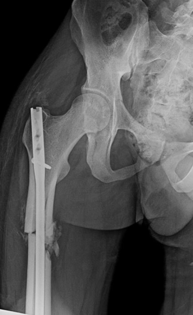
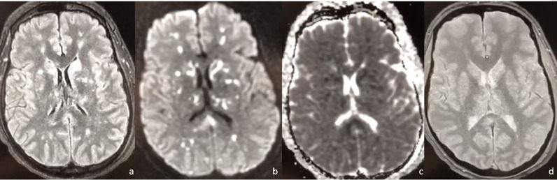
Cerebral fat embolism is a rare and potentially fatal condition that may occur following a long bone fracture. Its characterized by respiratory, neurological, and mucocutaneous signs. Isolated severe brain syndrome remains exceptional. We report a 21-year-old male patient admitted for the cerebral manifestation of a fat embolism syndrome due to a fracture of long bone after a traffic accident injury. Neurological deterioration after a free interval was seen with generalized tonic-clonic seizures. MRI of the brain was indicated which showed numerous multifocal hyperintensities involving the deep white matter of both hemispheres producing a "starfield" appearance. This pattern of cytotoxic cerebral edema, with lesions in the white matter rather than the grey matter, is indicative of the subacute stage of fat embolism. The patient was treated with comprehensive support in the intensive care unit, he returned to normal neurological function and was discharged after 3 weeks of hospitalization.
Keywords: Cerebral fat embolism; MRI; long bone fracture.
© 2021 The Authors. Published by Elsevier Inc. on behalf of University of Washington.
Figures
References
-
- Suh SI, Seol HY, Seo WK, Keun-Sik Hong, Sun U Kwon, et al. Cerebral fat embolism: susceptibility-weighted magnetic resonance imaging. Arch Neurol. 2009;66:1170. - PubMed
-
- Rutman AM, Rapp EJ, Hippe DS, Baoanh BS, Mossa-Basha Mahmud MD, et al. T2*-weighted and diffusion magnetic resonance imaging differentiation of cerebral fat embolism from diffuse axonal injury. J Comput Assist Tomogr. 2017;41:877–883. - PubMed
Publication types
LinkOut - more resources
Full Text Sources
