

Florid Mesothelial Hyperplasia Associated with Abdominal Wall Endometriosis Mimicking Invasive Carcinoma
- PMID: 34877024
- PMCID: PMC8645372
- DOI: 10.1155/2021/3439700
Florid Mesothelial Hyperplasia Associated with Abdominal Wall Endometriosis Mimicking Invasive Carcinoma
Abstract
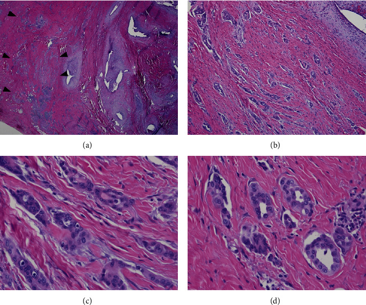
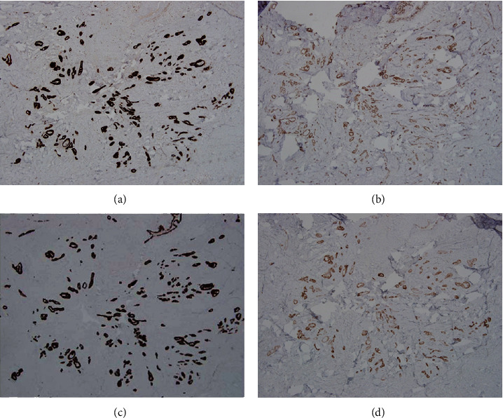
Florid mesothelial hyperplasia typically occurs in the pelvis, abdomen, or chest associated with an underlying neoplastic or inflammatory process. These lesions are of clinical significance because they can mimic a neoplasm. Early reports were published in the 1970s, but only a few case series of such lesions have been published in the gynecologic pathology literature. Here, we report a case of florid mesothelial hyperplasia with an infiltrative growth pattern, mimicking an invasive carcinoma. The lesion was associated with endometriosis forming a mass lesion in the abdominal wall. Histologically, tubular arrangements and nests of mesothelial cells, some with artifactual slit-like spaces, formed a stellate lesion adjacent to endometrial glands and stroma. Cytologic atypia was mild and reactive, and positive immunostaining for calretinin, WT-1, and cytokeratin 5 identified the lesion as mesothelial and benign. We describe in detail the histologic findings in this case and review the pertinent literature. We discuss the clinically importance of this diagnostic pitfall and the path to arriving at the correct diagnosis.
Copyright © 2021 Edgar G. Fischer and Shweta Agarwal.
Conflict of interest statement
The authors declare that they have no conflicts of interest.
Figures
Similar articles
-
Florid mesothelial hyperplasia associated with ovarian tumors: a potential source of error in tumor diagnosis and staging.Int J Gynecol Pathol. 1993 Jan;12(1):51-8. doi: 10.1097/00004347-199301000-00007. Int J Gynecol Pathol. 1993. PMID: 8418078
-
Florid mesothelial hyperplasia of the tunica vaginalis mimicking malignant mesothelioma: a clinicopathologic study of 12 cases.Am J Surg Pathol. 2014 Jan;38(1):54-9. doi: 10.1097/PAS.0b013e31829ab20e. Am J Surg Pathol. 2014. PMID: 24061516
-
[Urothelial hyperplastic lesion with endophytic growth pattern: a clinicopathologic study].Zhonghua Bing Li Xue Za Zhi. 2011 May;40(5):319-23. Zhonghua Bing Li Xue Za Zhi. 2011. PMID: 21756826 Chinese.
-
Selected topics in peritoneal pathology.Int J Gynecol Pathol. 2014 Jul;33(4):393-401. doi: 10.1097/PGP.0000000000000146. Int J Gynecol Pathol. 2014. PMID: 24901399 Review.
-
The pathology of endometriosis: a survey of the many faces of a common disease emphasizing diagnostic pitfalls and unusual and newly appreciated aspects.Adv Anat Pathol. 2007 Jul;14(4):241-60. doi: 10.1097/PAP.0b013e3180ca7d7b. Adv Anat Pathol. 2007. PMID: 17592255 Review.
Cited by
-
A diagnostic challenge of primary serous borderline tumor involving the peritoneum: A case report.Int J Surg Case Rep. 2025 Aug;133:111527. doi: 10.1016/j.ijscr.2025.111527. Epub 2025 Jun 16. Int J Surg Case Rep. 2025. PMID: 40578237 Free PMC article.
-
Crosslinking Surgical Oncology and the Assessments of Hernia Sac Tissues With Malignant Transformations.Cureus. 2025 May 18;17(5):e84317. doi: 10.7759/cureus.84317. eCollection 2025 May. Cureus. 2025. PMID: 40530201 Free PMC article.
References
Publication types
LinkOut - more resources
Full Text Sources
Research Materials
