

Imaging Surveillance of the Reconstructed Breast in a Subset of Patients May Aid in Early Detection of Breast Cancer Recurrence
- PMID: 34877066
- PMCID: PMC8645461
- DOI: 10.25259/JCIS_113_2021
Imaging Surveillance of the Reconstructed Breast in a Subset of Patients May Aid in Early Detection of Breast Cancer Recurrence
Abstract
Objectives: The purpose of this study is to determine the biological markers more frequently associated with recurrence in the reconstructed breast, to evaluate the detection method, and to correlate recurrent breast cancers with the detection method.
Material and methods: An institutional review board-approved retrospective study was conducted at a single institution on 131 patients treated with mastectomy for primary breast cancer followed by breast reconstruction between 2005 and 2012. Imaging features were correlated with clinical and pathologic findings.
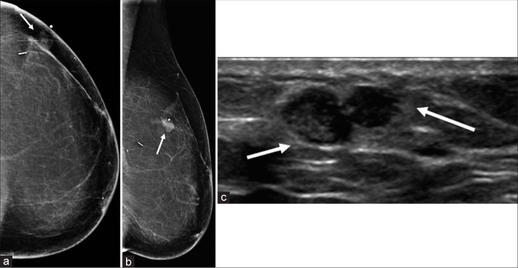
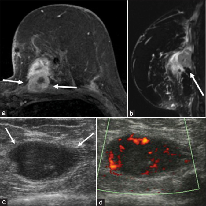
Results: Of the 131 patients who met our inclusion criteria, 40 patients presented with breast cancer recurrence. The most common histopathologic type of primary breast cancer was invasive ductal carcinoma in 82.5% (33/40) of patients. Triple-negative breast cancer was the most common biological marker with 42.1% (16/38) of cases. Clinically, 70% (28/40) of the recurrences presented as palpable abnormalities. Of nine patients who underwent mammography, a mass was seen in eight patients. Of the 35 patients who underwent ultrasound evaluation, an irregular mass was found in 48.6% (17/35) of patients. Nine patients with recurrent breast cancer underwent breast MRI, and MRI showed an irregular enhancing mass in four patients, an oval mass in four patients, and skin and trabecular thickening in one patient. About 55% of patients with recurrent breast cancer were found to have distant metastases.
Conclusion: Patients at higher risk for locoregional recurrence may benefit from imaging surveillance in order to detect early local recurrences.
Keywords: Biological markers; Breast cancer recurrence; Deep recurrences; Reconstructive surgery.
© 2021 Published by Scientific Scholar on behalf of Journal of Clinical Imaging Science.
Conflict of interest statement
Gary Whitman is a member of the Editorial Board of the Journal of Clinical Imaging Science.
Figures
References
-
- US. Breast Cancer Statistics. 2021. Available from: https://www.breastcancer.org/symptoms/understand_bc/statistics [Last accessed on 2021 May 20]
-
- ACR Appropriateness Criteria? Imaging after Mastectomy and Breast Reconstruction American College of Radiology. 2021. Available from: https://www.acsearch.acr.org/docs/3155410/narrative [Last accessed on 2020 Jul 21]
LinkOut - more resources
Full Text Sources
Miscellaneous