Review
. 2021 Nov 24;3(1):20210011.
doi: 10.1259/bjro.20210011. eCollection 2021.

Imaging findings of vitamin deficiencies: are they forgotten diseases?

Affiliations
Review

Imaging findings of vitamin deficiencies: are they forgotten diseases?

Akitoshi Inoue et al. BJR Open. .

Abstract

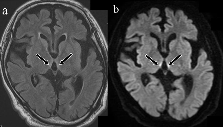
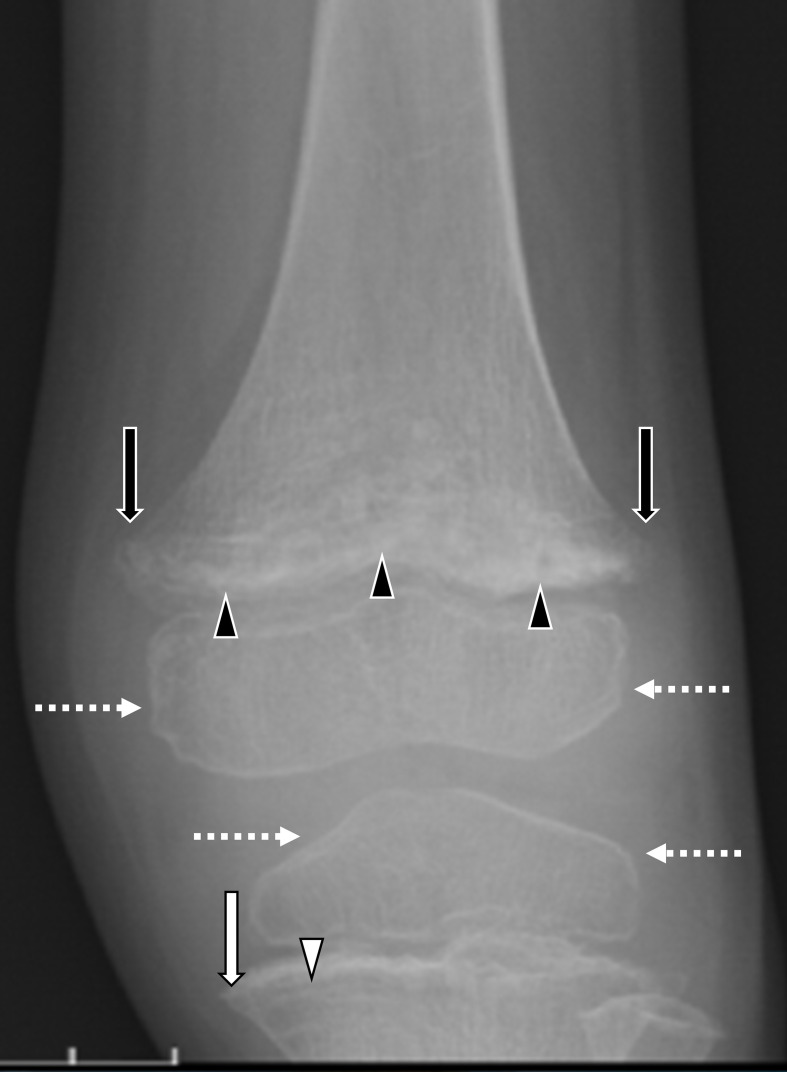
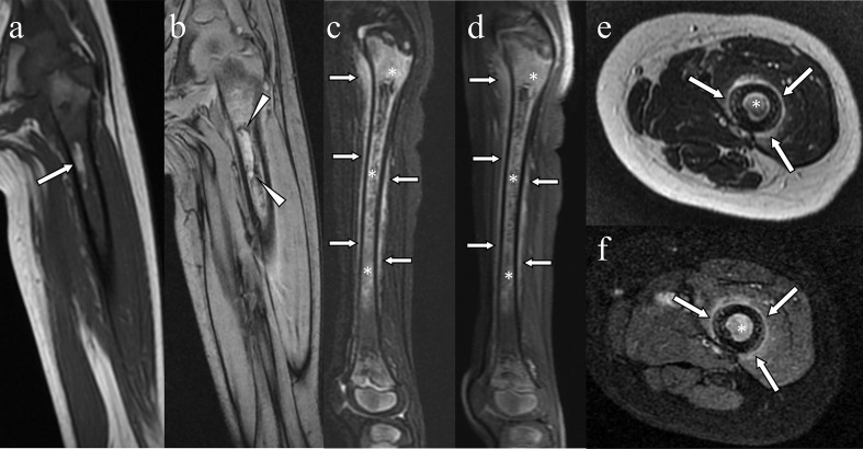
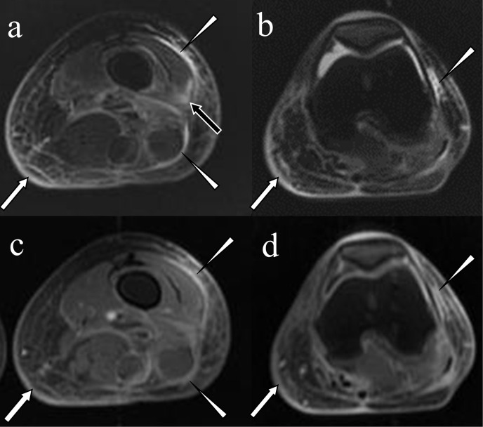
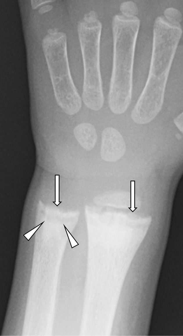
Vitamin deficiency is rare in modern industrialised countries; however, it still occurs in patients with specific backgrounds, such as those with extremely unbalanced diets, those with alcoholism and those who have undergone gastrointestinal surgery. Imaging examinations that demonstrate classic findings confirm the clinical diagnosis of vitamin deficiency and help monitor response to treatment. Because vitamin deficiencies are not prevalent, the diagnosis might not be straightforward. Therefore, imaging should be performed in cases of suspected vitamin deficiency. Radiologists should be familiar with characteristic imaging findings of vitamin deficiency and should survey an affected patient's background and blood vitamin levels. Because symptoms of vitamin deficiency are quickly improved by vitamin replacement, early diagnosis is essential. This pictorial review provides imaging findings for deficiencies in vitamins B1 (Wernicke encephalopathy and wet beriberi), B12 (subacute combined degeneration), C (scurvy), D (rickets) and K (bleeding tendency).


Figures

Figure 1.
Figure 1.
Wernicke encephalopathy in alcoholism. A 54-year-old male was found lying on the ground. He exhibited severe disturbance of consciousness, and his Glasgow Coma Scale score was 3. He was hospitalised to manage his alcoholism. Laboratory tests revealed a decreased vitamin B1 level (23 ng ml−1 [normal range: 24–66 ng ml−1]). The symmetrical hyperintensity lesions around the third ventricle were observed on fluid-attenuated inversion recovery imaging (a: arrows). These lesions were more conspicuous on diffusion-weighted imaging (b: arrows).
Figure 2.
Figure 2.
Wernicke encephalopathy with cortical involvement. An 82-year-old female who received a large infusion as a treatment for acute pancreatitis and hyponatraemia demonstrated disturbance of consciousness. Osmotic encephalopathy was suspected because of the hyponatraemia and infusion therapy. Fluid-attenuated inversion recovery imaging (a, b) and diffusion-weighted imaging (c, d) revealed symmetrical lesions of hyperintensity in the bilateral postcentral gyrus (a, c: arrows), corpora quadrigemina (b, d: arrowheads) and brain parenchyma around the third ventricle (b, d: arrows), which are highly suggestive of Wernicke encephalopathy. Her vitamin B1 level was decreased (12 ng ml−1 [normal range: 24–66 ng ml−1]).
Figure 3.
Figure 3.
Wet beriberi. A 70-year-old male presented with dyspnoea, nausea and appetite loss in the emergency room. He presented with normal blood pressure (101/71 mmHg) but tachycardia (heart rate: 107 bpm). Arterial blood gas analysis revealed acidosis (pH: 7.256 [normal range: 7.36–7.44]), a decreased pressure of CO2 (10 mm Hg [normal range: 35–45 mm Hg]), a decreased base excess (−20 mEq/L [normal range: −2—+2 mEq l−1 ]), an increased lactate level (12 mmol l−1 [normal range:<5 mmol l−1]), and an unremarkable arterial partial pressure of oxygen (104 mm Hg [normal range:>80 mm Hg]), which means metabolic acidosis due to elevated lactic level. His cardiac index was 3.0, and his pulmonary capillary wedge pressure was 23 mm Hg, which signified Forrester Class II heart failure. Laboratory tests revealed a decreased vitamin B1 level (17 ng ml−1 [normal range: 24–66 ng ml−1]). CT demonstrated bilateral pleural effusion (asterisks), cardiomegaly and a collection of pericardial fluid effusion that was more highly attenuated than pleural effusion (arrows).
Figure 4.
Figure 4.
Subacute combined degeneration of the spinal cord. A 72-year-old male who had undergone total gastrectomy for gastric cancer 11 years earlier presented with bilateral numbness in the upper extremities. Sagittal T 2WI showed a high-intensity lesion at the level of C4 to C5 (a: arrow), and axial T 2WI showed a symmetrical high-intensity lesion in the posterior column, known as the ‘inverted V’ sign (b: arrow). His vitamin B12 level (130 pg ml−1) was lower than the normal range (249–938 pg ml−1). T 2WI, T 2 weighted imaging.
Figure 5.
Figure 5.
Paediatric case of scurvy demonstrated on radiography. A 4-year-old boy with autistic spectrum disorder suffered from left thigh pain. Anteroposterior radiographs demonstrated the heterogeneous and irregular appearance of the growth plate (black arrowheads) and metaphyseal beaking (Pelkan spur: black arrows) in the femur and a sclerotic cortical rim around osteopaenic epiphyseal ossification centres of the femur and tibia (Wimberger ring sign: broken white arrows). In the tibia, a dense metaphyseal band (Frankel line; arrow) and a lucent band (scurvy line; white arrowhead) are visible. Laboratory examination revealed lower vitamin C level (0.2 µg ml−1 [range: 4.7—17.8 µg ml−1]).
Figure 6.
Figure 6.
Paediatric case of scurvy (same case as in Figure 5). A spotty hyperintensity lesion in the diffuse hypointense bone marrow of the femur was visible on coronal T 1 weighted magnetic resonance imaging (a: arrow). The rim-like hypointensity around the lesion (arrowheads) was suggestive of haemosiderin around the bone marrow lesion on coronal T 2* weighted imaging (b: arrowheads). The bone marrow was diffusely hyperintense and contained a heterogeneous hypointense area (asterisks) in association with hyperintensity along the periosteum (arrows), as observed on sagittal fat-suppressed T 2 weighted imaging (c) and contrast enhanced fat suppressed T 1 weighted imaging (d). The contrast enhancements with hyperintensity were seen in the bone marrow and periosteum on axial T 2 weighted imaging (e: arrows) and on contrast-enhanced fat-suppressed T 1 weighted imaging (f: arrows).
Figure 7.
Figure 7.
Adult case of scurvy. A 40-year-old female who had schizophrenia, a manifestation of which was an unbalanced diet, presented with a swollen left lower extremity with bleeding spots. Fat-suppressed T 2 weighted images demonstrated linear hyperintensity on the fascia (a; arrowheads) and the lateral retinaculum of the patella (b; arrowhead), an area of intramuscular hyperintensity (a; black arrow) and thickened skin in association with reticular hyperintensity in the subcutaneous tissue (a and b; white arrows). On fat-suppressed T 1 weighted imaging after intravenous gadolinium injection, contrast enhancement was observed in the fascia (c; arrowheads) and the lateral retinaculum of the patella (d; arrowhead) and thickened skin (c and d; arrows). Laboratory examination demonstrated an extremely low vitamin C level (0.6 µg ml−1 [normal range: 4.7—17.8 µg ml−1]).
Figure 8.
Figure 8.
Rickets in the knee. An 18-month-old boy who was exclusively breastfed lost his appetite after starting nursery school. His mother was aware of the difference of the length of his lower extremities. Posteroanterior radiograph demonstrated indistinct metaphyseal margins in the femur, tibia and fibula (fraying; arrowheads) and the widening of metaphyseal ends in the femur and tibia (splaying; black arrows). Healing stage was suggested due to the provisional zone of calcification although actual onset was unknown. His activated vitamin D level (0.87 pg ml−1) was lower than the paediatric normal range (20–70 pg ml−1).
Figure 9.
Figure 9.
Rickets in the wrist. A 3-year-old boy who was an extremely picky eater and had a cognitive delay. He always stayed at home and rarely played outside. Anteroposterior radiograph of the left wrist showed concave deformity of the growth plate of the ulna (cupping: arrowheads) and an indistinct metaphyseal margin (fraying: arrows) in the radius and ulna. His activated vitamin D level (<5 pg ml−1) was lower than the paediatric normal range (20–70 pg ml−1).
Figure 10.
Figure 10.
Intracranial haemorrhage caused by vitamin K deficiency. A 2-month-old boy presented with disturbance of consciousness and decreased feeding. Head computed tomography demonstrated subdural haematoma around the falx and posterior area (a; white arrows), subarachnoid haemorrhage on the surface of the right temporal lobe (b; circle) and intraparenchymal haemorrhage in the temporal lobe (b; black arrow). He was examined the causes of bleeding tendency and diagnosed with biliary atresia.

References

    1. Ota Y, Capizzano AA, Moritani T, Naganawa S, Kurokawa R, Srinivasan A. Comprehensive review of Wernicke encephalopathy: pathophysiology, clinical symptoms and imaging findings. Jpn J Radiol 2020; 38: 809–20. doi: 10.1007/s11604-020-00989-3 - DOI - PubMed
    1. Zhong C, Jin L, Fei G. Mr imaging of nonalcoholic Wernicke encephalopathy: a follow-up study. AJNR Am J Neuroradiol 2005; 26: 2301–5. - PMC - PubMed
    1. Yamamura M, Murai H, Kaneko S, Usui S, Furusho H, Takamura M. Case report: pericardial effusion with constrictive physiology in a patient with wet beriberi. Nutr J 2016; 15: 37. doi: 10.1186/s12937-016-0156-y - DOI - PMC - PubMed
    1. Essa E, Velez MR, Smith S, Giri S, Raman SV, Gumina RJ. Cardiovascular magnetic resonance in wet beriberi. J Cardiovasc Magn Reson 2011; 13: 41: 41. doi: 10.1186/1532-429X-13-41 - DOI - PMC - PubMed
    1. Kuno T, Nakamura H, Endo Y, Saito K, Yamazaki H, Motoda H, et al. Clinical history and colliquative myocytolysis are keys to the diagnosis of shoshin beriberi. Case Rep Pathol 2014; 2014: 506072 10.1155/2014/506072 - DOI - PMC - PubMed