Review
. 2021 Nov 24;3(1):20210034.
doi: 10.1259/bjro.20210034. eCollection 2021.

Contrast-enhanced mammography: what the radiologist needs to know

Affiliations
Review

Contrast-enhanced mammography: what the radiologist needs to know

Lidewij M F H Neeter et al. BJR Open. .

Abstract

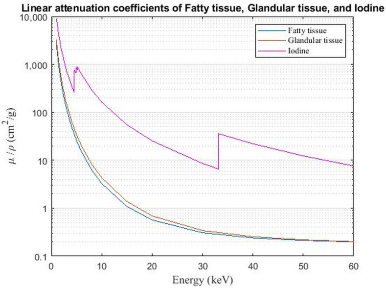
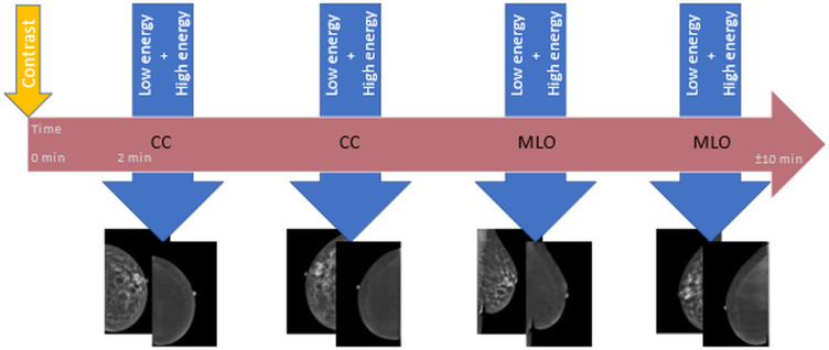
Contrast-enhanced mammography (CEM) is a combination of standard mammography and iodinated contrast material administration. During the last decade, CEM has found its place in breast imaging protocols: after i.v. administration of iodinated contrast material, low-energy and high-energy images are retrieved in one acquisition using a dual-energy technique, and a recombined image is constructed enabling visualisation of areas of contrast uptake. The increased incorporation of CEM into everyday clinical practice is reflected in the installation of dedicated equipment worldwide, the (commercial) availability of systems from different vendors, the number of CEM examinations performed, and the number of scientific articles published on the subject. It follows that ever more radiologists will be confronted with this technique, and thus be required to keep up to date with the latest developments in the field. Most importantly, radiologists must have sufficient knowledge on how to interpret CEM images and be acquainted with common artefacts and pitfalls. This comprehensive review provides a practical overview of CEM technique, including CEM-guided biopsy; reading, interpretation and structured reporting of CEM images, including the accompanying learning curve, CEM artefacts and interpretation pitfalls; indications for CEM; disadvantages of CEM; and future developments.


Conflict of interest statement

Conflicts of interest: RA received institutional grant and consulting fee from GE Healthcare. JW received institutional grants and speaker’s fees from AGFA, Bayer Healthcare, Bard Medical, GE Healthcare, Optimed, Philips Healthcare, and Siemens Healthineers. ML received several research grant and speaker’s fees from GE Healthcare, Hologic, Bayer, and Guerbet. The other authors of this manuscript declare no relationships with any companies, whose products or services may be related to the subject matter of the article.

Figures

Figure 1.
Figure 1.
Principle of iodine-based contrast enhancement. Mass attenuation coefficients of fatty tissue, glandular tissue, and iodine are shown. The iodine curve shows a steep elevation in attenuation at 33.2 keV, which is the k-edge of iodine. Differences in attenuation between breast tissue and iodinated contrast material are larger beyond the k-edge of iodine. Thus in high energy images (44–49 kVp), the differences in attenuation are larger than in low-energy images (26–30 kVp). Image processing of low- and high-energy images subsequently results in recombined images, showing contrast enhancement overlay.
Figure 2.
Figure 2.
Contrast-enhanced mammographic images in a 67-year-old female recalled from the breast cancer screening program because of a new, spiculated mass in the right breast. A-D. Low energy images. E-H. Recombined images. Images were acquired of the right and left breast in craniocaudal (CC) and mediolateral oblique (MLO) views. The mass in the right breast is visible on low-energy images in both CC and MLO views (arrows in A and C). The recombined images of the right breast show enhancement of the lesion in both CC and MLO views (arrows in E and G). Histopathological results showed an invasive breast cancer of no special type, Grade 2, size 1.4 cm.
Figure 3.
Figure 3.
Diagram of image acquisition for contrast-enhanced mammography. The horizontal arrow represents the time window of 10 min in which a full (at least four views) contrast-enhanced mammography examination must be performed in order to be considered of diagnostic value. The iodine-based contrast agent is administered at time point zero (small vertical arrow), 2 min prior to the acquisition of the first view. Per view, one low energy and one high energy image are acquired within one compression (larger vertical arrows). The order of views may differ. After image processing, low energy and recombined images are retrieved for clinical assessment.
Figure 4.
Figure 4.
Enhancing fibroadenoma. A,B. Contrast-enhanced mammographic of right breast in mediolateral oblique view in a 63-year-old female recalled from screening because of a new ill-defined and partly obscured mass. A. Low-energy image showing the suspect mass (arrow in A). B. Corresponding recombined image in which the suspect lesion is showing enhancement (arrow in B). The lines visible in the caudal part of the breast (circle) are the result of slight motion between the low- and high-energy image acquisition, the ripple artefact. Histopathological results showed a classic fibroadenoma.
Figure 5.
Figure 5.
Contrast-enhanced mammographic images in a 55-year-old female recalled from screening because of a new mass in the left breast. A. Low-energy image in mediolateral oblique view shows an ill-defined round mass (arrow in A). B. Corresponding recombined image. At the site of the suspect lesion a subtle ‘eclipse sign’ is visible, implicating a cyst (arrow in B). No screen-detected interval breast cancer has been reported in the 18-month follow-up period. The ripple artefact is also visible on the recombined image (circle).
Figure 6.
Figure 6.
Contrast-enhanced mammographic images in craniocaudal view in a 63-year-old female. A. Low-energy image of the right breast. B. The rim artefact is shown in the recombined image (small arrows). In addition, the skin line enhancement artefact is visible in the anteromedial part of the breast (larger arrow). No suspicious findings were reported.
Figure 7.
Figure 7.
CEM-guided biopsy in a 61-year-old patient with palpable lesion in right breast (IDC, not shown) and additional contralateral (left breast) finding on diagnostic CEM. A,B. Low-energy (LE) and recombined image (RE) of left breast in craniocaudal view. There is a 6 mm mass enhancement at 12 o’clock, with no ultrasound correlation and not enough references on 2D/3D in order to favour a conventional mammographic-guided biopsy. C-E. The procedure of CEM-guided biopsy is similar to a standard stereotactic biopsy (one scout and a pair of angled stereotactic images) with the additional step of contrast media injection 2 min before compression and first imaging. Like a routine CEM, each acquisition is composed of one low-energy (LE) and one high-energy (HE) exposure. The inclusion of the enhancing lesion is confirmed with a recombined scout view (0°), followed by the two angled views. Another pair of stereotactic angled images ( ± 15°) is sometimes acquired, previous to fire-forward, in order to confirm that the target was reached. F. Final CC view after clip placement. Histopathological results showed an invasive lobular carcinoma in the left breast.

References

    1. Dibden A, Offman J, Duffy SW, Gabe R. Worldwide review and meta-analysis of cohort studies measuring the effect of mammography screening programmes on incidence-based breast cancer mortality. Cancers 2020; 12: 976. doi: 10.3390/cancers12040976 - DOI - PMC - PubMed
    1. Boyd NF, Guo H, Martin LJ, Sun L, Stone J, Fishell E, et al. . Mammographic density and the risk and detection of breast cancer. N Engl J Med 2007; 356: 227–36. doi: 10.1056/NEJMoa062790 - DOI - PubMed
    1. Nazari SS, Mukherjee P. An overview of mammographic density and its association with breast cancer. Breast Cancer 2018; 25: 259–67. doi: 10.1007/s12282-018-0857-5 - DOI - PMC - PubMed
    1. Tagliafico AS, Bignotti B, Rossi F, Signori A, Sormani MP, Valdora F, et al. . Diagnostic performance of contrast-enhanced spectral mammography: systematic review and meta-analysis. Breast 2016; 28: 13–19. doi: 10.1016/j.breast.2016.04.008 - DOI - PubMed
    1. Zhu X, Huang J-M, Zhang K, Xia L-J, Feng L, Yang P, et al. . Diagnostic value of contrast-enhanced spectral mammography for screening breast cancer: systematic review and meta-analysis. Clin Breast Cancer 2018; 18: e985–95. doi: 10.1016/j.clbc.2018.06.003 - DOI - PubMed