. 2021 Dec 2;3(1):obab033.
doi: 10.1093/iob/obab033. eCollection 2021.

Environmental Thermal Stress Induces Neuronal Cell Death and Developmental Malformations in Reptiles

Affiliations

Environmental Thermal Stress Induces Neuronal Cell Death and Developmental Malformations in Reptiles

Thomas J Sanger et al. Integr Org Biol. .

Abstract

Every stage of organismal life history is being challenged by global warming. Many species are already experiencing temperatures approaching their physiological limits; this is particularly true for ectothermic species, such as lizards. Embryos are markedly sensitive to thermal insult. Here, we demonstrate that temperatures currently experienced in natural nesting areas can modify gene expression levels and induce neural and craniofacial malformations in embryos of the lizard Anolis sagrei. Developmental abnormalities ranged from minor changes in facial structure to significant disruption of anterior face and forebrain. The first several days of postoviposition development are particularly sensitive to this thermal insult. These results raise new concern over the viability of ectothermic species under contemporary climate change. Herein, we propose and test a novel developmental hypothesis that describes the cellular and developmental origins of those malformations: cell death in the developing forebrain and abnormal facial induction due to disrupted Hedgehog signaling. Based on similarities in the embryonic response to thermal stress among distantly related species, we propose that this developmental hypothesis represents a common embryonic response to thermal insult among amniote embryos. Our results emphasize the importance of adopting a broad, multidisciplinary approach that includes both lab and field perspectives when trying to understand the future impacts of anthropogenic change on animal development.


Figures

Fig. 1
Fig. 1
Temperature-induced developmental malformations in brown anole embryos. There is a precipitous decline in embryo survival (A, dark gray) and a concomitant increase in the rates of structural malformation (A, light gray) between 33°C and 36°C. At 36°C, there is a decrease in body size (B, 27°C; C, 36°C). Compared with embryos incubated at 27°C (D), we observed brachycephaly (E, F), mandibulary prognathism (E), and facial clefting (F) in embryos raised at elevated temperatures. The temperatures that can induce these malformations are observed in natural nest sites today (G–J). Incubation regimes based on field observations can induce bilateral (G) or unilateral (H) facial clefting after only 8 h at 36°C. Compared with embryos incubated at temperatures similar to shady nest sites (J), embryos that experience only one hour heat shock of 39°C exhibit noticeable changes in facial length (J; dashed line serves as a reference at eye level, solid line highlights the tip of the snout). Each scale bar is 1 mm.
Fig. 2
Fig. 2
Temperature profiles of putative A. sagrei nest sites. Nest sites with high sun exposure (three highest Lux sites in red) exhibit considerably more variation in temperature than those in the shade (three lowest Lux sites in blue; black dots represent data from remaining 15 data loggers). Gray box represents the critical thermal window of A. sagrei embryos revealed by our incubation experiments. Time 0 represents 12:00 am.
Fig. 3
Fig. 3
Craniofacial development of A. sagrei. The timing of craniofacial development in A. sagrei is consistent with the time that Shh may be disrupted by thermal stress. At oviposition (A), maxillary (Mx) and mandibulary (Mn) processes are present. The bifurcated midline facial prominences do not appear until day 3 (B, arrows). Over the next several days (C, days 6–7; D, days 8–9), the facial prominences grow and begin to fuse with the midline facial prominences. By days 11–12, a recognizable, forward-facing snout is present. (Tl, telencephalon; Np, nasal pit; A, B scale bar = 1 mm; C, D, E scale bar = 2 mm).
Fig. 4
Fig. 4
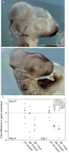
Undifferentiated neural crest cells in early Anolis embryos. (A) At oviposition, HNK1 positive cells (dark staining) are observed in the most anterior portion of the presumptive facial region (arrow) and within the developing trigeminal nerve (cranial nerve V; V). This pattern is maintained throughout the first 48 h of postoviposition development (B). At this time, HNK1 positive cells remain in the presumptive facial region, although the trigeminal nerve is further developed (Vmax = maxillary branch of V; Vmand = mandibulary branch of V; and Vopth = ophthalmic branch of V). (C) Our RT-qPCR experiment of the undifferentiated neural crest marker, Sox10, reveals that elevated incubation temperatures lead to reduced transcriptional activity or a reduced number of neural crest cells. Although there are not statistically significant differences in mean expression level on the day of oviposition, lower levels of Sox10 are observed in some individuals immediately following thermal stress (i.e., Day 0). These differences become exacerbated among all individuals the following day (i.e., Day 1), even though the thermal insult was already removed. Statistical results of the RT-qPCR experiment are summarized in Table 2.
Fig. 5
Fig. 5
The expression and function of Shh in A. sagrei. At oviposition, Shh is expressed within the ventral telencephalon (A; B, arrow). Shh is also expressed within the limb bud (Flb, forelimb; Hlb, hindlimb) and notochord at this time (A). Approximately 36–48 h after oviposition (C), two transitory domains of Shh mark the future position of facial prominences. Panel (C) shows the superior aspect of the oral epithelium prior to the formation of the midline facial prominences. Scale bars equal 1 mm.
Fig. 6
Fig. 6
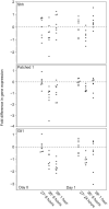
RT-qPCR results for members of the Hedgehog signaling pathway. Quantification of gene expression of the Hedgehog pathway in embryos incubated under different thermal profiles reveals modification in signaling in embryos incubated under elevated temperatures. The most notable changes in Hedgehog signaling are observed on Day 0, immediately after thermal stress has impacted the embryo. In these embryos, there are pronounced differences in the expression levels of Patched1 and Gli1. On Day 1, after the period that thermal stress has been removed, there are no statistical differences in Hedgehog gene expression between embryos incubated at relatively low and high temperatures, although there are differences between thermal stress treatments. Statistical results are summarized in Table 2.
Fig. 7
Fig. 7
Functional analysis of Shh in the developing head of A. sagrei. Increasing concentrations of cyclopamine lead to a reduction in midline facial tissue and shortening of the face. At 50 μM concentrations, clefting between the maxillary and midline facial prominences is visible in some embryos. Concentrations above 70 μM lead to the complete ablation of midline facial tissue (pers. obs.). The small anteriorly facing projections between the eyes are the remainder of the olfactory processes with a thin ectodermal covering.
Fig. 8
Fig. 8
Proliferation and cell death in thermally stressed A. sagrei embryos. Embryos incubated at 27°C (A) show widespread proliferation and low levels of cell death in the telencephalon. In contrast, embryos incubated at 36°C (B) show a wide domain of cell death in the ventral telencephalon (arrows). Quantification of these patterns (C: closed circles represent individuals incubated at 27°C and open circles 36°C) demonstrates that the number of apoptotic cells increases over the first 48 h of development (N = 6 for each time and treatment; *P < 0.05 Kruskal–Wallis nonparametric test).
Fig. 9
Fig. 9
A developmental model of embryonic thermal stress. Our developmental model predicts that thermal stress leads to cell death in the developing telencephalon, specifically in the region associated with Shh secretion. Modified levels of Hedgehog signaling then disrupt facial induction. Individual variation in the amount of cell death leads to varying levels of Shh secretion and the spectrum of phenotypes, from normal to severe, observed in our incubation experiments.

References

    1. Abayarathna T, Webb JK.. 2020. Effects of incubation temperatures on learning abilities of hatchling velvet geckos. Anim Cogn 23:613–20. - PubMed
    1. Abzhanov A, Tabin CJ.. 2004. Shh and Fgf8 act synergistically to drive cartilage outgrowth during cranial development. Dev Biol 273:134–48. - PubMed
    1. Ahlgren SC, Bronner-Fraser M.. 1999. Inhibition of Sonic hedgehog signaling in vivo results in craniofacial neural crest cell death. Curr Biol 9:1304–14. - PubMed
    1. Altizer S, Ostfeld RS, Johnson PTJ, Kutz S, Harvell CD. 2013. Climate change and infectious diseases: from evidence to a predictive framework. Science 341:514–9. - PubMed
    1. Amiel JJ, Tingley R, Shine R. 2011. Smart moves: effects of relative brain size on establishment success of invasive amphibians and reptiles. PLoS One 6:e18277. - PMC - PubMed

LinkOut - more resources