. 2021 Nov 14;24(12):103434.
doi: 10.1016/j.isci.2021.103434. eCollection 2021 Dec 17.

STING regulates peripheral nerve regeneration and colony stimulating factor 1 receptor (CSF1R) processing in microglia

Affiliations

STING regulates peripheral nerve regeneration and colony stimulating factor 1 receptor (CSF1R) processing in microglia

Giulio Morozzi et al. iScience. .

Abstract

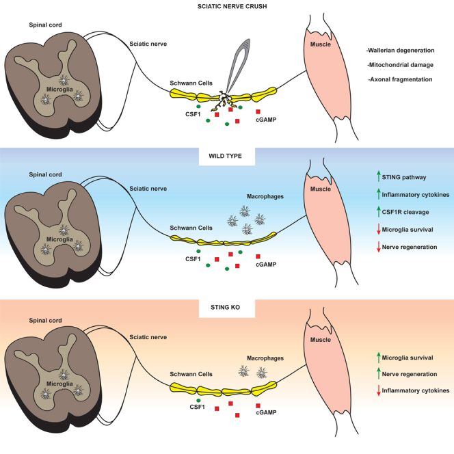
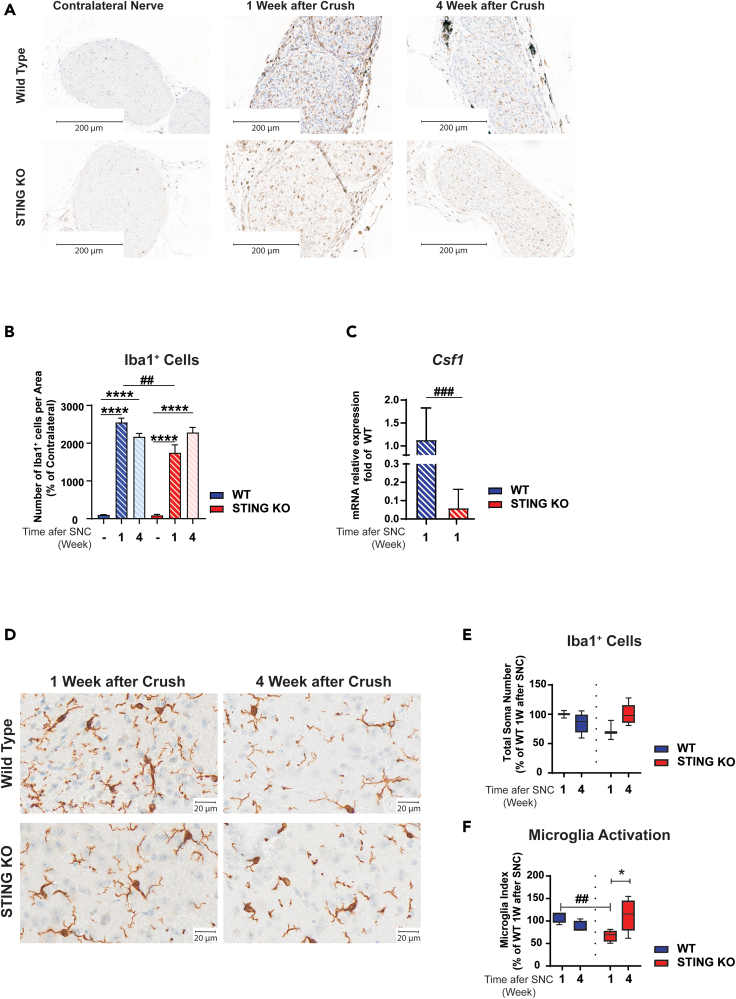
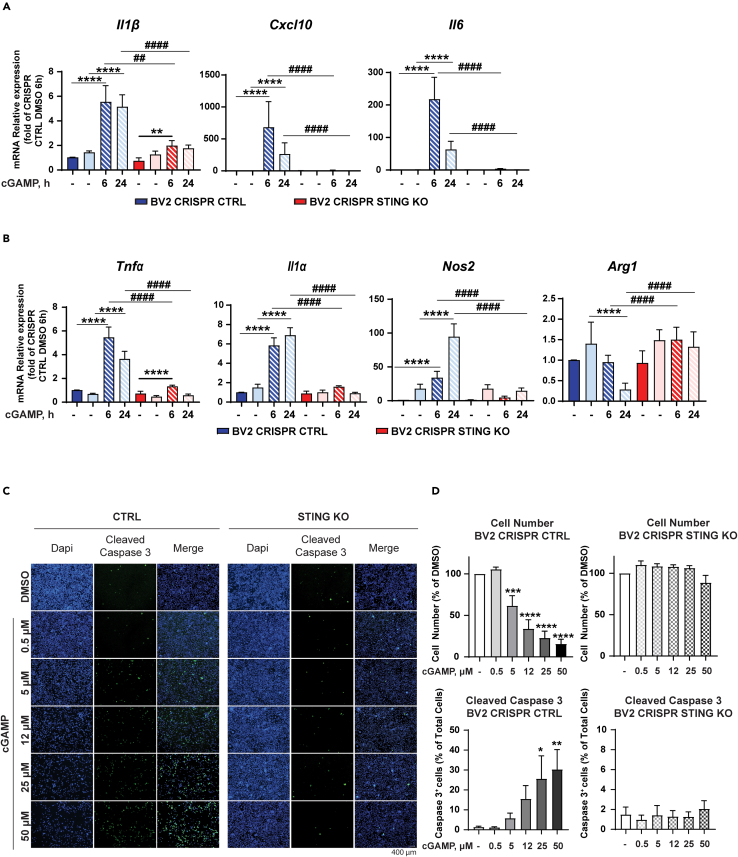
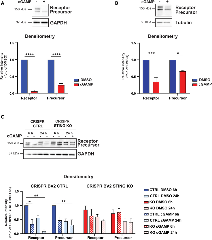
Inflammatory responses are crucial for regeneration following peripheral nerve injury (PNI). PNI triggers inflammatory responses at the site of injury. The DNA-sensing receptor cyclic GMP-AMP synthase (cGAS) and its downstream effector stimulator of interferon genes (STING) sense foreign and self-DNA and trigger type I interferon (IFN) immune responses. We demonstrate here that following PNI, the cGAS/STING pathway is upregulated in the sciatic nerve of naive rats and dysregulated in old rats. In a nerve crush mouse model where STING is knocked out, myelin content in sciatic nerve is increased resulting in accelerated functional axon recovery. STING KO mice have lower macrophage number in sciatic nerve and decreased microglia activation in spinal cord 1 week post injury. STING activation regulated processing of colony stimulating factor 1 receptor (CSF1R) and microglia survival in vitro. Taking together, these data highlight a previously unrecognized role of STING in the regulation of nerve regeneration.

Keywords: Cell biology; Immunology; Molecular biology; Neuroscience.


Conflict of interest statement

All authors are employees and some are shareholders of Novartis.

Figures

None
Graphical abstract
Figure 1
Figure 1
Differentially expressed pathways in young and old rats following sciatic nerve crush Gene expression in the distal nerve stump was assessed at day 1, 7, 14, and 21 after subjecting young and old adult rats (2 and 19 months, respectively) to sciatic nerve crush injury. Gene expression levels in the uninjured, contralateral nerve at day 7 were used as control. (A) Volcano plots depicting significantly enriched gene ontology pathways at day 1 and 7 in distal vs. contralateral nerve, as determined by GSEA. Highlighted are three gene sets linked to inflammation (GO:0006954), innate immune response (GO:0045087), and ensheathment of neurons (GO:0007272). Statistical significance was defined as BH-adjusted pvalue below or equal to 0.05 (dotted horizontal line). Dot size reflects gene set size. (B and C) Longitudinal gene expression changes for genes in the three highlighted ontology gene sets, and the cGAS/STING pathway reveals a resolution of injury-induced effect by day 21 in young animals, contrasting the sustained injury effect and differential gene expression in old rats. Gene expression values (log2 transformed rpkm) were normalized to mean expression level in the contralateral nerve. Heatmap rows and columns are representing distinct genes and individual animals at different time points post SNC, respectively.
Figure 2
Figure 2
Absence of STING accelerates nerve recovery in mice subjected to sciatic SNC Young and old C57BL/6J-Tmem173gt/J (STING KO) and C57BL/6J (WT) mice were subjected to SNC. (A) Schematic representation of in-vivo studies' design. We performed toe spread analysis and CMAP measurement at the indicated time points after SNC. Mice were sacrificed at day 7, 35, and 56 post SNC. Sciatic nerves and gastrocnemius muscles were excised for RNA and protein extraction. (B) Ablation of STING was confirmed by immunoblotting in gastrocnemius muscle 1 week post SNC. GAPDH was used as loading control. The graph shows the quantification of immunoreactive bands by densitometry. (∗p < 0.05, ∗∗p < 0.001, two-way ANOVA). (C and D) Toe spread and CMAP analysis was performed as indicated in A in young (C) and old animals (D). Blue and red values around the boxes indicate percentage of mice at each score. Toe spread scores were 0 - no spreading, 1 - intermediate spreading, 2 - full toe spreading. ∗∗p < 0.01, ∗∗∗p < 0.001, ∗∗∗∗p < 0.0001 versus contralateral leg that had a score of 2 at all-time points (Mann-Whitney test). CMAP data are mean amplitude ± SEM; the dotted line corresponds to baseline measurements performed one week before SNC. Six mice/group for 16-month-old mice (C),and eight mice/group for 14-week-old mice (D). ∗p < 0.05, ∗∗∗p < 0.001 (two-way ANOVA).
Figure 3
Figure 3
Ablation of STING increases the expression of the myelin marker MBP C57BL/6J-Tmem173gt/J (STING KO) and C57BL/6J (WT) mice were subjected to SNC. (A) Schematic representation of sciatic nerve, with segments proximal (R4) and distal (R1) to the crush site and respective sectioning points. The crush site is indicated in yellow in the mid segment (R2/R3). (B) Representative images of WT and STING KO sciatic nerve cross sections stained with anti-NF200 (axons, in green) and anti-MBP (myelin, in red) antibodies in region R2/R3 of the sciatic nerve at 1 or 4 weeks post SNC. The R2/R3 region of the contralateral non-crushed sciatic nerve is included for comparison. Fluorescent microscope images were taken at 20× magnification and scale bars are 50 μm. (C) Quantification of myelin fluorescent signal intensity. Only myelin associated with an axon was evaluated. Data represent myelin intensity (% of WT) ± SEM. # p<0.05 versus KO, Brown-Forsythe, and Welch ANOVA. (D) Quantification of axon number and area. Five mice/group and genotype. Mice were 6 months old at the beginning of the experiment. Data represent the mean ± SEM. ∗∗p<0.01, ∗∗∗p<0.001, ∗∗∗∗p<0.0001, one-way ANOVA.
Figure 4
Figure 4
STING KO decreases macrophage number in sciatic nerve and microglia activation in spinal cord following nerve crush C57BL/6J-Tmem173gt/J (STING KO) and C57BL/6J (WT) mice were subjected to SNC. (A and D) Representative images of Iba1 immunostaining of WT and STING KO sciatic nerve (A) and spinal cord (D) at 1 or 4 weeks post SNC. Scale bars are 200 μm in A and 20 μm in D; n = five mice/group and genotype. (B and E) Mice were 6 months old at the beginning of the experiment. Quantitative analysis of Iba1+ cells at the R2/R3 region in sciatic nerve (B) and spinal cord (lumbar region L5-L6) (E). (F) The microglia activation index is the ratio of the distal area and the soma plus the proximal area. Five mice/group. Data represent the mean ± SEM. (C) mRNA abundance of Csf1 was analyzed by qRT-PCR in sciatic nerve from 16-month-old mice subjected to SNC. Data represent mean fold increase of WT animals ±SEM, n = 6. ∗p<0.05, ∗∗p<0.01, ∗∗∗p<0.001, ∗∗∗∗p < 0.0001; (C) t test and (B, E, and F) two-way ANOVA. ∗No crush versus crush: #WT versus STING KO.
Figure 5
Figure 5
cGAMP-induced apoptosis and activation of BV2 microglial cells is STING dependent (A and B) BV2 STING KO and control (CTRL) cells were treated with the indicated concentrations of cGAMP or DMSO for 6 and 24 h. Analysis of inflammatory cytokines (A) or markers of microglia activation (B) was performed by qRT-PCR. (C) Representative immunofluorescence images are shown. (D) Cell number and cell death were evaluated by DAPI and cleaved caspase-3 staining (D), respectively. Scale bars are 400 μm. The experiment was repeated 3 times, and the data are expressed as means ± SEM (∗p < 0.05, ∗∗p<0.01, ∗∗∗p < 0.001, ∗∗∗∗p<0.0001; two-way ANOVA). (A and B) Data represent means ± SEM. ∗p < 0.05, ∗∗p<0.01, ∗∗∗p < 0.001, ∗∗∗∗p<0.0001 versus. ∗ DMSO versus cGAMP treatment. # BV2 CRISPR CTRL versus CRISPR BV2 STING KO.
Figure 6
Figure 6
cGAMP downregulates CSF1R expression in microglial cells via STING (A and B) BV2 cells (A) and iPSC-derived microglia (B) were treated with 50 μM cGAMP or DMSO for 24 h. CSF1R protein expression was analyzed by immunoblotting. The graph shows the quantification of the different immune-reactive bands of CSF1R by densitometry. GAPDH and tubulin were used as loading controls. The experiment was repeated 3 times. Data are mean band intensity ±SEM. ∗p<0.05, ∗∗p < 0.01, ∗∗∗p < 0.001, ∗∗∗∗p < 0.0001 (two-way ANOVA). (C) STING ablation prevents CSF1R proteolysis induced by cGAMP treatment. BV2 STING KO and control (CTRL) cells were treated with 50 μM cGAMP for the indicated time. The graph shows the quantification of the different immunoreactive bands of CSF1R by densitometry. GAPDH was used as a loading control. Data are mean band intensity ±SEM. ∗p<0.05, ∗∗p < 0.01, ∗∗∗p < 0.001, ∗∗∗∗p < 0.0001, (two-way ANOVA).

References

    1. Arnold W.D., Sheth K.A., Wier C.G., Kissel J.T., Burghes A.H., Kolb S.J. Electrophysiological motor unit number estimation (MUNE) measuring compound muscle action potential (CMAP) in mouse hindlimb muscles. J. Vis. Exp. 2015 doi: 10.3791/52899. - DOI - PMC - PubMed
    1. Arthur-Farraj P.J., Latouche M., Wilton D.K., Quintes S., Chabrol E., Banerjee A., Woodhoo A., Jenkins B., Rahman M., Turmaine M., et al. c-Jun reprograms Schwann cells of injured nerves to generate a repair cell essential for regeneration. Neuron. 2012;75:633–647. - PMC - PubMed
    1. Baccarini M., Dello Sbarba P., Buscher D., Bartocci A., Stanley E.R. IFN-gamma/lipopolysaccharide activation of macrophages is associated with protein kinase C-dependent down-modulation of the colony-stimulating factor-1 receptor. J. Immunol. 1992;149:2656–2661. - PubMed
    1. Barrette B., Calvo E., Vallieres N., Lacroix S. Transcriptional profiling of the injured sciatic nerve of mice carrying the Wld(S) mutant gene: Identification of genes involved in neuroprotection, neuroinflammation, and nerve regeneration. Brain Behav. Immun. 2010;24:1254–1267. - PubMed
    1. Beckmann N., Giorgetti E., Neuhaus A., Zurbruegg S., Accart N., Smith P., Perdoux J., Perrot L., Nash M., Desrayaud S., et al. Brain region-specific enhancement of remyelination and prevention of demyelination by the CSF1R kinase inhibitor BLZ945. Acta Neuropathol.Commun. 2018;6:9. - PMC - PubMed