. 2021 Nov 3:12:100091.
doi: 10.1016/j.mbplus.2021.100091. eCollection 2021 Dec.

Protection and rebuilding of the endothelial glycocalyx in sepsis - Science or fiction?

Affiliations

Protection and rebuilding of the endothelial glycocalyx in sepsis - Science or fiction?

Carolin Christina Drost et al. Matrix Biol Plus. .

Abstract

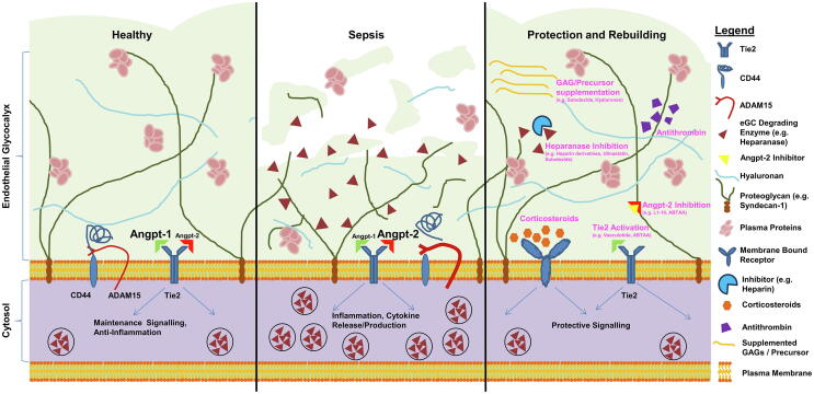
The endothelial glycocalyx (eGC), a delicate carbohydrate-rich structure lining the luminal surface of the vascular endothelium, is vital for maintenance of microvascular homeostasis. In sepsis, damage of the eGC triggers the development of vascular hyperpermeability with consecutive edema formation and organ failure. While there is evidence that protection or rebuilding of the eGC might counteract sepsis-induced vascular leakage and improve outcome, approved therapeutics are not yet available. This narrative review aims to outline possible therapeutic strategies to ameliorate organ dysfunction caused by eGC impairment.

Keywords: Glycocalyx; Glycosaminoglycan; Heparanase; Perfused boundary region; Sepsis.


Conflict of interest statement

The authors declare that they have no known competing financial interests or personal relationships that could have appeared to influence the work reported in this paper.

Figures

Fig. 1
Fig. 1
Protection and rebuilding of the endothelial glycocalyx (eGC): a schematic overview: The scheme summarizes potential approaches to prevent or ameliorate sepsis-induced eGC damage. Left: intact eGC with intact angiopoietin (Angpt)/Tie2 signalling promoting a quiescent state in healthy vessels. Middle: In sepsis, the Angpt-1/Angpt-2 ratio switches to Tie2 deactivation, which facilitates enzymatic degradation of the eGC via release of heparanase. Another eGC degrading mechanism is cleavage of the CD44 ectodomain, an important anchor for hyaluronan. Right: overview on possible therapeutic approaches to counteract eGC damage in sepsis.
Fig. 2
Fig. 2
Visualization and measurement of the endothelial glycocalyx (eGC): (A) Representative image of intravital microscopy in mouse cremaster model with FITC-dextran exclusion assay. The difference between bright field image vessel diameter and diameter of FITC-dextran (150 kDa), which cannot enter the eGC, allows estimation of eGC height in vivo. Image adapted from Yang et al. , modified. (B) Electron microscopy image of in vivo perfused/fixated and lanthanum-stained eGC in isolated rat aorta. Image adapted from Wiesinger et al. , modified. (C) Immunofluorescence wheat germ agglutinin (WGA)-staining of the eGC on endothelial cells with cross-sectional slide (indicated by the dashed line) along the stack. Image adapted from Drost et al. , modified. (D) Force versus distance curve generated upon nanoindentation on endothelial cells with atomic force microscopy. When indenting into the eGC the cantilever is deflected and linear fitting of the corresponding first slope allows deductions on eGC height. When indenting deeper the cantilever reaches the cell cortex resulting in the second slope as indicated in the schematic. Image adapted from Wiesinger et al. [38], modified.
Fig. 3
Fig. 3
Analysis of the sublingual microvasculature and illustration of PBR measurement: (A) Overview on the workflow of performing sublingual microscopy in humans. Parameters such as the perfused boundary region (PBR), an inverse estimate of the eGC thickness, are calculated by the software post-hoc. Image adapted from Rovas et al. , modified. (B) Representative image acquired with the GlycoCheckTM system using side-stream darkfield imaging. The software automatically discards invalid segments (yellow lines). Image adapted from Rovas et al. , modified. (C) Schematic illustration of the PBR in healthy vascular conditions with intact eGC (low PBR) in contrast to compromised vasculature with damaged eGC (high PBR). (For interpretation of the references to colour in this figure legend, the reader is referred to the web version of this article.)

References

    1. Fleischmann C., Scherag A., Adhikari N.K.J., Hartog C.S., Tsaganos T., Schlattmann P., Angus D.C., Reinhart K. Assessment of global incidence and mortality of hospital-treated sepsis current estimates and limitations. Am. J. Respir. Crit. Care Med. 2016;193(3):259–272. doi: 10.1164/rccm.201504-0781OC. - DOI - PubMed
    1. PRISM Investigators K.M., Rowan D.C., Angus M., Bailey A.E., Barnato R., Bellomo R.R., Canter T.J., Coats A., Delaney E., Gimbel R.D., Grieve D.A., Harrison A.M., Higgins B., Howe D.T., Huang J.A., Kellum P.R., Mouncey E., Music S.L., Peake F., Pike M.C., Reade M.Z., Sadique M., Singer D.M. Yealy, early, goal-directed therapy for septic shock — a patient-level meta-analysis. N. Engl. J. Med. 2017;376:2223–2234. doi: 10.1056/nejmoa1701380. - DOI - PubMed
    1. Marik P., Bellomo R. A rational approach to fluid therapy in sepsis. Br. J. Anaesth. 2016;116(3):339–349. doi: 10.1093/bja/aev349. - DOI - PubMed
    1. Clajus C., Lukasz A., David S., Hertel B., Lichtinghagen R., Parikh S.M., Simon A., Ismail I., Haller H., Kümpers P. Angiopoietin-2 is a potential mediator of endothelial barrier dysfunction following cardiopulmonary bypass. Cytokine. 2012;60(2):352–359. doi: 10.1016/j.cyto.2012.04.002. - DOI - PMC - PubMed
    1. Bae J.-S., Lee W., Son H.-N., Lee Y.-M., Kim I.-S. Anti-transforming growth factor β-induced protein antibody ameliorates vascular barrier dysfunction and improves survival in sepsis. Acta Physiol. 2014;212(4):306–315. doi: 10.1111/apha.12398. - DOI - PubMed

LinkOut - more resources