

Evaluation of squamous cell carcinoma antigen 1 expression in oral squamous cell carcinoma (tumor cells and peritumoral T-lymphocytes) and verrucous carcinoma and comparison with normal oral mucosa
- PMID: 34878006
- PMCID: PMC8653806
- DOI: 10.1590/1678-7757-2021-0374
Evaluation of squamous cell carcinoma antigen 1 expression in oral squamous cell carcinoma (tumor cells and peritumoral T-lymphocytes) and verrucous carcinoma and comparison with normal oral mucosa
Abstract
Background: Squamous cell carcinoma antigen (SCCA) is used as a prognostic marker for recurrence of squamous cell carcinoma in various sites, including head and neck. Studies suggest that its high serum levels are correlated to some clinical features, such as nodal metastasis. However, it is still unknown if high SCCA in patients with SCCA tissue expression in tumor cells are related to peripheral T-lymphocytes. Therefore, we did this study to evaluate SCCA expression in squamous cell carcinoma and verrucous carcinoma and to compare it with normal oral mucosa, also investigating the correlation between serum-based and tissue-based antigen levels.
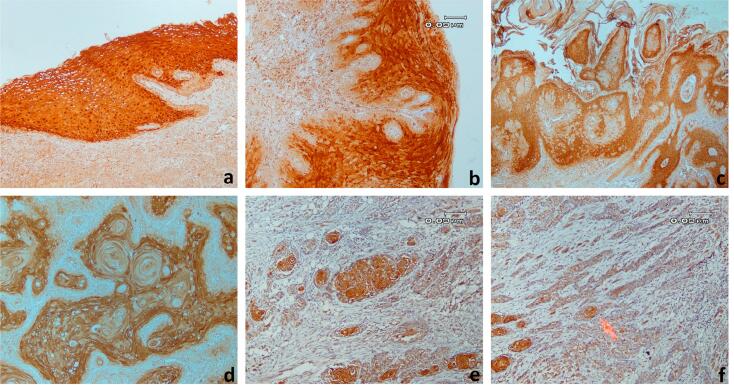
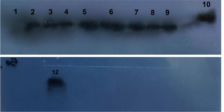
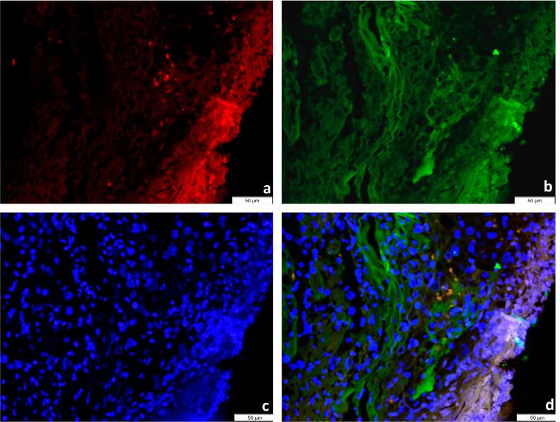
Methodology: In this study, the immunohistochemistry (IHC) technique was used to determine the SCCA1 expression pattern in 81 specimens divided into 3 groups, including oral squamous cell carcinoma, verrucous carcinoma, and normal oral mucosa. Serum-based and tissue-based antigen levels of 20 oral squamous cell carcinoma cases were compared by the western blot assay. SCCA expression was also evaluated and compared in both tumor cells and peripheral T-lymphocytes by the immunofluorescence assay.
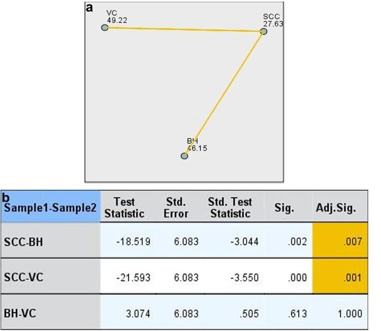
Results: Our results showed that the SCCA levels in SCC specimens were significantly lower than in verrucous carcinoma and normal and hyperplastic oral mucosa specimens. We found no correlation between the IHC expression of SCCA and serum levels. SCCA was well expressed in both tumor cells and peripheral T-lymphocytes.
Conclusion: Decreasing SCCA in SCC specimens suggested that SCC tumor cells may affect more than the serum levels of SCCA in some patients. In addition, expression of SCCA in peripheral T-lymphocytes showed that both tumor cells and T-lymphocytes may cause serum SCCA.
Conflict of interest statement
All authors declare to have no conflict of interests.
Figures
References
-
- Chi AC, Day TA, Neville BW, et al. Oral cavity and oropharyngeal squamous cell carcinoma: an update. CA Cancer J Clin. 2015;65(5):401–421. doi: 10.3322/caac.21293. - DOI - PubMed
- Chi AC, Day TA, Neville BW. Oral cavity and oropharyngeal squamous cell carcinoma: an update. CA Cancer J Clin. 2015;65(5):401-21. doi: 10.3322/caac.21293 - PubMed
-
- Howlander N, Noone A, Krapcho M, Miller D, Bishop K, Kosary C. SEER cancer statistics review: 1975-2014. Bethesda: National Cancer Institute; 2017.
- Howlander N, Noone A, Krapcho M, Miller D, Bishop K, Kosary C. SEER cancer statistics review: 1975-2014. Bethesda: National Cancer Institute; 2017.
-
- Rivera C, Venegas B. Histological and molecular aspects of oral squamous cell carcinoma (Review) Oncol Lett. 2014;8(1):7–11. doi: 10.3892/ol.2014.2103. - DOI - PMC - PubMed
- Rivera C, Venegas B. Histological and molecular aspects of oral squamous cell carcinoma (Review). Oncol Lett. 2014;8(1):7-11. . doi: 10.3892/ol.2014.2103 - PMC - PubMed
-
- Mishra A, Verma M. Cancer biomarkers: are we ready for the prime time? Cancers (Basel) 2010;2(1):190–208. doi: 10.3390/cancers2010190. - DOI - PMC - PubMed
- Mishra A, Verma M. Cancer biomarkers: are we ready for the prime time? Cancers (Basel). 2010;2(1):190-208. doi: 10.3390/cancers2010190 - PMC - PubMed
-
- Kato H, Torigoe T. Radioimmunoassay for tumor antigen of human cervical squamous cell carcinoma. Cancer. 1977;40(4):1621–1628. doi: 10.1002/1097-0142(197710)40:4<1621::aid-cncr2820400435>3.0.co;2-i. - DOI - PubMed
- Kato H, Torigoe T. Radioimmunoassay for tumor antigen of human cervical squamous cell carcinoma. Cancer. 1977;40(4):1621-8. doi: 10.1002/1097-0142(197710)40:4<1621::aid-cncr2820400435>3.0.co;2-i - PubMed
Publication types
MeSH terms
Substances
LinkOut - more resources
Full Text Sources
Medical
Molecular Biology Databases
Research Materials
