. 2021 Dec 8;13(623):eabh1962.
doi: 10.1126/scitranslmed.abh1962. Epub 2021 Dec 8.

Combining a CAR and a chimeric costimulatory receptor enhances T cell sensitivity to low antigen density and promotes persistence

Affiliations

Combining a CAR and a chimeric costimulatory receptor enhances T cell sensitivity to low antigen density and promotes persistence

Afroditi Katsarou et al. Sci Transl Med. .

Erratum in

Abstract

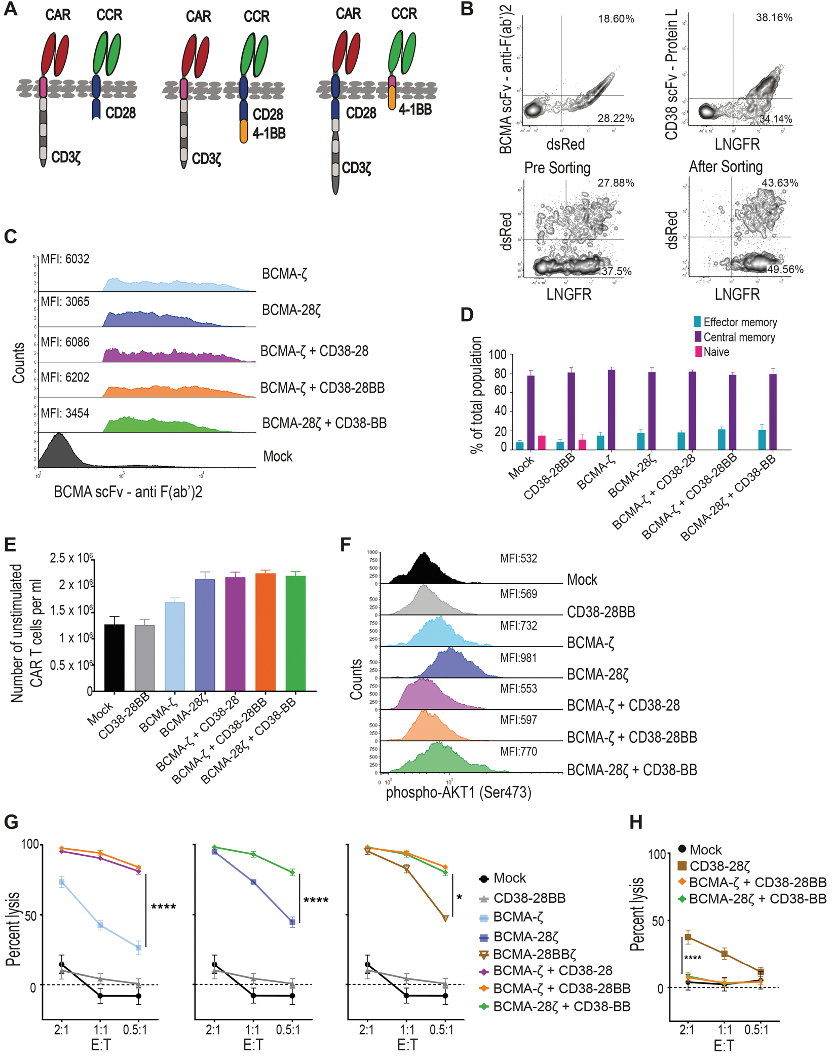
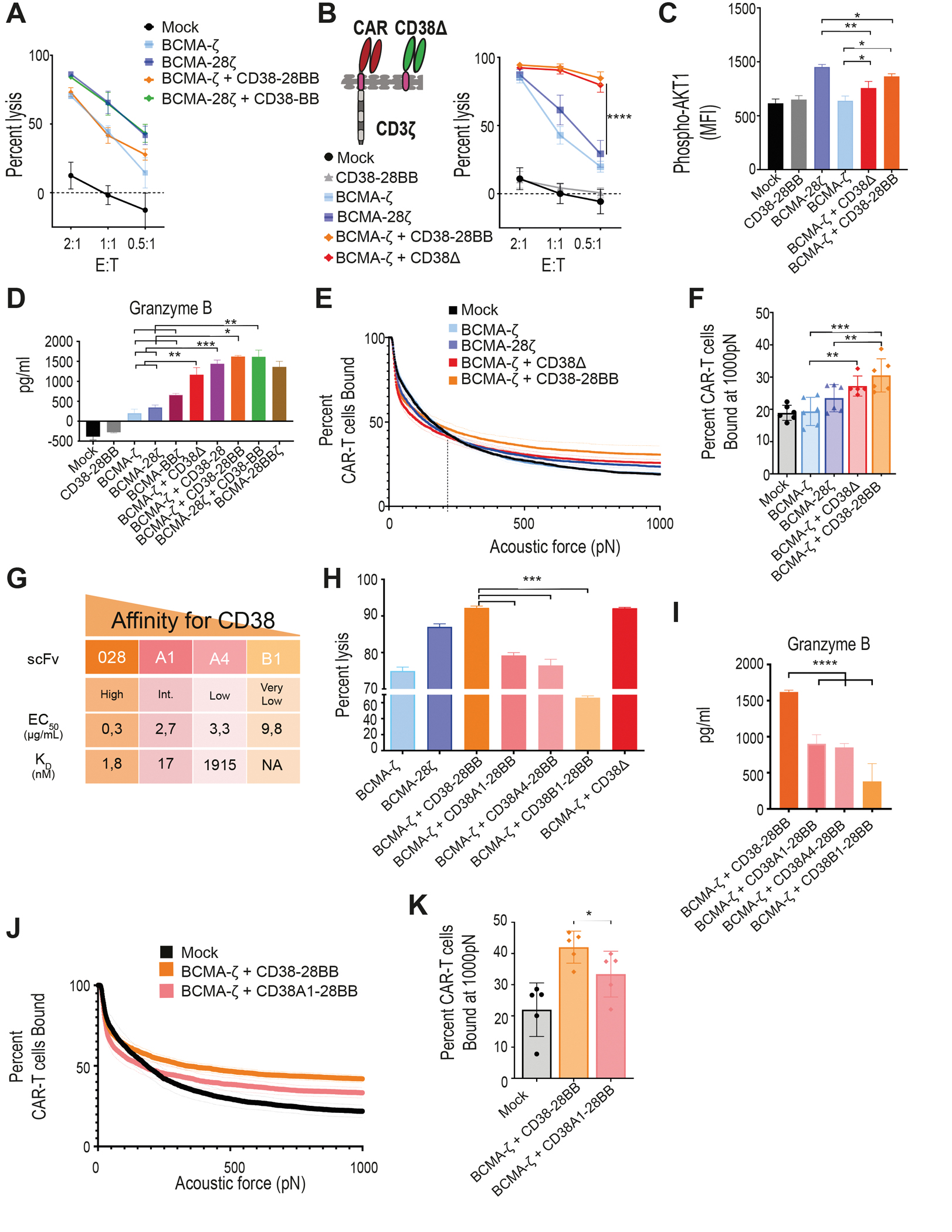
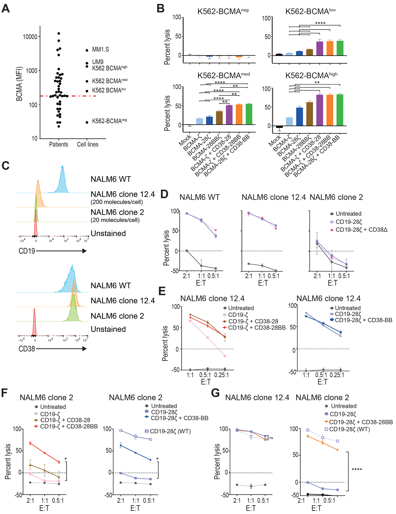
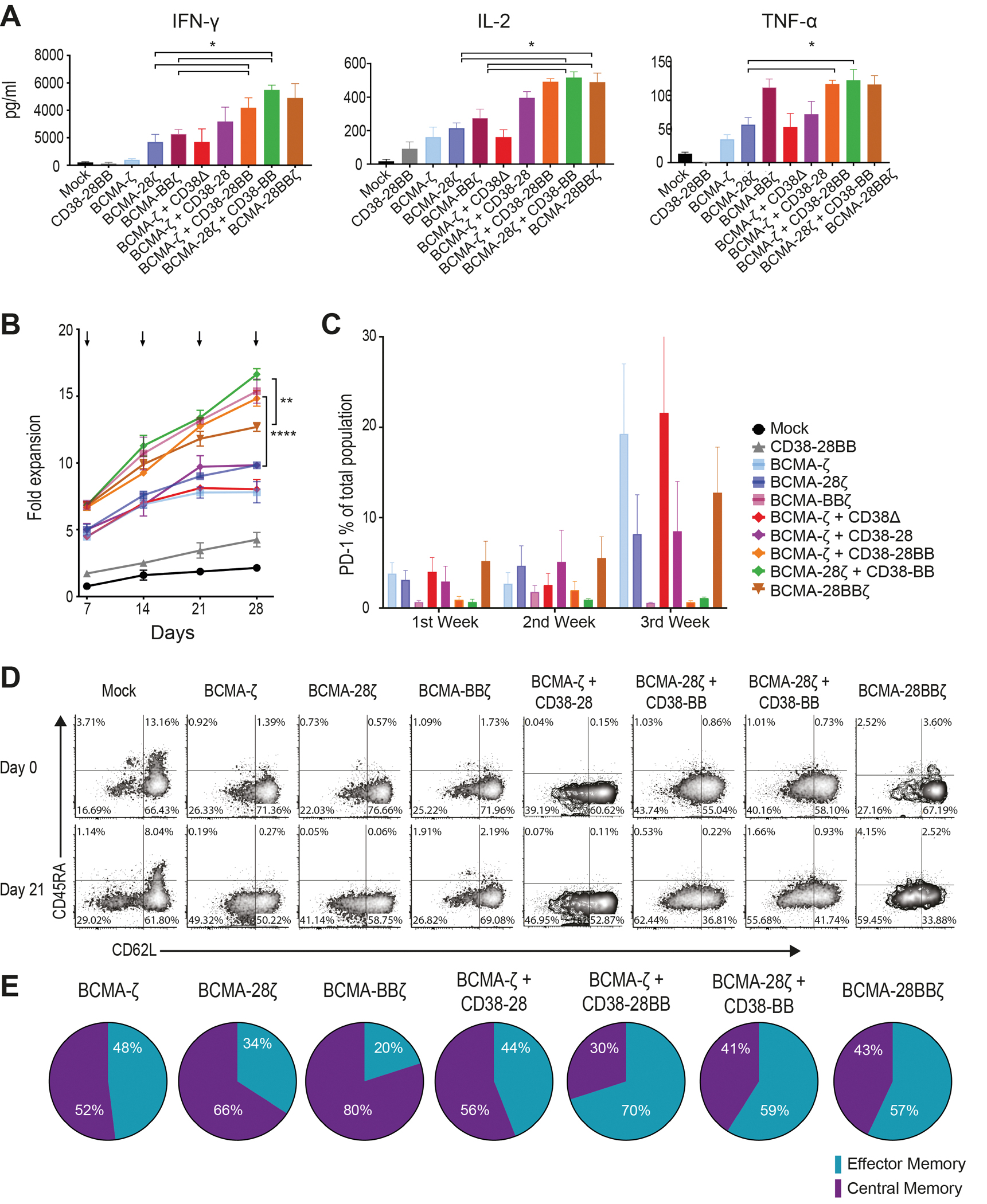
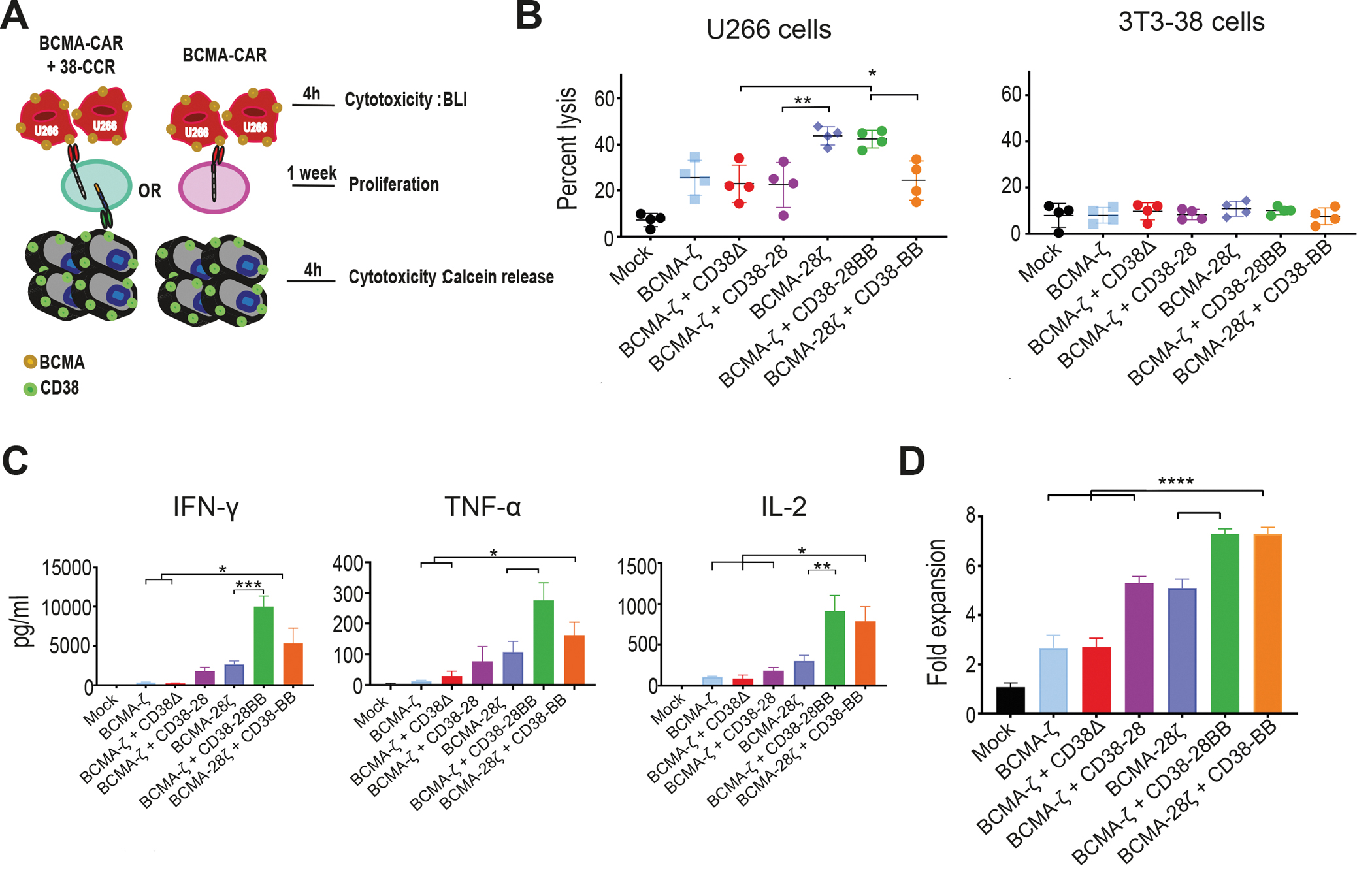
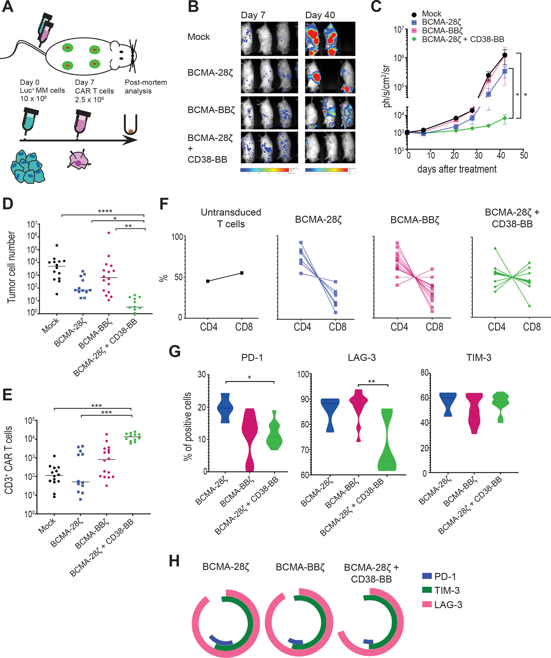
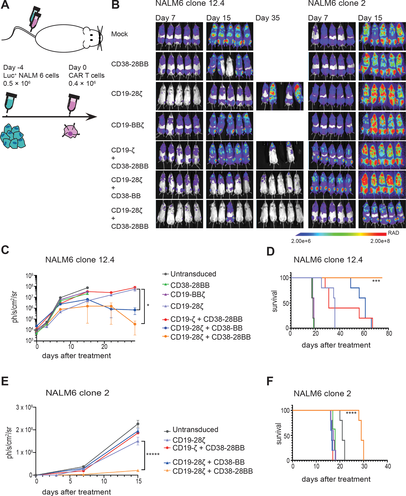
Despite the high remission rates achieved using T cells bearing a chimeric antigen receptor (CAR) against hematogical malignancies, there is still a considerable proportion of patients who eventually experience tumor relapse. Clinical studies have established that mechanisms of treatment failure include the down-regulation of target antigen expression and the limited persistence of effective CAR T cells. We hypothesized that dual targeting mediated by a CAR and a chimeric costimulatory receptor (CCR) could simultaneously enhance T cell cytotoxicity and improve durability. Concomitant high-affinity engagement of a CD38-binding CCR enhanced the cytotoxicity of BCMA-CAR and CD19-CAR T cells by increasing their functional binding avidity. In comparison to second-generation BCMA-CAR or CD19-CAR T cells, double-targeted CAR + CD38-CCR T cells exhibited increased sensitivity to recognize and lyse tumor variants of multiple myeloma and acute lymphoblastic leukemia with low antigen density in vitro. In addition, complimentary costimulation by 4-1BB and CD28 endodomains provided by the CAR and CCR combination conferred increased cytokine secretion and expansion and improved persistence in vivo. The cumulatively improved properties of CAR + CCR T cells enabled the in vivo eradication of antigen-low tumor clones, which were otherwise resistant to treatment with conventional CAR T cells. Therefore, multiplexing targeting and costimulation through the combination of a CAR and a CCR is a powerful strategy to improve the clinical outcomes of CAR T cells by enhancing cytotoxic efficacy and persistence, thus preventing relapses of tumor clones with low target antigen density.


Conflict of interest statement

Competing interests: J.M-S. holds unrelated patent on CAR T cell technologies. N.W.C.J.v.d.D. has received research support from Janssen Pharmaceuticals, AMGEN, Celgene, Novartis, and BMS, and serves on advisory boards for Janssen Pharmaceuticals, AMGEN, Celgene, BMS, Novartis, Takeda, Bayer, and Servier. S.Z. has received research support from Celgene, Takeda and Janssen Pharmaceuticals and serves in advisory boards for Celgene, Takeda, Janssen and Amgen. T.M. has received research support from Janssen Research and Development, Celgene, Onkimmune and Genmab. I.S., Y.R.P., E.M., and R.M.R. are employed by LUMICKS. H.Y. and J.d.B. are employed by Kuros Biosciences BV. D.A. has received research support from Celgene, Pharmacyslics and Kite Pharma and serves in advisory boards for Celgene, Juno, Partners Tx, Karyopharm, BMS, Aviv MedTech Ltd., Takeda, Legend BioTech, and Chugai. M.S. receives research funding from Fate Therapeutics, Takeda Pharmaceuticals and Atara Biotherapeutics and holds patents on CAR T cell technologies. R.W.J.G. has received research support from Janssen Research and Development. M.T. holds unrelated patent on CAR T cell technologies. The rest of the authors have no conflicts of interest to declare.

Figures

Figure 1.
Figure 1.. Combinatorial tumor targeting strategy with a CAR and a CCR results in enhanced cytotoxicity.
(A) Schematic representations of dual targeted CAR+CCR strategies are shown. First generation CARs were combined with the CD38-CCR construct bearing either CD28 (CD38-28) or both CD28 and 4-1BB costimulatory domains (CD38-28BB). Second generation CARs were combined with the CD38-CCR that contains only the 4-1BB signaling domain (CD38-BB). (B) Representative flow cytometry plots illustrating BCMA-CAR/dsRed and CD38-CCR/LNGFR expression of double transduced T cells are shown. BCMA-CAR expression was measured with an F(ab’)2 Fragment Goat Anti-Mouse antibody. CD38-CCR expression was measured after binding of Protein L. (C) Mean fluorescence intensity (MFI) of BCMA-CAR expression on transduced T cells is shown. MFI values are reported. Mock indicates mock-transduced T cells. (D) Percentage of CAR T cells that have a naïve (CD62L+CD45RA+), central memory (CD62L+CD45RA), or an effector memory (CD62LCD45RA) phenotype are shown (n=4 per group). (E) The absolute number of unstimulated CAR T cells per ml is shown at five days after transduction (n=4 per group). (F) AKT1 phosphorylation (phosho-AKT1) in unstimulated CAR T cells was assessed by flow cytometry. Representative results from one donor are shown. (G) Lysis of luciferase expressing MM1.S (BCMA+CD38+) cells is shown. Tumor cell killing was measured in a 16 hour bioluminescence (BLI) assay (n=4 per group) at the indicated effector:target (E:T) ratios. (H) Calcein-loaded 3T3 cells transduced to express CD38 were co-cultured at indicated E:T ratios with either double-targeting BCMA-CAR+CD38-CCR T cells or single targeting CD38-28ζ CAR T cells. Tumor cell killing was measured after 4 hours (n=4 per group). Statistical analysis in (G) and (H) was performed by two-way ANOVA and subsequent multiple comparison, corrected by Turkey test. *p<0.05, ****p<0.0001.
Figure 2.
Figure 2.. CD38-CCR engagement results in enhanced cytotoxicity of CAR T cells by increasing functional avidity.
(A) Lysis of luciferase expressing U266 cells (BCMA+CD38) was measured in a 16 hour BLI assay (n=4 per group) at the indicated E:T ratios. (B) A first generation BCMA-CAR was combined with the CD38Δ construct lacking the intracellular costimulatory tail. BCMA-ζ+CD38Δ T cells and the BCMA-ζ+CD38-28BB T cells were incubated with MM1.S cells and lysis was determined in a 16 hour BLI assay (n=4) at the indicated E:T ratios. Statistical analysis was performed with a two-way ANOVA and subsequent multiple comparison, corrected by Turkey test. ****p<0.0001. (C) MFI of AKT1 phosphorylation (pAKT1) is shown in BCMA-CAR+CD38-CCR T cells activated by stimulation with UM9 cells for 15 minutes as assessed by flow cytometry (n=4 per group). Statistical analysis was performed by one-way ANOVA and subsequent multiple comparison, corrected by Turkey test. *p<0.05, **p<0.01. (D) Granzyme B secretion by CAR T cells co-cultured with ΜΜ1.S (E:T ratio 1:1) for 16 hours is shown (n=4 per group). Statistical analysis was performed by paired t-test. *p<0.05, **p<0.01, ***p<0.001. (E) The strength of interaction between single- and double-targeting T cells and MM1.S target cells was measured. Percentage of total CAR T cells remaining bound to target cells as the acoustic force ramp is applied from 0 to 1000 pN are shown. The dotted line refers to the threshold beyond which all measurement are considered specific binding. (F) The percentage of CAR T cells remaining bound to MM1.S target cells at 1000 pN is shown; data are presented as mean ±SEM from 6 pooled measurements; p-values were calculated using a paired t-test. **p<0.01, ***p<0.001. (G) Affinity characteristics are shown for anti-CD38 antibodies used to generate scFvs. The surface plasmon resonance–determined dissociation constant (KD value, nmol/L) and half-effective concentration (EC50) when titrated on CHO-CD38 cells (μg/mL), described in (29). (H) MM1.S cells were co-cultured at a 1:1 ratio with BCMA-CAR T cells co-expressing CD38-CCRs with gradually lower affinities for CD38 or CD38Δ. Tumor cell lysis was measured in a BLI assay (n=4 per group). Statistical analysis was performed with a paired t-test. (I) Cell supernatants from (H) were harvested to measure granzyme B secretion by ELISA (n=4 per group). Statistical analysis was performed with a paired t-test. ***p<0.001. (J) The strength of interaction between BCMA-ζ + CD38-28BB (EC50 0.3) or BCMA-ζ+CD38A1-28BB (EC50 2.7) T cells and MM1.S target cells is shown as the percentage of total CAR T cells remaining bound to target cells as the acoustic force ramp is applied from 0 to 1000 pN. Dotted line refers to the threshold beyond which all measurement are considered specific binding. (K) The percentage of CAR T cells remaining bound to MM1.S target cells at 1000 pN is shown; data are presented as mean ±SEM from 5 pooled measurements and p-values were calculated using a paired t-test. *p<0.05.
Figure 3.
Figure 3.. CAR and CCR combinations restore the cytotoxic capacity of T cells against low-antigen expressing tumor variants.
(A) Primary malignant plasma cells and cell lines were analyzed for BCMA expression by flow cytometry. Dot plots depict the MFI of BCMA found on primary MM tumor cells from 49 patients, in comparison with MM cell lines and K562 cells transduced to express BCMA in variant concentrations. The red line depicts median MFI for primary MM cells. (B) K562 cells with different BCMA expression were co-cultured with first, second, or third generation BCMA-CAR T cells and double BCMA-CAR+CD38-CCR T cells (BCMA-ζ+CD38-28BB or BCMA-28ζ+CD38-BB) at 1:1 effector to target ratio. Cell lysis was determined at 16 hours by BLI (n=3 per group). Statistical analysis performed with a two-way ANOVA and subsequent multiple comparison, corrected by Turkey test. **p<0.01, ****p<0.0001. (C) Histograms of CD19 and CD38 expression are shown for the NALM6 acute lymphoblastic leukemia cell line (WT) and on two NALM6 clones (clone 12.4 and clone 2). The red histogram shows the unstained control. (D) Specific lysis of NALM6 WT, clone 12–4 (200 molecules per cell), and NALM6 clone 2 (20 molecules per cell) is shown when cell lines were co-cultured with CD19-28ζ or double CD19-28ζ+CD38Δ T cells. (E) Specific lysis of NALM6 clone 12-4 (200 molecules per cell) is shown when cocultured with CD19-ζ, double CD19-ζ+CD38-28, or double CD19-ζ+CD38-28BB T cells (left) or with CD19-28ζ or double CD19-28ζ+CD38-BB (right) T cells. (F) Specific lysis of NALM6 clone 2 is shown when co-cultured with CD19-ζ, double CD19-ζ+CD38-28, or double CD19-ζ+CD38-28BB T cells (left) or with CD19-28ζ or double CD19-28ζ+CD38-BB (right) T cells. The open squares represent the lysis activity of the CD19 wild type NALM6 cell line when treated with CD19-28ζ CAR T cells. Statistical analysis was performed with using two-way ANOVAs and subsequent multiple comparison, corrected by Turkey test. *p<0.05. (G) Specific cytolysis of NALM6 clone 12-4 (200 molecules per cell) and NALM6 clone 2 (20 molecules per cell) is shown when cells were cocultured with CD19-28ζ or double CD19-28ζ+CD38-28BB T cells at indicated E:T ratios. The open squares represent the lysis activity of the CD19 WT NALM6 cell line when treated with CD19-28ζ CAR T cells. Statistical analysis was performed using two-way ANOVA and subsequent multiple comparison, corrected by Turkey test. ****p<0.0001.
Figure 4.
Figure 4.. Co-expression of a CCR enhances the expansion of CAR T cells.
(A) Cytokine secretion is shown in supernatants collected from co-cultures with ΜΜ1.S (E:T ratio 1:1) for 16 hours (n=4 per group). Statistical analysis was performed by paired t-tests. *p<0.05. (B) CAR T cell proliferation is shown following weekly antigen-specific stimulations with irradiated tumor cells. Black arrows indicate addition of irradiated tumor cells. The fold of the expansion of the CAR+ T cells is indicated on the y-axis. The data represent the mean±SEM of 4 experiments with different donors. Statistical analysis was performed using a two-way ANOVA and subsequent multiple comparison, corrected by Turkey test. **p<0.01, ****p<0.0001. (C) PD-1 expression is shown for T cells from the proliferation assay in (B) (n=4 donors). (D) Flow cytometry density plots of phenotypic profile of each group from (B) are shown at day 0 (before expansion) and at day 21. Cells were characterized as naive (CD45RA+/CD62L+), central memory (CD45RA/CD62L+), effector memory (CD45RA/CD62L) or effector (CD45RA+/CD62L). (E) Percentage of CAR T cells from (B) that have a central memory or an effector memory phenotype at day 21 (n=4 donors).
Figure 5.
Figure 5.. CD38-CCR can signal through in-trans binding of the antigen expressed on accessory cells without off-tumor toxicity.
(A) A schematic representation of the coculture cytotoxicity/proliferation assay is shown. Luciferase-positive BCMA+CD38 U266 cells were co-cultured with calcein-loaded 3T3-38+ cells and single- or double-targeting T cells were added in the co-culture. Lysis of U266 cells was determined by BLI and lysis of 3T3-CD38 cells by calcein release assay. (B) U266 cells and calcein-loaded 3T3-CD38 cells were cocultured at a 3:1 E:T ratio with mock, BCMA-ζ, BCMA-ζ+CD38Δ, BCMA-ζ+CD38-28, BCMA-28ζ, BCMA-28ζ+CD38-BB or BCMA-ζ+CD38-28BB CAR T cells and cell lysis was measured after 4 hours by BLI and a Calcein-AM release assay, respectively (n=4 per group). Statistical analysis was performed by a paired t-test. *p<0.05, **p<0.01. (C) Cell supernatants from (B) were harvested to measure cytokine secretion with a flow cytometry–based assay (n=4 per group). Statistical analysis was performed with paired t-tests. *p<0.05, **p<0.01, ***p<0.001. (D) Irradiated BCMA+CD38 U266 cells were co-cultured with irradiated 3T3-38+ cells. Mock, BCMA-ζ, BCMA-ζ+CD38Δ, BCMA-ζ+CD38-28, BCMA-28ζ, BCMA-28ζ+CD38-BB or BCMA-ζ+CD38-28BB CAR T cells were added in the co-culture and the expansion of CAR T cells was measured 7 days later. Statistical analysis was performed with one-way ANOVAs and subsequent multiple comparison, corrected by Turkey test. ****p<0.0001.
Figure 6.
Figure 6.. BCMA-CAR+CD38-CCR T cells show enhanced in vivo anti-tumor function, improved persistence, and reduced expression of exhaustion markers.
(A) A schematic representation of the MM scaffold-based xenograft murine model is shown. 10 × 106 Luc-GFP UM9 cells were administrated intravenously. Mock-transduced, BCMA-28ζ, BCMA-BBζ or BCMA-28ζ+CD38-BB CAR T cells were infused intravenously 7 days later. Tumor burden was measured weekly by BLI. (B) Representative BLI images of two time points are shown, with the pixel intensity represented in color. (C) Average tumor burden of mice was quantified by BLI and is depicted as units of photons per second per square centimeter per steradian (ph/sec/cm2/sr) (n=4 mice per group). Statistical analysis was performed using a two-way ANOVA and subsequent multiple comparison, corrected by Turkey test. *p<0.05. (D to H) Post-mortem scaffolds were harvested from each mouse and dissociated. Single-cell suspensions were counted, stained, and measured by flow cytometry. (D) Absolute UM9 tumor cell (GFP+/CD38+) numbers in the scaffolds are shown. Each dot represents one scaffold. (E) Absolute CAR T cell numbers in the scaffolds are shown. Each dot represents one scaffold. (F) CD4 and CD8 CAR T cell percentages in each scaffold are shown. (G) Violin plots show the expression of PD-1, LAG-3, and TIM-3 on CAR T cells isolated from scaffolds. (H) Co-expression of inhibitory receptors of (G) are presented. Statistical comparisons in (D), (E), and (G) were performed by Kruskal-Wallis test between the indicated groups; ns, not significant. *p<0.05, **p<0.01, ***p<0.001, ****p<0.0001.
Figure 7.
Figure 7.. The CD19-CAR+CD38-CCR combination elicits improved in vivo anti-tumor function against tumor variants with very low antigen density.
(A) A schematic representation of the ALL in vivo model is shown. 0.5 × 10⁶ FFLuc-GFP NALM6 clone 12.4 or NALM6 clone 2 cells were administrated intravenously. Untransduced, CD38-28BB, CD19-ΒΒζ, CD19-28ζ, CD19-ζ+CD38-28BB, CD19-28ζ+CD38-BB or CD19-28ζ+CD38-28BB CAR T cells were infused intravenously 4 days later. Tumor burden was measured weekly by BLI. (B) Representative images of three time points are shown with the pixel intensity represented in color. (C) Average tumor burden of mice injected with FFLuc-GFP NALM6 clone 12.4 cells is shown over time. Tumor burden is depicted as units of photons per second per square centimeter per steradian (ph/sec/cm2/sr) (n=5 mice per group). (D) Survival of mice injected with NALM6 clone 12-4 is shown. (E) Average tumor burden of mice injected with FFLuc-GFP NALM6 clone 2 cells is shown over time. Tumor burden is depicted as units of photons per second per square centimeter per steradian (ph/sec/cm2/sr); n=5 mice per group. (F) Survival of mice injected with NALM6 clone 2 is shown. Statistical analysis of tumor burden (C and E) was performed with two-way ANOVAs and subsequent multiple comparison, corrected by Turkey test. *p<0.05. Statistical analysis of survival (D and F) was performed with log-rank tests. ***p<0.001, ****p<0.0001.

References

    1. van der Stegen SJ, Hamieh M, Sadelain M, The pharmacology of second-generation chimeric antigen receptors. Nat Rev Drug Discov 14, 499–509 (2015). - PMC - PubMed
    1. Maude SL, Laetsch TW, Buechner J, Rives S, Boyer M, Bittencourt H, Bader P, Verneris MR, Stefanski HE, Myers GD, Qayed M, De Moerloose B, Hiramatsu H, Schlis K, Davis KL, Martin PL, Nemecek ER, Yanik GA, Peters C, Baruchel A, Boissel N, Mechinaud F, Balduzzi A, Krueger J, June CH, Levine BL, Wood P, Taran T, Leung M, Mueller KT, Zhang Y, Sen K, Lebwohl D, Pulsipher MA, Grupp SA, Tisagenlecleucel in Children and Young Adults with B-Cell Lymphoblastic Leukemia. N Engl J Med 378, 439–448 (2018). - PMC - PubMed
    1. Park JH, Riviere I, Gonen M, Wang X, Senechal B, Curran KJ, Sauter C, Wang Y, Santomasso B, Mead E, Roshal M, Maslak P, Davila M, Brentjens RJ, Sadelain M, Long-Term Follow-up of CD19 CAR Therapy in Acute Lymphoblastic Leukemia. N Engl J Med 378, 449–459 (2018). - PMC - PubMed
    1. Majzner RG, Mackall CL, Clinical lessons learned from the first leg of the CAR T cell journey. Nat Med 25, 1341–1355 (2019). - PubMed
    1. Fry TJ, Shah NN, Orentas RJ, Stetler-Stevenson M, Yuan CM, Ramakrishna S, Wolters P, Martin S, Delbrook C, Yates B, Shalabi H, Fountaine TJ, Shern JF, Majzner RG, Stroncek DF, Sabatino M, Feng Y, Dimitrov DS, Zhang L, Nguyen S, Qin H, Dropulic B, Lee DW, Mackall CL, CD22-targeted CAR T cells induce remission in B-ALL that is naive or resistant to CD19-targeted CAR immunotherapy. Nat Med 24, 20–28 (2018). - PMC - PubMed

Publication types