. 2021 Dec 10;7(50):eabk0568.
doi: 10.1126/sciadv.abk0568. Epub 2021 Dec 8.

Diurnal metabolic control in cyanobacteria requires perception of second messenger signaling molecule c-di-AMP by the carbon control protein SbtB

Affiliations

Diurnal metabolic control in cyanobacteria requires perception of second messenger signaling molecule c-di-AMP by the carbon control protein SbtB

Khaled A Selim et al. Sci Adv. .

Abstract

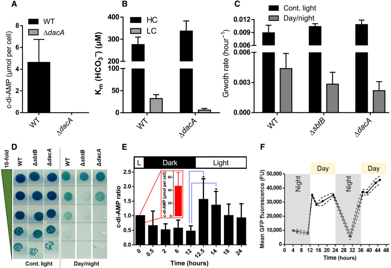
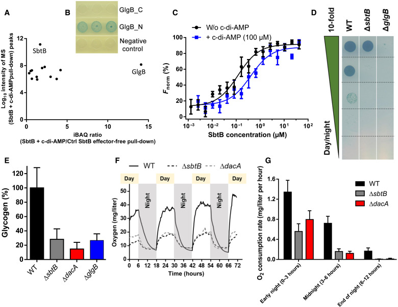
Because of their photosynthesis-dependent lifestyle, cyanobacteria evolved sophisticated regulatory mechanisms to adapt to oscillating day-night metabolic changes. How they coordinate the metabolic switch between autotrophic and glycogen-catabolic metabolism in light and darkness is poorly understood. Recently, c-di-AMP has been implicated in diurnal regulation, but its mode of action remains elusive. To unravel the signaling functions of c-di-AMP in cyanobacteria, we isolated c-di-AMP receptor proteins. Thereby, the carbon-sensor protein SbtB was identified as a major c-di-AMP receptor, which we confirmed biochemically and by x-ray crystallography. In search for the c-di-AMP signaling function of SbtB, we found that both SbtB and c-di-AMP cyclase–deficient mutants showed reduced diurnal growth and that c-di-AMP–bound SbtB interacts specifically with the glycogen-branching enzyme GlgB. Accordingly, both mutants displayed impaired glycogen synthesis during the day and impaired nighttime survival. Thus, the pivotal role of c-di-AMP in day-night acclimation can be attributed to SbtB-mediated regulation of glycogen metabolism.


Figures

Fig. 1.
Fig. 1.. Identification of SbtB as a major c-di-AMP receptor protein in cyanobacteria.
(A) ITC analysis shows that SbtB binds c-di-AMP in an anticooperative manner with Kd values as indicated. Top: The raw ITC data in the form of the heat produced during the titration of 33.3 μM SbtB (trimeric concentration) with 0.5 mM c-di-AMP. Bottom: The binding isotherms and the best-fit curves according to the three sequential binding site model. (B) SDS–polyacrylamide gel electrophoresis analysis of c-di-AMP pull-down elution fraction and Western blot detection of SbtB, using α-SbtB antibodies. Samples were analyzed with quantitative MS-based proteomics analysis. Identified proteins are sorted by their scores. NAD+, nicotinamide adenine dinucleotide; ATPase, adenosine triphosphatase; ABC, ATP-binding cassette; NUDIX hydrolases cleave nucleoside diphosphates linked to any (“x”) moiety. (C to F) Structural and binding properties of the ScSbtB protein. (C) Overall architecture of the trimeric SbtB:c-di-AMP complex with nucleotide-binding pockets located in the intersubunit clefts and shown in ribbon representation with different color for each monomer. (D) The electron density of c-di-AMP is shown as an Fo-Fc omit map contoured at 2.5 σ. (E) Superposition of ScSbtB:c-di-AMP (brown) with ScSbtB:AMP (pink; PDB: 5O3R), yielding an root mean square deviation of 0.33 Å and showing that the T-loop in the SbtB:c-di-AMP complex is partially ordered and adopts a different conformation than in the SbtB:AMP structure. (F) Close-up of the c-di-AMP binding site with relevant residues for nucleotide binding shown as sticks, and H bonds indicated by thin lines. (E) Inset: Highlighting the superposition of the nucleotide binding sites, with residues specific for c-di-AMP binding labeled in blue and those for AMP in orange.
Fig. 2.
Fig. 2.. Physiological characterization of ΔsbtB and ΔdacA mutants.
(A) c-di-AMP concentration shown in μmol per cell within vegetative photoautotrophic growing Synechocystis sp. PCC 6803 WT (black bar) and the di-adenylate cyclase deficient mutant ΔdacA (gray bar; undetectable). (B) Bicarbonate affinity represented by the Km (HCO3) values of Synechocystis WT and the ΔdacA mutant under either high carbon (HC; black bars) or low carbon (LC; gray bars) regimes. (C) Specific growth rate of Synechocystis WT, ΔsbtB, and ΔdacA cells under either continuous light (black bars) or a 12-hour diurnal rhythm (gray bars). (D) Growth test by drop plate assay of Synechocystis WT, ΔsbtB, and ΔdacA cells as indicated under either continuous light (left) or a 12-hour diurnal rhythm (right). Cells were normalized to an optical density at 750 nm (OD750) of 1.0 and serial diluted in 10-fold steps (top to bottom; depicted by a green triangle). (E) Relative c-di-AMP concentration within Synechocystis WT cells throughout a 12-hour diurnal rhythm. Statistically significant differences (P < 0.05) are indicated by asterisk (*) for the transition from the end of the night phase (12 hours) to early day-phase (12.5 and 14 hours). Values are means ± SD; n = 5 to 6 independent measurements. The c-di-AMP was not detectable within ΔdacA cells. The x axis shows the time in hours; the y axis shows the relative amount of c-di-AMP normalized to the first time point at the end of the day phase (indicated by 0.0 hours). (E) Inset: c-di-AMP concentration shown in micromoles per cell for the first measurable time point (0.0 hours). (F) Mean of in vivo SbtB-sfGFP expression throughout a 12-hour diurnal rhythm, as indicated. The x axis shows the time in hours; the y axis shows the mean GFP fluorescence in fluorescence units (FU).
Fig. 3.
Fig. 3.. Regulation of glycogen metabolism via c-di-AMP dependent SbtB signaling.
(A) Streptavidin magnetic bead-based pull-downs using strep-tagged ScSbtB protein in the absence or presence of c-di-AMP. The c-di-AMP enriched SbtB-GlgB interaction. (B) BACTH assay was performed using E. coli cells expressing T25-SbtB fusion together with either C-terminal (GlgB_C) or N-terminal (GlgB_N) T18-GlgB fusion, or empty Cya-T18 (negative control). SbtB-GlgB interaction is evidenced by appearance of a blue color on X-Gal reporter plates (middle). (C) MST analysis of the SbtB-GlgB interaction in either presence (blue line) or absence (black line) of 100 μM c-di-AMP, as indicated. The y axis shows the relative, normalized fluorescence units. (D) Growth test by drop plate assay of Synechocystis WT, ΔsbtB, and ΔglgB cells, as indicated in a 12-hour diurnal rhythm. Cells were normalized to an OD750 of 1.0 and serial diluted in 10-fold steps (up to down). (E) Relative glycogen levels of Synechocystis WT (black bar), ΔsbtB (gray bar), ΔdacA (red bar), and ΔglgB (blue bar) cells in the midday of a 12-hour diurnal rhythm. The glycogen content was normalized to 100% of WT cells. (F) Photosynthetic oxygen production and respiration of Synechocystis WT (black line) in comparison to ΔsbtB (black, dashed line) and ΔdacA (gray, dashed line) throughout a 12-hour diurnal rhythm for 72 hours, as indicated. The y axis shows the oxygen levels in parts per million (milligrams per liter). (G) Oxygen consumption rates in milligrams per liter per hour based on the data from (F). Oxygen consumption rates for WT (black bars), ΔsbtB (gray bars), and ΔdacA (red bars) were calculated for the early night (first 3 hours), midnight (next 3 to 6 hours), and the end of the night (last 6 to 12 hours).
Fig. 4.
Fig. 4.. Model of regulation of day-night switch of glycogen metabolism via c-di-AMP sensing by SbtB.
During the day, cyanobacteria use an active carbon concentrating mechanism, which composes of several Ci uptake systems (among them the HCO3 transporter SbtA), and the carboxysome, where HCO3 is dehydrated to CO2 by carbonic anhydrase (CA) and then CO2 fixation occurs by RubisCO. Via the activity of Calvin-Benson (CBB) cycle, a part of the newly fixed carbon is redirected toward synthesis of carbon storage compound (glycogen). Simultaneously, the concentration of the second messenger nucleotide c-di-AMP increases in the day due to di-adenylate cyclase (DacA) activity. The soluble fraction of SbtB protein, not sequestered by SbtA, interacts with c-di-AMP and promotes glycogen synthesis by interacting with the glycogen-branching enzyme GlgB. After nightfall, c-di-AMP concentration decreases, and the catabolism of glycogen, which produced in the day, is the resource for nighttime survival.

Similar articles

Cited by

References

    1. Welkie D. G., Rubin B. E., Diamond S., Hood R. D., Savage D. F., Golden S. S., A hard day's night: Cyanobacteria in diel cycles. Trends Microbiol. 27, 231–242 (2019). - PMC - PubMed
    1. Eelderink-Chen Z., Bosman J., Sartor F., Dodd A. N., Kovács Á. T., Merrow M., A circadian clock in a nonphotosynthetic prokaryote. Sci. Adv. 7, eabe2086 (2021). - PMC - PubMed
    1. Welkie D. G., Rubin B. E., Chang Y. G., Diamond S., Rifkin S. A., LiWanga A., Golden S. S., Genome-wide fitness assessment during diurnal growth reveals an expanded role of the cyanobacterial circadian clock protein KaiA. Proc. Natl. Acad. Sci. U.S.A. 115, E7174–E7183 (2018). - PMC - PubMed
    1. Wan N., DeLorenzo D. M., He L., You L., Immethun C. M., Wang G., Baidoo E. E. K., Hollinshead W., Keasling J. D., Moon T. S., Tang Y. J., Cyanobacterial carbon metabolism: Fluxome plasticity and oxygen dependence. Biotechnol. Bioeng. 114, 1593–1602 (2017). - PubMed
    1. Makowka A., Nichelmann L., Schulze D., Spengler K., Wittmann C., Forchhammer K., Gutekunst K., Glycolytic Shunts Replenish the Calvin–Benson–Bassham Cycle as Anaplerotic Reactions in Cyanobacteria. Mol. Plant 13, 471–482 (2020). - PubMed