. 2021 Dec 8;15(12):e0010027.
doi: 10.1371/journal.pntd.0010027. eCollection 2021 Dec.

A MEKK1 - JNK mitogen activated kinase (MAPK) cascade module is active in Echinococcus multilocularis stem cells

Affiliations

A MEKK1 - JNK mitogen activated kinase (MAPK) cascade module is active in Echinococcus multilocularis stem cells

Kristin Stoll et al. PLoS Negl Trop Dis. .

Abstract

Background: The metacestode larval stage of the fox-tapeworm Echinococcus multilocularis causes alveolar echinococcosis by tumour-like growth within the liver of the intermediate host. Metacestode growth and development is stimulated by host-derived cytokines such as insulin, fibroblast growth factor, and epidermal growth factor via activation of cognate receptor tyrosine kinases expressed by the parasite. Little is known, however, concerning signal transmission to the parasite nucleus and cross-reaction with other parasite signalling systems.

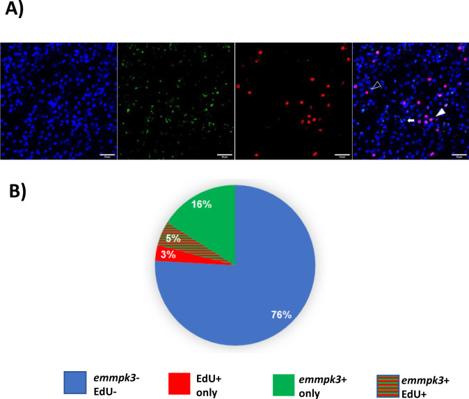
Methodology/principal findings: Using bioinformatic approaches, cloning, and yeast two-hybrid analyses we identified a novel mitogen-activated kinase (MAPK) cascade module that consists of E. multilocularis orthologs of the tyrosine kinase receptor interactor Growth factor receptor-bound 2, EmGrb2, the MAPK kinase kinase EmMEKK1, a novel MAPK kinase, EmMKK3, and a close homolog to c-Jun N-terminal kinase (JNK), EmMPK3. Whole mount in situ hybridization analyses indicated that EmMEKK1 and EmMPK3 are both expressed in E. multilocularis germinative (stem) cells but also in differentiated or differentiating cells. Treatment with the known JNK inhibitor SP600125 led to a significantly reduced formation of metacestode vesicles from stem cells and to a specific reduction of proliferating stem cells in mature metacestode vesicles.

Conclusions/significance: We provide evidence for the expression of a MEKK1-JNK MAPK cascade module which, in mammals, is crucially involved in stress responses, cytoskeletal rearrangements, and apoptosis, in E. multilocularis stem cells. Inhibitor studies indicate an important role of JNK signalling in E. multilocularis stem cell survival and/or maintenance. Our data are relevant for molecular and cellular studies into crosstalk signalling mechanisms that govern Echinococcus stem cell function and introduce the JNK signalling cascade as a possible target of chemotherapeutics against echinococcosis.


Conflict of interest statement

The authors have declared that no competing interests exist.

Figures

Fig 1
Fig 1. Sequence features of EmMEKK1.
(A) EmMEKK1 domain structure. Shown in blue are the locations of the RING domain (R) and the serine/threonine kinase domain (S/TKD). (B) Amino acid sequence alignment of the RING domains of different MEKK1 orthologs of E. multilocularis (EmMEKK1; this work), Dugesia japonica (DjMEKK1; DDBJ/EMBL/GenBank accession no.: BBA10910) and H. sapiens (HsMEKK1; Q13233). Sites of perfect alignment (*) as well as groups of strong (;) or weak (.) similarity are marked below the alignment. Highly conserved cysteine residues of the RING zink-finger are additionally marked by a dot above the alignment. (C) Amino acid sequence alignment of serine/threonine kinase domains of different MEKK1 orthologs. Sequence origins and decorations are as in (B).
Fig 2
Fig 2. Expression of emmekk1 in metacestode vesicles.
A) WISH on vesicle germinal layer. Shown are from left to right: nuclear DAPI staining (blue), EdU detection (red), emmekk1 WISH (green), and merge of all channels. Full triangles indicate cells with double staining for EdU and the emmekk1-probe, open triangle indicates EdU+ alone, arrow indicates emmekk1+ alone. Bar represents 20 μm. B) Summary of the WISH results. Values are given in percent for isolates Ingrid (upper panel) and J2012 (lower panel). The color code is indicated below.
Fig 3
Fig 3. Upstream interaction partners of EmMEKK1.
A) Summary of Y2H interaction experiments with putative upstream interaction partners. N-terminal and C-terminal regions of EmMEKK1 are indicated to the left as regions encompassing the RING- and the serine/threonine kinase domain, respectively, fused to Gal4-AD or Gal4-BD as indicated. Possible interaction partners tested in this study are indicated above as AD- and BD-fusions. Empty vector controls are marked by “e”. No interaction (-) or interactions under high stringency conditions (+++) are indicated. nd = not determined. B) Y2H experiment showing the interaction between EmMEKK1 and EmGrb2. Left plate, SD-Leu-Trp for transfection control; right plate, SD-Leu-Trp-His-Ade for high stringency interaction. 1, positive control (T-antigen-AD x p53-BD; 2, negative control (T-antigen-AD x lamC-BD); 3, MEKK1-3‘-AD x empty-BD; 4, MEKK1-5‘-AD x empty-BD; 5, EmBrb2-AD x empty BD; 6, EmGrb2-AD x MEKK1-3‘-BD; 7, EmGrb2-AD x MEKK1-5‘-BD. C) Amino acid sequence alignment between EmGrb2 (above) and human Grb2 (HsGrb2). Identical residues are marked by “*” and biochemically similar residues by “:”. SH3-domains are indicated in blue, the SH2 domain in red.
Fig 4
Fig 4. Interaction between EmMEKK1 and EmMKK3.
A) Phylogenetic tree of Echinococcus MAPKK (as characterized in this work) and human MAPKK. CLUSTALW alignment was performed with full-length amino acid sequences of E. multilocularis EmMKK1 (accession no. FN434110), EmMKK2 (FN434111), EmMKK3 (MW358635), EmMKK4 (MW358636), and EmMKK5 (MW358637) as well as human (Hs) MKK1 (Q02750), MKK2 (P36507), MKK3 (P46734), MKK4 (P45985), MKK5 (Q13163), MKK6 (P52564), and MKK7 (O14733). Node distances are indicated to the right. Specificities of human enzymes to MAPK of the Erk-branch (TEY), the JNK-branch (TPY) and the p38-branch (TGY) are indicated in red. B) Y2H experiment showing the interaction between EmMEKK1 and EmMKK3. Left plate, SD-Leu-Trp for transfection control; right plate, SD-Leu-Trp-His with 7,5 mM 3-Amino-1,2,4-triazol. 1, positive control (T-antigen-AD x p53-BD); 2, negative control (T-antigen-AD x lamC-BD); 3, EmMEKK1-5‘-AD x empty-BD; 4, EmMKK3-BD x empty-AD; 5, EmMEKK1-5‘-AD x EmMKK3-BD.
Fig 5
Fig 5. Sequence features and interactions of EmMPK3.
A) Y2H experiment showing the interaction between EmMKK3 and EmMKK2 with EmMPK3. Left plate, SD-Leu-Trp for transfection control; right plate, SD-Leu-Trp-His with 7,5 mM 3-Amino-1,2,4-triazol. 1, positive control (T-antigen-AD x p53-BD); 2, negative control (T-antigen-AD x lamC-BD); 3, EmMKK2-AD x EmMPK3-BD; 4, EmMKK3-AD x EmMPK3-BD; 5, EmMKK2-AD x empty-BD; 6, EmMKK3-AD x empty-BD; 7, EmMPK3-BD x empty-AD. B) Amino acid sequence alignment of JNK of different origin. Compared are the sequences of E. multilocularis EmMPK3 (this work; accession no. MW358638), human JNK1 (HsJNK1; P45983), and Schmidtea mediterranea JNK (SmedJNK; AHL18082.1). Sites of perfect alignment (*) as well as groups of strong (:) or weak (.) similarity are marked below the alignment. The JNK-typical TPY motif of the activation loop is shown by a line above the alignment. Residues of the hydrophobic cleft that interact with SP600125 are marked by ‚+‘ above the alignment.
Fig 6
Fig 6. Expression of emmpk3 in metacestode vesicles.
A) WISH on vesicle germinal layer. Shown are from left to right: nuclear DAPI staining (blue), emmpk3 WMISH (green), EdU detection (red), and merge of all channels. Examples cells are marked by full triangle (Edu+, emmpk3+), open triangle (Edu+ only) or arrow (emmpk3+ only). Bar represents 20 μm. B) Summary of the WISH results. Values are given in percent for cells which are negative for EdU and for emmpk3 (blue), EdU+ cells only (red), emmpk3+ cells only (green), and double stained cells EdU+/emmpk3+ (red/green).
Fig 7
Fig 7. Effect of SP600125 on metacestode vesicles and parasite stem cells.
A) Effect on metacestode vesicles. Upper panel: Microscopic images of in vitro cultivated metacestode vesicles after incubation with 25 μM SP600125. left: control without inhibitor; middle: 25 μM SP600125 for 6 days; right: 25 μM SP600125 for 13 days. Lesions in the germinal layer are marked by black triangles. B) Effect of SP600125 on percentage of EdU+ cells in germinal layer. Vesicles had been incubated for 13 days in the presence of SP600125 at concentrations as indicated. Left: dose-dependent effect of SP600125 on number of EdU+ cells in the germinal layer. Statistical analysis of three biological replicates (***, p<0,0001). Right: Examples of EdU+ cells in germinal layer of vesicles incubated with 0 μM (control) and 25 μM SP600125 as indicated. C) Effect of SP600125 on vesicle formation from primary cells. Primary cell cultures had been incubated in the presence of SP600125 at indicated concentrations for 19 days and fully mature vesicles were counted. Statistical analysis of three biological replicates. ns, non significant; *, p = 0,02; **, p<0,008.
Fig 8
Fig 8. Model for MAPK cascade interactions in E. multilocularis.
Depicted are E. multilocularis MAPK, MAPKK (MKK), and MAPKKK (MKKK) as well es upstream regulatory factors identified in this work and previous studies [–17]. Interactions which are verified by Y2H analyses are shown as solid black arrows, those which are presumed are indicated by dotted black arrows. Red asterix indicates that EmMPK2 is a constitutively active MAPK [17]. RTK, receptor tyrosine kinase.

Similar articles

Cited by

References

    1. Romig T, Deplazes P, Jenkins D, Giraudoux P, Massolo A, Craig PS, et al.. Ecology and life cycle patterns of Echinococcus species. Adv Parasitol. 2017;95:213–314. doi: 10.1016/bs.apar.2016.11.002 - DOI - PubMed
    1. Koziol U, Rauschendorfer T, Zanon Rodríguez L, Krohne G, Brehm K. The unique stem cell system of the immortal larva of the human parasite Echinococcus multilocularis. Evodevo. 2014;5:10. doi: 10.1186/2041-9139-5-10 - DOI - PMC - PubMed
    1. Brehm K, Koziol U. Echinococcus-Host interactions at cellular and molecular levels. Adv Parasitol. 2017;95:147–212. doi: 10.1016/bs.apar.2016.09.001 - DOI - PubMed
    1. Thompson RCA. Biology and systematics of Echinococcus. Adv Parasitol. 2017;95:65–109. doi: 10.1016/bs.apar.2016.07.001 - DOI - PubMed
    1. Koziol U, Jarero F, Olson PD, Brehm K. Comparative analysis of Wnt expression identifies a highly conserved developmental transition in flatworms. BMC Biol. 2016;14:10. doi: 10.1186/s12915-016-0233-x - DOI - PMC - PubMed

MeSH terms