

Glomerulotubular pathology in dogs with subclinical ehrlichiosis
- PMID: 34879085
- PMCID: PMC8654155
- DOI: 10.1371/journal.pone.0260702
Glomerulotubular pathology in dogs with subclinical ehrlichiosis
Abstract
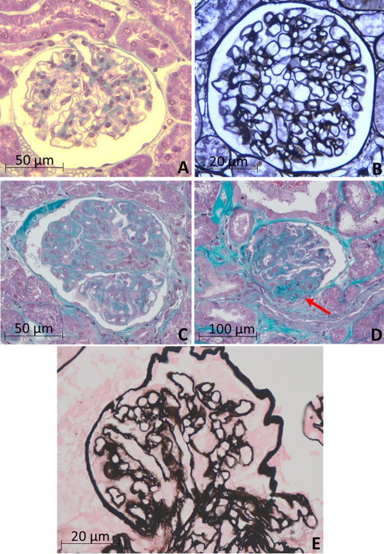
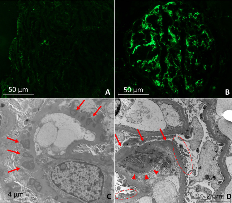
Subclinical stage of ehrlichiosis is characterized by absence of clinical or laboratory alterations; however, it could lead to silent glomerular/tubular changes and contribute significantly to renal failure in humans and animals. The aim of this study was to evaluate glomerular and tubular alterations in dogs with subclinical ehrlichiosis. We evaluated renal biopsies of 14 bitches with subclinical ehrlichiosis and 11 control dogs. Samples were obtained from the left kidney, and the tissue obtained was divided for light microscopy, immunofluorescence, and transmission electron microscopy. Abnormalities were identified by light microscopy in 92.9% of dogs with ehrlichiosis, but not in any of the dogs of the control group. Mesangial cell proliferation and synechiae (46.1%) were the most common findings, but focal segmental glomerulosclerosis and ischemic glomeruli (38.4%), focal glomerular mesangial matrix expansion (30.7%), mild to moderate interstitial fibrosis and tubular atrophy (23%), and glomerular basement membrane spikes (23%) were also frequent in dogs with ehrlichiosis. All animals with ehrlichiosis exhibited positive immunofluorescence staining for immunoglobulins. Transmission electron microscopy from dogs with ehrlichiosis revealed slight changes such as sparse surface projections and basement membrane double contour. The subclinical phase of ehrlichiosis poses a higher risk of development of kidney damage due to the deposition of immune complexes.
Conflict of interest statement
The authors have declared that no competing interests exist.
Figures
References
-
- Troy GC, Forrester SD. Canine Ehrlichiosis. In: Greene CE. Infectious disease of the dog and cat. Philadelphia: WB. Saunders;1990. pp. 404–418.
-
- Waner T, Keysary A, Bark H, Sharabani E, Harrus S. Canine monocytic ehrlichiosis–an overview. ISR J Vet Med. 1999; 54: 103–107.
-
- Neer TM, Harrus S. Canine Monocytotropic Ehrlichiosis and Neorickettiosis (E. canis, E. chaffeensis, E. ruminatium, N. sennetsu, and N. risticii) infections. In: Greene C. Infectious Diseases of the Dog and Cat. Philadelphia, Saunders Company; 2006. pp. 203–216.
-
- Borin S, Crivelenti LZ, Ferreira FA. Epidemiological, clinical, and hematological aspects of 251 dogs naturally infected with Ehrlichia spp morulae. Arq Bras Med Vet Zootec. 2009; 61: 566–571.
Publication types
MeSH terms
Substances
LinkOut - more resources
Full Text Sources
