. 2022 Feb;42(2):175-188.
doi: 10.1161/ATVBAHA.121.317197. Epub 2021 Dec 9.

Ischemic-Trained Monocytes Improve Arteriogenesis in a Mouse Model of Hindlimb Ischemia

Affiliations

Ischemic-Trained Monocytes Improve Arteriogenesis in a Mouse Model of Hindlimb Ischemia

Gustavo Falero-Diaz et al. Arterioscler Thromb Vasc Biol. 2022 Feb.

Abstract

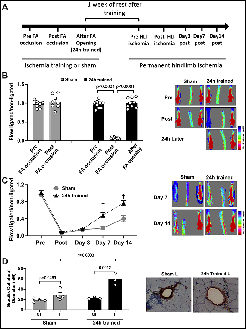
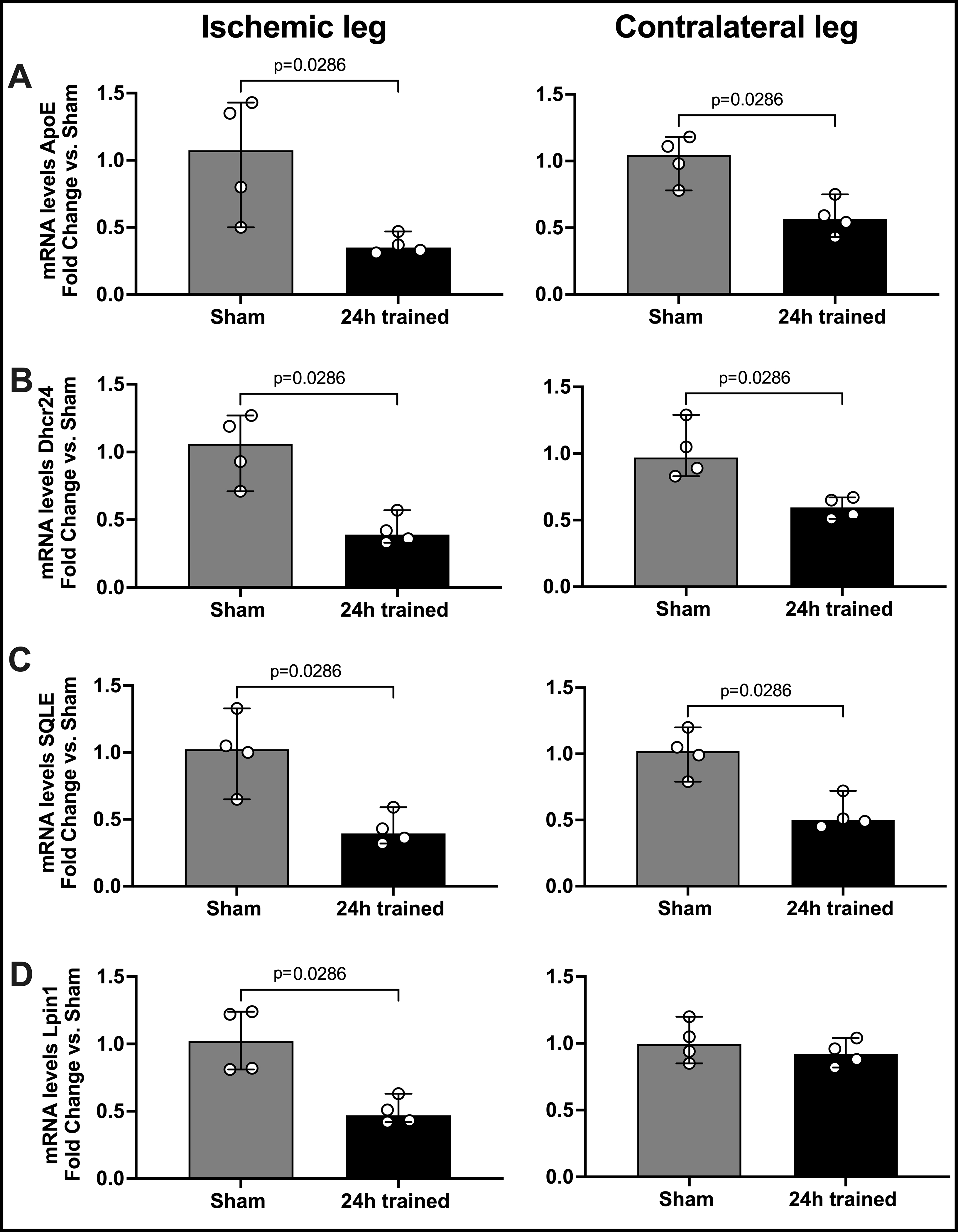
Objective: Monocytes, which play an important role in arteriogenesis, can build immunologic memory by a functional reprogramming that modifies their response to a second challenge. This process, called trained immunity, is evoked by insults that shift monocyte metabolism, increasing HIF (hypoxia-inducible factor)-1α levels. Since ischemia enhances HIF-1α, we evaluate whether ischemia can lead to a functional reprogramming of monocytes, which would contribute to arteriogenesis after hindlimb ischemia.

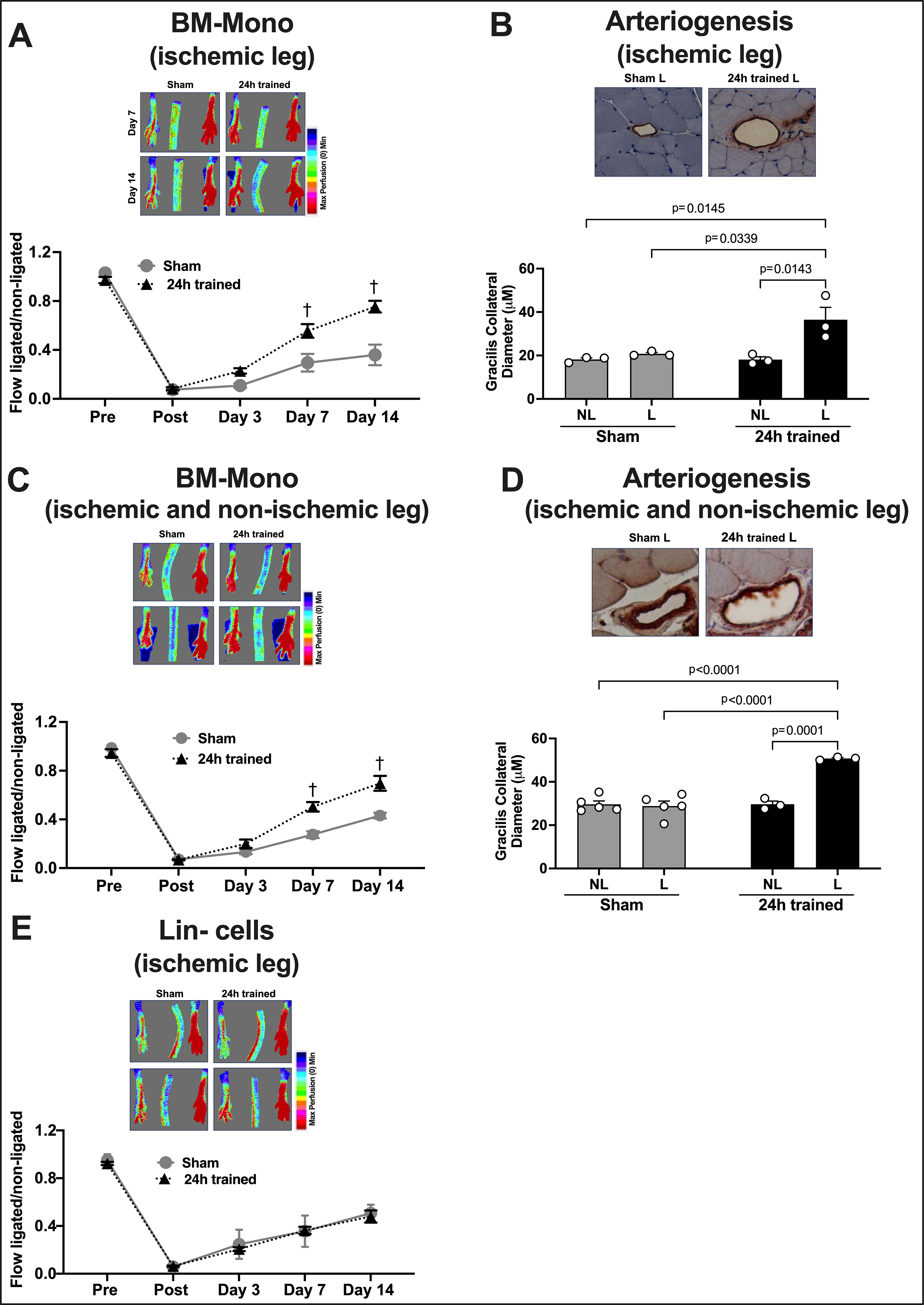
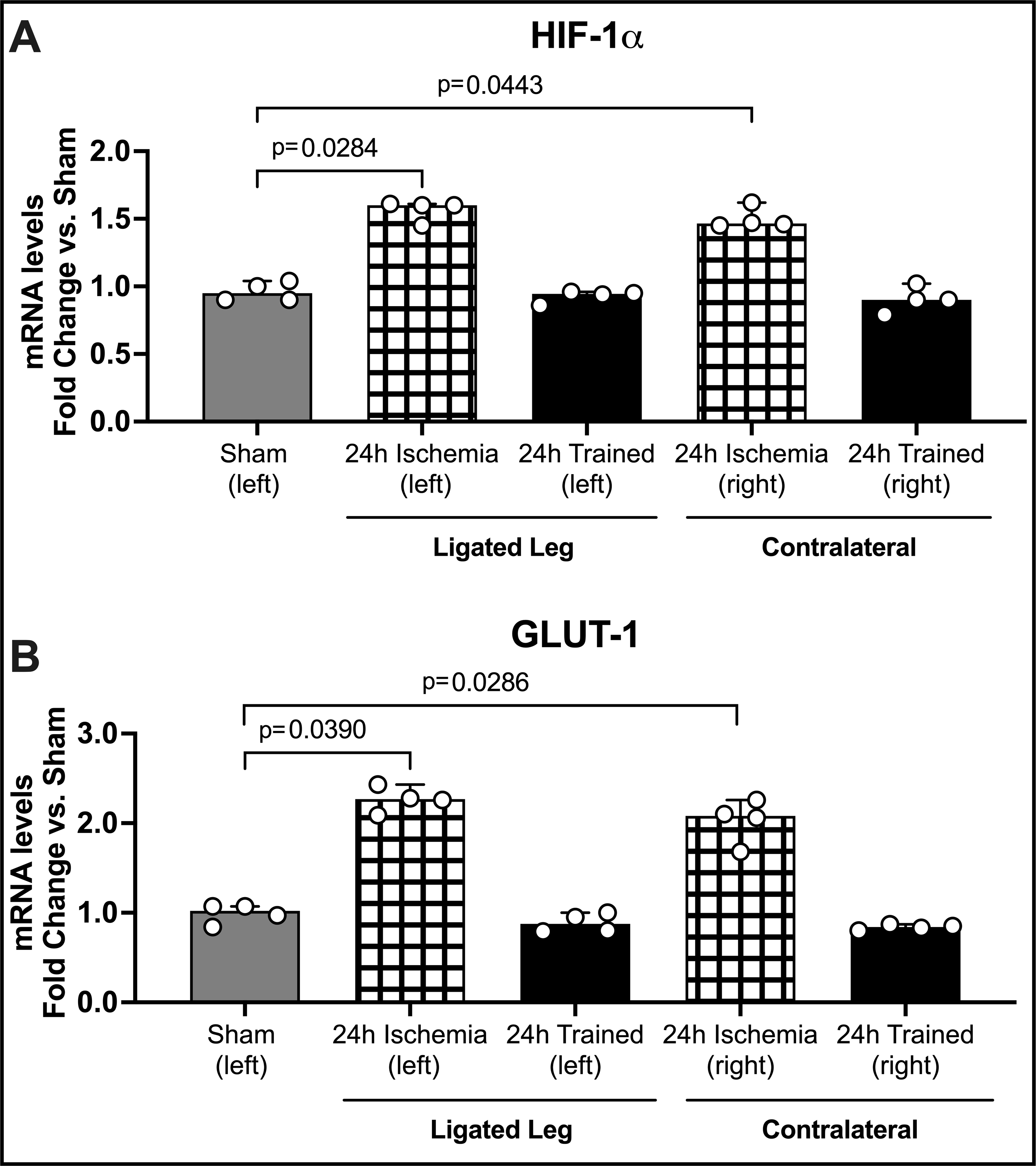
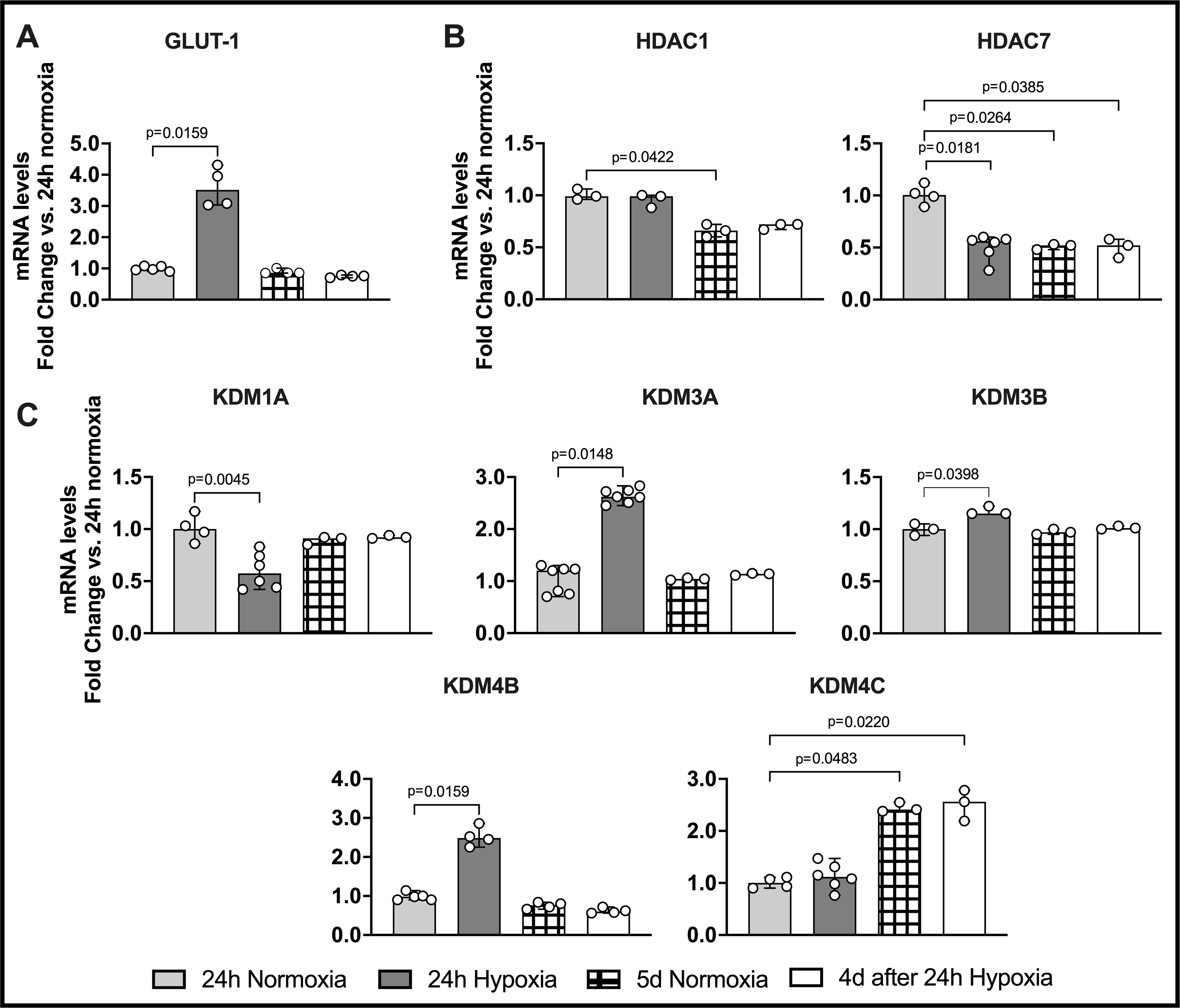
Methods and results: Mice exposed to ischemia by 24 hours (24h) of femoral artery occlusion (24h trained) or sham were subjected to hindlimb ischemia one week later; the 24h trained mice showed significant improvement in blood flow recovery and arteriogenesis after hindlimb ischemia. Adoptive transfer using bone marrow-derived monocytes (BM-Mono) from 24h trained or sham donor mice, demonstrated that recipients subjected to hindlimb ischemia who received 24h ischemic-trained monocytes had remarkable blood flow recovery and arteriogenesis. Further, ischemic-trained BM-Mono had increased HIF-1α and GLUT-1 (glucose transporter-1) gene expression during femoral artery occlusion. Circulating cytokines and GLUT-1 were also upregulated during femoral artery occlusion.Transcriptomic analysis and confirmatory qPCR performed in 24h trained and sham BM-Mono revealed that among the 15 top differentially expressed genes, 4 were involved in lipid metabolism in the ischemic-trained monocytes. Lipidomic analysis confirmed that ischemia training altered the cholesterol metabolism of these monocytes. Further, several histone-modifying epigenetic enzymes measured by qPCR were altered in mouse BM-Mono exposed to 24h hypoxia.

Conclusions: Ischemia training in BM-Mono leads to a unique gene profile and improves blood flow and arteriogenesis after hindlimb ischemia.

Keywords: bone marrow; hindlimb; ischemia; lipids; monocytes.


Figures

Figure 1.
Figure 1.. Ischemia Training by FA Occlusion Followed by Permanent Hindlimb Ischemia 1 Week Later.
(A) The timeline illustrates the experiments performed in this figure. (B) LDI demonstrates that blood flow is maintained after the dissection of the FA in the sham group, while in the 24h trained group, blood flow is significantly decreased after FA occlusion and completely restored immediately after FA opening. (C) LDI demonstrates that blood flow recovery after permanent hindlimb ischemia in the 24h trained group was significantly improved by days 7 and 14 (n=8). (D) Arteriogenesis of the gracillis collaterals showed a remarkable improvement in the 24h trained group. FA: femoral artery, HLI: hindlimb ischemia, NL: non-ligated, L: ligated. The data were analyzed by 2-way ANOVA, followed by Sidak multiple comparison tests. Errors bars represent SEM (†p<0.01 vs. same timepoint - Sham).
Figure 2.
Figure 2.. Adoptive Transfer of BM-Mono or Bone Marrow Mononuclear Lin Cells.
Cells from donor mice were injected systemically in recipient mice subjected to hindlimb ischemia the previous day. Monocyte adoptive transfer was performed using BM-Mono from the ischemic leg only (A) or from both legs (B). LDI demonstrates a significant improvement of blood flow recovery in recipient mice who received BM-Mono from the ischemic leg (n=10) (A) and from both legs (n=10) (C); however, no improvement in blood flow recovery was observed in recipient mice who received ischemic-trained Lin cells (24h trained) compared to recipients with sham Lin cells (n=9) (E). Remarkably effective arteriogenesis of gracillis collaterals was observed in recipient mice who received BM-Mono from the ischemic leg (B) and from both legs (D). NL: non-ligated, L: ligated. The data were analyzed by 2-way ANOVA, followed by Sidak multiple comparison tests. Errors bars represent SEM (†p<0.01 vs. same timepoint - Sham).
Figure 3.
Figure 3.. Gene Expression in Sham, 24h Ischemia, and 24h Trained Monocytes.
BM-Mono were isolated from 24h ischemia (when FA was still occluded after 24h of ligation); 24h trained (when FA was occluded for 24h but already opened for 2 days); and sham. For gene quantification (A) HIF-1α and (B) GLUT-1 qPCR was performed. Both genes were significantly up-regulated in the 24h ischemia group (median; 95% CI) by Kruskal-Wallis test followed by Dunn’s multiple comparisons test. Surprisingly, the same pattern occurred in the contralateral leg, suggesting a systemic effect of FA occlusion. GLUT-1, which is known to be targeted by HIF-1α, was more than two-fold up-regulated (± 0.13).
Figure 4.
Figure 4.. Gene Expression in Mouse BM-Mono Exposed to Hypoxia.
Mouse BM-Mono were exposed or not to 24h hypoxia followed by 4 days in normoxia. qPCR was performed to quantify the gene expression of GLUT-1 and several epigenetic enzymes. (A) GLUT-1 expression was up-regulated in 24h hypoxia. Histone-modifying epigenetic enzymes were significantly down-regulated (B) HDAC7 and (C) KDM1A, while others were up-regulated (KDM3A, KDM3B, and KDM4B) in 24h hypoxia as compared to 24h normoxia. Several histone-modifying epigenetic enzymes were also significantly regulated in 5d normoxia and 4d after 24h hypoxia groups. The data were analyzed by Kruskal-Wallis test followed by Dunn’s multiple comparisons test. Values are median and 95% CI.
Figure 5.
Figure 5.. Confirmatory qPCR in Sham and 24h trained BM-Mono.
BM-Mono total RNA was isolated from sham and 24h trained groups (ischemic and contralateral leg) for gene expression quantification. Based on the transcriptomic analysis (Table S1 supplement demonstrated that 15 genes were significantly regulated in ischemic-trained BM-Mono with FDR<0.05), quantification of four genes involved in lipid metabolism, Apoe (A), Dhcr24 (B), SQLE (C), and Lpin1 (D), was performed. Here we demonstrate that Dhcr24, SQLE and Lpin1 were significantly down-regulated in the 24h trained BM-Mono from ischemic leg and three of them in the contralateral leg (median; 95% CI) by Mann-Whitney test.

Comment in

References

    1. Norgren L, Hiatt WR, Dormandy JA, Nehler MR, Harris KA, Fowkes FGR. Inter-Society Consensus for the Management of Peripheral Arterial Disease (TASC II). J Vasc Surg. 2007;45(1 SUPPL.). doi:10.1016/j.jvs.2006.12.037 - DOI - PubMed
    1. Fung E, Helisch A. Macrophages in collateral arteriogenesis. Front Physiol. 2012;3 SEP. doi:10.3389/fphys.2012.00353 - DOI - PMC - PubMed
    1. Scholz D, Ziegelhoeffer T, Helisch A, et al. Contribution of arteriogenesis and angiogenesis to postocclusive hindlimb perfusion in mice. J Mol Cell Cardiol. 2002;34:775–787. - PubMed
    1. Stabile E, Susan Burnett M, Watkins C, et al. Impaired arteriogenic response to acute hindlimb ischemia in CD4-knockout mice. Circulation. 2003;108:205–210. doi:10.1161/01.CIR.0000079225.50817.71 - DOI - PubMed
    1. Kinnaird T, Stabile E, Burnett MS, et al. Marrow-Derived Stromal Cells Express Genes Encoding a Broad Spectrum of Arteriogenic Cytokines and Promote In Vitro and In Vivo Arteriogenesis Through Paracrine Mechanisms. Circ Res. 2004;94:678–685. doi:10.1161/01.RES.0000118601.37875.AC - DOI - PubMed

Publication types