. 2021 Dec 14;118(50):e2114171118.
doi: 10.1073/pnas.2114171118.

The hippocampus as the switchboard between perception and memory

Affiliations

The hippocampus as the switchboard between perception and memory

Matthias S Treder et al. Proc Natl Acad Sci U S A. .

Abstract

Adaptive memory recall requires a rapid and flexible switch from external perceptual reminders to internal mnemonic representations. However, owing to the limited temporal or spatial resolution of brain imaging modalities used in isolation, the hippocampal-cortical dynamics supporting this process remain unknown. We thus employed an object-scene cued recall paradigm across two studies, including intracranial electroencephalography (iEEG) and high-density scalp EEG. First, a sustained increase in hippocampal high gamma power (55 to 110 Hz) emerged 500 ms after cue onset and distinguished successful vs. unsuccessful recall. This increase in gamma power for successful recall was followed by a decrease in hippocampal alpha power (8 to 12 Hz). Intriguingly, the hippocampal gamma power increase marked the moment at which extrahippocampal activation patterns shifted from perceptual cue toward mnemonic target representations. In parallel, source-localized EEG alpha power revealed that the recall signal progresses from hippocampus to posterior parietal cortex and then to medial prefrontal cortex. Together, these results identify the hippocampus as the switchboard between perception and memory and elucidate the ensuing hippocampal-cortical dynamics supporting the recall process.

Keywords: gamma power; hippocampus; intracranial EEG; memory; recall.


Conflict of interest statement

The authors declare no competing interest.

Figures
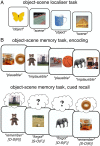
Fig. 1.
Fig. 1.
Experimental paradigm. (A) In a perceptual localizer session, participants saw trial-unique images of objects and scenes and indicated the category of the given image. This part served as an independent training dataset for multivariate pattern analyses. (B) The main experiment employed an object–scene memory task, consisting of an encoding phase (Top) and a cued recall phase (Bottom). During encoding, participants saw trial-unique object–scene pairs and indicated whether the given combination was plausible or implausible. During cued recall, participants were given either the object or the scene image as the cue and were asked to recall the paired target (scene or object image, respectively). The key conditions were 1) trials in which participants indicated they did remember the target image (“Remember” trials) and 2) trials in which participants indicated they did not remember the target image (“Forgot” trials). Labels below denote the cue-target (memory) status of trials. O = object, S = scene, R = remember, F = forgot.
Fig. 2.
Fig. 2.
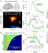
Hippocampal recall effects. (A) Hippocampal contacts across participants shown on a normalized sagittal (Left) and horizontal (Right) brain template. (B) Results from a time-frequency analysis (P < 0.05, corrected), contrasting “Remember” vs. “Forgot” trials and revealing a cluster in the high gamma range (55 to 110 Hz, peak at 85 Hz) with power increases for “Remember” trials, followed by a cluster in the alpha band (2 to 29 Hz, peak at 10 Hz) with power decreases for “Remember” trials. For an unthresholded map, see SI Appendix, Fig. S2A. (C) Power time courses for “Remember” (green) and “Forgot” (gray) trials in the gamma (Top, significant from 0.61 to 1.71 s) and alpha (Bottom, significant from 0.84 to 2.41 s) ranges. Lines show condition means ± SEM of condition differences across participants. For time courses encompassing the entire cluster frequency range, see SI Appendix, Fig. S2B. (D) Hippocampal gamma power (80 to 90 Hz) across time from −0.5 s to RT across all “Remember” trials (pooled across participants), sorted based on trial-specific RT (white line). Dashed vertical line indicates median peak latency across all trials (720 ms). For an RT-locked representation, see SI Appendix, Fig. S2D. (E) Response-locked hippocampal gamma power (80 to 90 Hz) for “Remember” (green) and “Forgot” (gray) trials, significant from −1.5 to −0.020 s (P < 0.05). Lines show condition means ± SEM of condition differences across participants.
Fig. 3.
Fig. 3.
Switch from cue to target representations. (A) Extrahippocampal contacts across participants shown on a normalized horizontal (Left), coronal (Middle), and sagittal (Right) brain template. (B) Object and scene evidence, based on a classifier trained on the “localizer” session, for O-S (blue) and S-O (red) trials, separately for “Forgot” (Left) and “Remember” (Right) trials (mean ± sem across participants). Within the first 500 ms, classifier evidence reflects the cue category. Only for “Remember” trials, classifier evidence then switches (dashed rectangle) to reflect the recalled target category. (C, Left) Overlay of the hippocampal recall effect (“Remember” vs. “Forgot” gamma t statistic, smoothed with a 100-ms sliding window) onto the extrahippocampal switch from cue to target representation for “Remember” trials. (Inset) Hippocampal contacts across patients. (C, Right) Object and scene evidence for “Remember” trials as in B, Right, but realigned to trial-by-trial hippocampal gamma peaks (time 0, vertical green line). Stars denote significant differences of classifier evidence for O-S trials vs. S-O trials (P < 0.05, corrected).
Fig. 4.
Fig. 4.
Brain-wide recall dynamics. Results from source-localized EEG alpha (8 to 12 Hz) power, comparing “Remember” vs. “Forgot” trials. (A) Voxel × time results (Pcluster < 0.05), revealing recall effects across the core retrieval network (including hippocampus, PPC, LTC, and vmPFC). Color reflects sum of significant t values across time. (B) Time-resolved results (proceeding in 100-ms steps), showing only time windows with significant effects (each map thresholded at Pcluster < 0.05). (C, Left) Regions of interest defined as the overlap of significant voxels resulting from the main voxel × time analysis (cf. A) and AAL masks for hippocampus (HIPP), medial PPC, and vmPFC. x coordinates refer to MNI space. (C, Right) t values for the comparison of “Remember” vs. “Forgot” from 0.5 to 1.6 s in each region. Dashed horizontal line marks the threshold for P < 0.05 (uncorrected).

References

    1. Lisman J. E., Relating hippocampal circuitry to function: Recall of memory sequences by reciprocal dentate-CA3 interactions. Neuron 22, 233–242 (1999). - PubMed
    1. Wallenstein G. V., Eichenbaum H., Hasselmo M. E., The hippocampus as an associator of discontiguous events. Trends Neurosci. 21, 317–323 (1998). - PubMed
    1. Goode T. D., Tanaka K. Z., Sahay A., McHugh T. J., An integrated index: Engrams, place cells, and hippocampal memory. Neuron 107, 805–820 (2020). - PMC - PubMed
    1. Teyler T. J., DiScenna P., The hippocampal memory indexing theory. Behav. Neurosci. 100, 147–154 (1986). - PubMed
    1. Marr D., Simple memory: A theory for archicortex. Philos. Trans. R. Soc. Lond. B Biol. Sci. 262, 23–81 (1971). - PubMed

Publication types