. 2021 Dec 8;12(1):7133.
doi: 10.1038/s41467-021-27467-2.

Engineering Bacillus subtilis for the formation of a durable living biocomposite material

Affiliations

Engineering Bacillus subtilis for the formation of a durable living biocomposite material

Sun-Young Kang et al. Nat Commun. .

Abstract

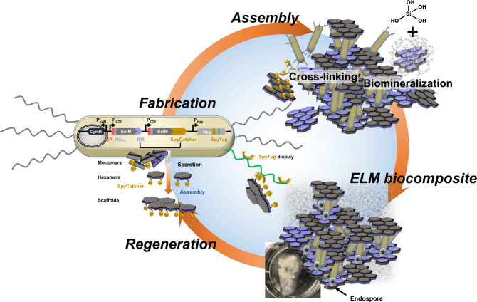
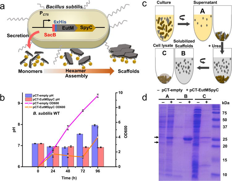
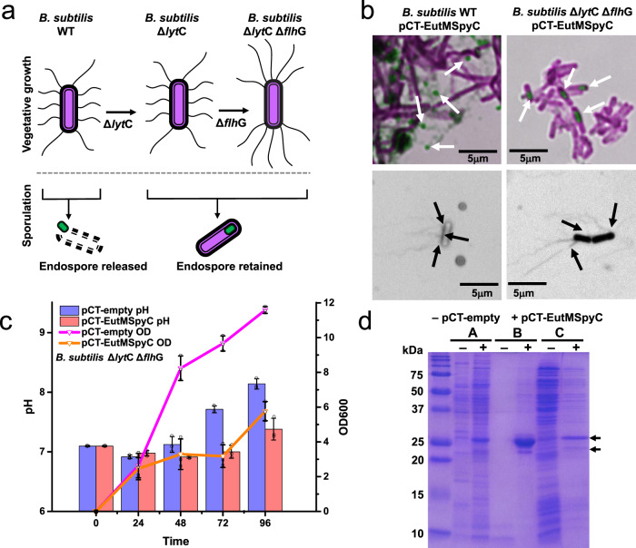
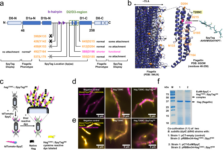
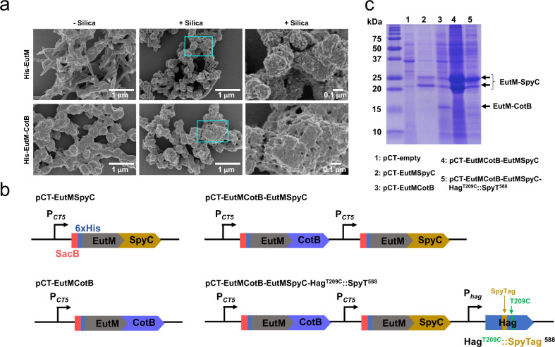
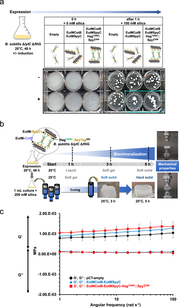
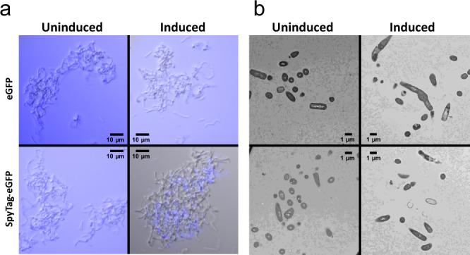
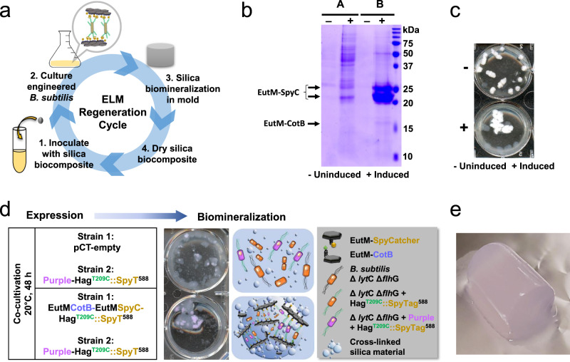
Engineered living materials (ELMs) are a fast-growing area of research that combine approaches in synthetic biology and material science. Here, we engineer B. subtilis to become a living component of a silica material composed of self-assembling protein scaffolds for functionalization and cross-linking of cells. B. subtilis is engineered to display SpyTags on polar flagella for cell attachment to SpyCatcher modified secreted scaffolds. We engineer endospore limited B. subtilis cells to become a structural component of the material with spores for long-term storage of genetic programming. Silica biomineralization peptides are screened and scaffolds designed for silica polymerization to fabricate biocomposite materials with enhanced mechanical properties. We show that the resulting ELM can be regenerated from a piece of cell containing silica material and that new functions can be incorporated by co-cultivation of engineered B. subtilis strains. We believe that this work will serve as a framework for the future design of resilient ELMs.


Conflict of interest statement

The authors declare no competing interests.

Figures

Fig. 1
Fig. 1. Formation of a living silica biocomposite material.
B. subtilis is designed to secrete a protein matrix from self-assembling EutM scaffold building blocks. Cells are engineered to display a SpyTag on clustered, polar flagella for covalent cross-linking via isopeptide bond formation with SpyCatcher domains fused to secreted scaffold building blocks. Scaffold building blocks are genetically fused with peptides to enhance silica polymerization on scaffolds. Finally, cells are engineered to retain spores as endospores. This allows cells to persist as structural components of the material and creates a durable biocomposite material that can be regenerated from a piece of silica material containing cells. (BM biomineralization peptide, SP, secretion signal peptide).
Fig. 2
Fig. 2. Establishment of self-assembling scaffold building block secretion.
a Self-assembling EutM scaffold building blocks containing a C-terminal SpyCatcher (SpyC) domain and a SacB secretion signal sequence followed by a His-tag are expressed and secreted by B. subtilis under the control of a cumate inducible promoter. b Growth and pH of cultures expressing EutMSpyC was followed for 96 h in SMM at 20 °C and compared to empty plasmid control cultures. Data are shown as mean values ± SD and error bars represent the standard deviations of three independent biological replicate cultures. Colored bars and lines represent mean values. Black symbols represent data points and error bar. c Self-assembling scaffolds settle with cells, requiring their solubilization from cell pellets by a gentle wash with 4 M urea (see Methods for details). d Protein expression and secretion after 48 h of cultivation were analyzed by SDS-PAGE where: (A) is the culture supernatant (10-fold concentrated by TCA precipitation), (B) urea supernatant after scaffold solubilization, and (C) resulting cell pellet after lysozyme treatment for analysis of remaining protein. Arrows indicate EutM-SpyCatcher bands. Expected sizes are 24.4 kDa and 21.1 kDa with and without SacB secretion signal peptide, respectively. The shown data is representative of three independent biological replicate cultures. Source data are provided as a Source Data file.
Fig. 3
Fig. 3. Development of a Bacillus strain for durable material fabrication.
a Construction of a B. subtilis ΔlytC ΔflhG strain deficient in spore release and exhibiting polar, clustered flagella was achieved by deleting the autolysin LytC and the flagella basal body protein FlhG. b Sporulation and flagella phenotypes of B. subtilis strains transformed with pCT-EutMSpyC were compared to confirm endospore and polar flagella phenotype under expression conditions. Representative samples show spore release for the wild-type strain (LB, 48 h, 20 °C shown) and endospore formation by the engineered strain (SMM, 72 h, 20 °C shown) (see also Supplementary Figs. 2, 3 and 6). Peritrichous flagella are observed for the wild-type strain (LB, 48 h, 20 °C shown) under all tested growth conditions through 72 h and flagellation decreases after 48 h (see also Supplementary Figs. 2 and 3). Clustered polar flagella are observed in the engineered strain through 48 h of growth (SMM, 48 h, 20 °C shown) (see also Supplementary Fig. 6). Samples were stained with either malachite green and safranin red (top) or RYU (bottom) and examined by light microscopy at 100x. Spore images had red replaced with magenta and flagella images were converted to grayscale. Images shown are representative three independent biological replicate cultures. c B. subtilis ΔlytC ΔflhG transformed with the pCT-empty (control) or pCT-EutMSpyC plasmid (SMM, 20 °C) show growth characteristics comparable to the B. subtilis wild-type strain in Fig. 2. Data are shown as mean values ± SD and error bars represent the standard deviations of three independent biological replicate cultures. Colored bars and lines represent mean values. Black symbols represent data points and error bar. d Expression and secretion of EutM-SpyCatcher by the engineered B. subtilis ΔlytC ΔflhG strain was verified in SMM medium at 20 °C after 48 h of induction by SDS-PAGE. Arrows indicate EutM-SpyCatcher bands that are not present in the empty plasmid control. A strong band corresponding to urea solubilized EutM-SpyCatcher scaffolds (B) from co-pelleted cells confirm comparable protein expression and secretion levels of the engineered and the wild-type strains (Fig. 2). Samples (A) and (C) corresponding to 10-fold concentrated culture supernatant and cell lysate after scaffold solubilization also show comparable protein bands to the wild-type strain. Shown data is representative of three independent biological replicate cultures. Source data are provided as a Source Data file.
Fig. 4
Fig. 4. Engineering of flagella for SpyTag display.
a A SpyTag sequence was inserted at different locations in the hypervariable regions of the flagellin protein (encoded by hag). Shown are conserved domains, including the solvent-exposed D2/D3 domains. The SpyTag insertions (orange & magenta labels indicate bp and residue locations, magenta denotes functional site) had different impacts on flagella phenotypes and SpyTag display in both B. subtilis 168 (WT) and B. subtilis ΔlytC ΔflhG expressing plasmid born hagT209C::SpyTagxxx. Except for less curved flagella observed for B. subtilis 168 compared to normal (indicated by *) flagella for B. subtilis ΔlytC ΔflhG, phenotypes were the same between the two strains. Several hag insertion mutants could not be transformed into either strain and seem to be lethal (black cross) when expressed in trans of the genomic hag copy. Gray cross: hag mutants could only be transformed into B. subtilis ΔlytC ΔflhG but recovered plasmids were mutated. No or some attachment: modified flagella could not or only be weakly labeled with tdTomato SpyCatcher. b Flagellar filament and flagellin structures show locations of the SpyTag insertions (orange & magenta, corresponding to labels in a) and T209C mutation (yellow box) for dye staining. SpyTag structure (PDB: 4MLI) and sequence are shown for reference. c Flagella assemble from genomic flagellin and plasmid born flagellin (HagT209CSpyTagxxx) subunits. Engineered flagellins are stained with a cysteine reactive dye (Alexa FluorTM 488 C5 maleimide) and functional SpyTag display was tested by growing cultures with purified tdTomato-SpyCatcher protein. d Fluorescence imaging of B. subtilis ΔlytC ΔflhG transformed with pCT-empty (control), pRBBm34-HagT209C or pRBBm34-HagT209C::SpyT588 show that expression of hagT209C::SpyTag588 results in shortened, clustered, polar flagella. Cells are stained and images false-colored as indicated. See Supplementary Fig. 9 for other SpyTag positions. e Functional SpyTag display by HagT209C::SpyTag588 was confirmed by growing strains in the presence of purified tdTomato-SpyCatcher. (Images generated for each strain in d and e are representative of cultures selected from three biological replicate cultures.) f In situ attachment of secreted EutM-SpyCatcher to SpyTags displayed on flagella of B. subtilis ΔlytC ΔflhG was verified by co-cultivation. SDS-PAGE analysis of extracted flagella shows a faint band with the expected size of 55 kDa for a EutM-SpyCatcher (21.1 kDa) and HagT209C::SpyTag588 (34.1 kDa) protein complex in co-cultures secreting EutM-SpyCatcher. Genomic flagellin (32.6 kDa) is the major protein. Shown data is from a single experiment. Source data are provided as a Source Data file.
Fig. 5
Fig. 5. Implementing silica biomineralization by scaffolds.
a Silica biomineralization by His-EutM and His-EutM-CotB scaffolds expressed and purified from E. coli was investigated by SEM following the incubation of 1 mg/mL protein without (left column) and with (center columns) 100 mM silica for 2 h at room temperature. Right columns show 10-fold magnification of marked regions. Images shown are representative of one set of purified proteins. b Different plasmids were constructed for EutM-CotB expression and secretion in B. subtilis alone or together with EutM-SpyCatcher as shown. For final ELM fabrication, a consolidated plasmid was designed for the co-expression of EutM-SpyCatcher and EutM-CotB with HagT209C::SpyTag588 in B. subtilis ΔlytC ΔflhG. Each gene is expressed by its own promoter (cumate inducible PCT5 promoter, native hag promoter). c Expression and secretion of EutM-CotB by B. subtilis ΔlytC ΔflhG was investigated and compared to EutM-SpyCatcher. Urea solubilized fractions were prepared from cultures transformed with different expression plasmids and grown as in Fig. 2. Except for pCT-EutMSpyC (lane 2), all urea fractions from the different recombinant cultures were concentrated 10-fold by TCA precipitation prior to SDS-PAGE analysis. Data shown are representative of three independent experiments. Source data are provided as a Source Data file.
Fig. 6
Fig. 6. Formation of living, silica biocomposites with increased rigidity.
a Generation of silica biocomposites was tested by cultivating B. subtilis ΔlytC ΔflhG transformed with plasmids (empty as control) for expression of the shown proteins and their functions. Cultures were first grown (SMM, 20 °C, 48 h) with (+ induced) and without (- uninduced) induction of protein expression. 5 mL cultures were transferred to 6-well plates (0 h, 0 mM silica) and incubated (20 °C, 100 rpm) for 1 h after addition of 100 mM silica (1 h, 100 mM). Experiments were performed with three biological culture replicates (see Supplementary Fig. 13). b 3D blocks of silica biocomposite materials were then fabricated from cultures of the same three strains grown under the same conditions with induction. After 48 h of growth, 200 mM silica was added into 1 mL cultures in syringes as molds and the aliquots of the cultures were cured at 20 °C or 25°C. At different time points, silica gel plugs were removed from their molds to evaluate their hardness. After 5 h of curing at 25 °C, solid gel plugs suitable for rheology testing were obtained. c Rheological properties of the cured gel plugs were measured using an extensional DMA Rheometer with 15 mm compression disks. A frequency sweep was performed with a gap of 4.5 mm and an oscillation strain of 1%. Three biological replicate gel blocks were measured for each strain. Variation was observed in the storage modulus (G’, solid line) and loss modulus (G”, dashed line). Data are shown as mean values ± SD and error bars represent the standard deviations of three independent biological samples. Source data are provided as a Source Data file.
Fig. 7
Fig. 7. Microscopy characterization of Bacillus silica biocomposites.
a The location of assembled scaffolds was tested by cultivating B. subtilis ΔlytC ΔflhG transformed with EutMCotB-EutMSpyC-HagT209C::SpyT588 plasmid (uninduced as control). Cultures were first grown (SMM, 20 °C, 48 h) with (induced) and without (uninduced) cumate induction of protein expression. 500 µL of culture was mixed with 0.125 mg eGFP or SpyTag-eGFP and mixed at RT for 30 min before acquiring DIC and GFP images which were merged and GFP channel colored blue. Induced cultures have EutM scaffolds assembled outside and between cells, with available binding sites for additional SpyTagged proteins. b 3D blocks of silica biocomposite materials were then formed from cultures of the same sample examined in a. 200 mM silica was added after 48 h of growth and 1 mL aliquots of the cultures were transferred to syringes as molds for curing at 25 °C. After 5 h of curing at 25 °C, solid gel plugs were cut into 2 mm3 pieces and prepared for thin sectioning. Cells were observed in clusters for both the uninduced and induced cultures, however only the induced culture contained clear zones around cells (see Supplementary Fig. 14a for a close-up). This zone corresponds to the SpyTag-eGFP labeled areas above and indicates there is interference with staining the scaffolds after silica is biomineralized. (Images shown are representative of two independent experiments.).
Fig. 8
Fig. 8. ELM biocomposite regeneration and proof-of-concept for material functionalization.
a Silica biocomposite material regeneration was tested by re-inoculating cultures with a piece of a silica plug fabricated from B. subtilis ΔlytC ΔflhG cultures co-expressing EutM-CotB and EutM-SpyCatcher and HagT209CSpyTag588, biomineralized with 200 mM silica (see Fig. 6) and cured for 24 h at 25 °C. Cultures were regrown (SMM, 20 °C, 48 h) with (+ induced) and without (- uninduced) cumate induction of protein expression. b Expression and secretion of EutM scaffold building blocks by the induced, regenerated cultures was confirmed by SDS-PAGE analysis of culture supernatant (A) and urea solubilized scaffolds (B). All fractions were concentrated 10-fold by TCA precipitation prior to SDS-PAGE analysis. c Silica biomineralization into a cross-linked biocomposite material was confirmed for the induced, regenerated cultures after 1 h at 20 °C with 100 mM silica in 6-well plates as in Fig. 6. d Proof-of-concept incorporation of additional functions into the silica ELM was demonstrated by the co-cultivation of two strains, one strain co-expresses scaffold building blocks and SpyTagged flagellin, while a second strain co-expresses a purple chromophore protein and SpyTagged flagellin. In the control co-culture, the plasmid for scaffold building block secretion in one strain was replaces with an empty plasmid. Strains were inoculated at a 1:1 ratio and cultures grown under inducing conditions for biomineralization as described above. Only co-cultures that co-expressed EutM-CotB and EutM-SpyCatcher formed an aggregated silica material with embedded purple cells. e A block of a purple, silica biocomposite (15 mm × 30 mm) was then fabricated from co-cultures of engineered strains co-expressing scaffold building blocks and the purple protein together with SpyTagged flagellin. A solid material was formed with 200 mM silica after curing for 24 h at 25 °C. All results shown are representative of three independent biological samples. Source data are provided as a Source Data file.

Similar articles

Cited by

References

    1. Chen AY, Zhong C, Lu TK. Engineering living functional materials. ACS Synth. Biol. 2015;4:8–11. - PMC - PubMed
    1. Tang, T.-C. et al. Materials design by synthetic biology. Nat. Rev. Mat. 6, 332–350 (2020).
    1. Srubar, W. V., 3rd. Engineered living materials: Taxonomies and emerging trends. Trends Biotechnol.39, 574–583 (2020). - PubMed
    1. Rivera-Tarazona, L. K., Campbell, Z. T. & Ware, T. H. Stimuli-responsive engineered living materials. Soft Matter17, 785–809 (2021). - PubMed
    1. Liu, X. et al. 3D printing of living responsive materials and devices. Adv. Mater30, 1704821 (2018). - PubMed

Publication types