A proteomics study identifying interactors of the FSHD2 gene product SMCHD1 reveals RUVBL1-dependent DUX4 repression
- PMID: 34880314
- PMCID: PMC8654949
- DOI: 10.1038/s41598-021-03030-3
A proteomics study identifying interactors of the FSHD2 gene product SMCHD1 reveals RUVBL1-dependent DUX4 repression
Abstract
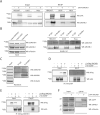
Structural Maintenance of Chromosomes Hinge Domain Containing 1 (SMCHD1) is a chromatin repressor, which is mutated in > 95% of Facioscapulohumeral dystrophy (FSHD) type 2 cases. In FSHD2, SMCHD1 mutations ultimately result in the presence of the cleavage stage transcription factor DUX4 in muscle cells due to a failure in epigenetic repression of the D4Z4 macrosatellite repeat on chromosome 4q, which contains the DUX4 locus. While binding of SMCHD1 to D4Z4 and its necessity to maintain a repressive D4Z4 chromatin structure in somatic cells are well documented, it is unclear how SMCHD1 is recruited to D4Z4, and how it exerts its repressive properties on chromatin. Here, we employ a quantitative proteomics approach to identify and characterize novel SMCHD1 interacting proteins, and assess their functionality in D4Z4 repression. We identify 28 robust SMCHD1 nuclear interactors, of which 12 are present in D4Z4 chromatin of myocytes. We demonstrate that loss of one of these SMCHD1 interacting proteins, RuvB-like 1 (RUVBL1), further derepresses DUX4 in FSHD myocytes. We also confirm the interaction of SMCHD1 with EZH inhibitory protein (EZHIP), a protein which prevents global H3K27me3 deposition by the Polycomb repressive complex PRC2, providing novel insights into the potential function of SMCHD1 in the repression of DUX4 in the early stages of embryogenesis. The SMCHD1 interactome outlined herein can thus provide further direction into research on the potential function of SMCHD1 at genomic loci where SMCHD1 is known to act, such as D4Z4 repeats, the inactive X chromosome, autosomal gene clusters, imprinted loci and telomeres.
© 2021. The Author(s).
Conflict of interest statement
The authors declare no competing interests.
Figures


Similar articles
-
Increased DUX4 expression during muscle differentiation correlates with decreased SMCHD1 protein levels at D4Z4.Epigenetics. 2015;10(12):1133-42. doi: 10.1080/15592294.2015.1113798. Epigenetics. 2015. PMID: 26575099 Free PMC article.
-
Sporadic DUX4 expression in FSHD myocytes is associated with incomplete repression by the PRC2 complex and gain of H3K9 acetylation on the contracted D4Z4 allele.Epigenetics Chromatin. 2018 Aug 20;11(1):47. doi: 10.1186/s13072-018-0215-z. Epigenetics Chromatin. 2018. PMID: 30122154 Free PMC article.
-
Smchd1 haploinsufficiency exacerbates the phenotype of a transgenic FSHD1 mouse model.Hum Mol Genet. 2018 Feb 15;27(4):716-731. doi: 10.1093/hmg/ddx437. Hum Mol Genet. 2018. PMID: 29281018 Free PMC article.
-
Genetic and epigenetic contributors to FSHD.Curr Opin Genet Dev. 2015 Aug;33:56-61. doi: 10.1016/j.gde.2015.08.007. Epub 2015 Sep 7. Curr Opin Genet Dev. 2015. PMID: 26356006 Free PMC article. Review.
-
A complex interplay of genetic and epigenetic events leads to abnormal expression of the DUX4 gene in facioscapulohumeral muscular dystrophy.Neuromuscul Disord. 2016 Dec;26(12):844-852. doi: 10.1016/j.nmd.2016.09.015. Epub 2016 Sep 19. Neuromuscul Disord. 2016. PMID: 27816329 Review.
Cited by
-
SMCHD1 maintains heterochromatin, genome compartments and epigenome landscape in human myoblasts.Nat Commun. 2025 Jul 26;16(1):6900. doi: 10.1038/s41467-025-62211-0. Nat Commun. 2025. PMID: 40715155 Free PMC article.
-
SMCHD1 has separable roles in chromatin architecture and gene silencing that could be targeted in disease.Nat Commun. 2023 Sep 25;14(1):5466. doi: 10.1038/s41467-023-40992-6. Nat Commun. 2023. PMID: 37749075 Free PMC article.
-
Update on the Molecular Aspects and Methods Underlying the Complex Architecture of FSHD.Cells. 2022 Aug 29;11(17):2687. doi: 10.3390/cells11172687. Cells. 2022. PMID: 36078093 Free PMC article. Review.
-
A discrete region of the D4Z4 is sufficient to initiate epigenetic silencing.Hum Mol Genet. 2025 Sep 3;34(18):1526-1540. doi: 10.1093/hmg/ddaf114. Hum Mol Genet. 2025. PMID: 40627547 Free PMC article.
References
-
- Padberg, G. W. Facioscapulohumeral Disease. Leiden University (1982).
Publication types
MeSH terms
Substances
Grants and funding
LinkOut - more resources
Full Text Sources
Miscellaneous

