. 2022 Jan;601(7892):274-279.
doi: 10.1038/s41586-021-04211-w. Epub 2021 Dec 8.

Structure and mechanism of the SGLT family of glucose transporters

Affiliations

Structure and mechanism of the SGLT family of glucose transporters

Lei Han et al. Nature. 2022 Jan.

Abstract

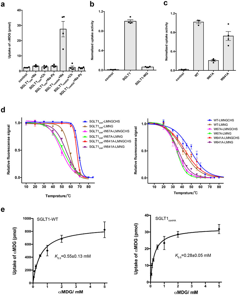
Glucose is a primary energy source in living cells. The discovery in 1960s that a sodium gradient powers the active uptake of glucose in the intestine1 heralded the concept of a secondary active transporter that can catalyse the movement of a substrate against an electrochemical gradient by harnessing energy from another coupled substrate. Subsequently, coupled Na+/glucose transport was found to be mediated by sodium-glucose cotransporters2,3 (SGLTs). SGLTs are responsible for active glucose and galactose absorption in the intestine and for glucose reabsorption in the kidney4, and are targeted by multiple drugs to treat diabetes5. Several members within the SGLT family transport key metabolites other than glucose2. Here we report cryo-electron microscopy structures of the prototypic human SGLT1 and a related monocarboxylate transporter SMCT1 from the same family. The structures, together with molecular dynamics simulations and functional studies, define the architecture of SGLTs, uncover the mechanism of substrate binding and selectivity, and shed light on water permeability of SGLT1. These results provide insights into the multifaceted functions of SGLTs.


Figures

Extended Data Fig. 1 ∣
Extended Data Fig. 1 ∣. Sequence alignments of selected SSS transporters.
The sequences of selected SSS transporters were aligned using Clustal Omega (http://www.uniprot.org/) and adjusted manually. The secondary structural elements of SGLT1 are indicated above the sequence alignment. The mutations in SGLT1con are highlighted.
Extended Data Fig. 2 ∣
Extended Data Fig. 2 ∣. Sequence alignments of SMCT homologs.
The sequences of selected SMCT homologs were aligned using Clustal Omega (http://www.uniprot.org/) and adjusted manually. The secondary structural elements of SMCT1 are indicated above the sequence alignment.
Extended Data Fig. 3 ∣
Extended Data Fig. 3 ∣. Glucose uptake of SGLT1 mutants
a, Uptake activities of SGLT1con and SGLT1conHA. SGLT1conHA is the same as SGLT1con, except that W660 and G661 of SGLT1con are reversed to H660 and A661. The uptake buffer contains different combinations of sodium (Na), choline (Ch), or phlorizin (Pz) as indicated. Uptake of αMDG is shown (mean ± SEM; n=4 biological replicates). b, Uptake activities of SGLT1 and SGLT1-WG mutant, in which H660 and A661 are substituted with W660 and G661 (mean ± SEM; n=4 biological replicates). c, Uptake activities of SGLT1 with mutations in the cholesterol-binding site (mean ± SEM; n=4 biological replicates). d, Thermostability of SGLT1con (left) or SGLT1 (right) and their variants under conditions with or without cholesteryl hemisuccinate (CHS) (mean ± SEM; n=3 biological replicates). e, The transport of αMDG by SGLT1 WT (left) and SGLT1conHA (right) in the presence of various concentrations of αMDG. Data were plotted according to the equation, U=Umax x [S]/ (K0.5+[S]) (mean ± SEM; n=3 biological replicates).
Extended Data Fig. 4 ∣
Extended Data Fig. 4 ∣. Cryo-EM sample preparation and data processing of SGLT1.
a, The elution profile of SGLT1con-Nb1 on a size-exclusion column. The insert shows SDS-PAGE analysis of the purified sample. Data are representative of five independent experiments with similar results. b, Representative cryo-EM micrograph of SGLT1con-Nb1 complex particles (from 15,039 micrographs with similar results). c, Selected 2D class averages of SGLT1con-Nb1 complex (from 100 classes with similar results). d, e, The workflow of classification and refinement. The overall nominal resolutions of the SGLT1con-Nb1 complex and the SGLT1conHA-Nb1 complex were determined by the ‘gold standard’ FSC curve using the FSC=0.143 criterion. f, Overlay of the SGLT1con model with the density map of SGLT1conHA.
Extended Data Fig. 5 ∣
Extended Data Fig. 5 ∣. Cryo-EM densities and refined models.
a, Local resolution of the cryo-EM map of the SGLT1conHA-Nb1 complex. b, Cryo-EM densities and model of SGLT1conHA transmembrane helices and extracellular loops. c, Local resolution of the cryo-EM map of the SGLT1con-Nb1 complex. d, Cryo-EM densities and model of SGLT1con transmembrane helices. e, Local resolution of the cryo-EM map of the SMCT1-Nb2 complex. f, Cryo-EM densities and model of SMCT1 transmembrane helices.
Extended Data Fig. 6 ∣
Extended Data Fig. 6 ∣. Functional characterization, cryo-EM sample preparation, and data processing of SMCT1.
a, Time course of pyruvate uptake by SMCT1 expressing oocytes (mean ± SEM; n=3 biological replicates). b, The transport of pyruvate by SMCT1 in the presence of various concentrations of pyruvate (mean ± SEM; n=3 biological replicates). c, The size-exclusion chromatography of SMCT1-Nb2 complex and SDS-PAGE analysis of the purified sample. Data are representative of five independent experiments with similar results. d, Representative cryo-EM micrograph of the SMCT1-Nb2 complex (from 8,823 micrographs with similar results). e, Selected 2D class averages of SMCT1-Nb2 complex particles (from 100 classes with similar results). f, The workflow of data processing on the SMCT1-Nb2 complex. The overall nominal resolutions of the SMCT1-Nb2 complex were determined by the ‘gold standard’ FSC curve using the FSC=0.143 criterion.
Extended Data Fig. 7 ∣
Extended Data Fig. 7 ∣. Structural features of SGLT1 and substrate selectivity.
a, The density map of the cholesteryl hemisuccinate binding site of SGLT1conHA. The cholesteryl hemisuccinate density map is indicated by dashed red oval. b, Structural overlay of SGLT1conHA (orange) and a dopamine transporter DAT (cyan, PDB: 4M48). The helices near the cholesterol binding site are shown as ribbons. c, The overall organization of the lid domain of SGLT1con. Disulfide bonds are indicated by dashed blue circles. d, Close view of the four disulfide bonds. e, For five simulations of cholesterol-bound SGLT1con, the heavy atom RMSD of cholesterol from its initial position is plotted over time. f, Conservation surface mapping of SGLT1. The conservation scores are calculated from 200 SGLT1 sequences using ConSurf. The cytosolic vestibule (middle) and extracellular cavity (right) are indicated by dashed yellow oval. g, Uptake activities of SGLT1 T287A variant (mean ± SEM; n=3 biological replicates. WT and control are the same as in Fig. 2c). h, αMDG (left) and galactose (right) uptake activities of SGLT1 variants with point mutations in the substrate binding pocket on residues that are different between SGLT1 and vSGLT (mean ± SEM; n=3 biological replicates).
Extended Data Fig. 8 ∣
Extended Data Fig. 8 ∣. Sodium-binding sites and SMCT1’s substrate binding site.
a, Structural overlay of SGLT1conHA (orange), SMCT1 (cyan), and SiaT (gray, PDB: 5NV9). The helices involved in sodium binding are shown as ribbons. The shift of TM helices from SiaT to SGLT1conHA or SMCT1-butyrate is depicted by red arrows. b, Sodium-binding sites compared between SGLT1conHA (yellow) or SMCT1 (yellow) and SiaT (gray). Residues in Na2 or Na3 sites are shown as sticks. c, Uptake activities of SGLT1 with mutations in the Na3-binding site. Uptake activities are normalized to WT (mean ± SEM; n=4 biological replicates). d, SMCT1-butyrate interaction shown as Ligplot+ diagram (yellow dashed lines, hydrogen bonds; spokes, hydrophobic interactions). e, Pyruvate uptake activities of SMCT1 mutants compared to WT (mean ± SEM; n=3 biological replicates).
Extended Data Fig. 9 ∣
Extended Data Fig. 9 ∣. Structural comparison among members of SSS family.
a, Superposition of the binding pockets of SMCT1 and apo SGLT1conHA. Transmembrane helices involved in forming the central pockets are labeled. The relative positions of substrates are indicated by 3D shapes: pink hexagon for glucose and orange oval for butyrate. SMCT1 and SGLT1conHA are colored in cyan and gray, respectively. b, Surface representation of the central cavity of SMCT1 (left) and SGLT1conHA (right). Substrates are placed in the binding sites, the extension of which is indicated by dashed yellow ovals. c, The sequence comparison of the substrate binding site residues. For the sugar-transporting branch, the positions equivalent to SGLT1’s sugar binding site residues are highlighted in orange. For the metabolite-transporting branch, the positions equivalent to SMCT1’s substrate binding site residues are highlighted in blue. d, Sliced view of SGLT1conHA (left panel) and SMCT1-butyrate (right panel). e, The superimposed SGLT1 (inward-facing) and SiaT (outward-facing). Zoomed-in view of regions that undergo considerable conformational changes are shown in blue boxes on the right (unrelated helices or loops are removed for clarity). The shift of TM helices between SiaT (gray) and SGLT1 (orange) is indicated by black arrows. From outward-open to inward-facing conformation, the N-terminal half of TM10 undergoes significant inward movement around a Gly-Pro-Pro motif at the center of the helix. Concomitantly, the short loop connecting TM9-TM10 and the C-terminal part of TM9 also moves inward. As a result, the N-terminal end of TM10 and the TM9-TM10 loop come into contact with TM2 and EL4 of the extracellular domain, which collapses the extracellular vestibule and stabilizes the closed conformation of the extracellular gate. Phe453TM10, at the end of TM10, thus moves into a position to contact other extracellular gate residues to shield the substrate-binding pocket from the extracellular solution. In association, TM11 and TM12 tilt away to accommodate the movement of TM10. On the intracellular side, TM5, together with TM4, tilts outward while TM8 and TM9 tilt away from TM1, TM5 and TM6. These movements open the intracellular entrance of the vestibule and widen the permeation pathway to enable substrate release. The increased distance between TM8 and TM1/TM5 is linked with the disruption of both Na2 and Na3 sites. Thus, Na+ binding is coupled to the conformational changes during state transitions and glucose transport. During state transitions, the extracellular lid domain is also expected to undergo significant conformational changes, which might help stabilize conformational states or give rise to distinct surface features to modulate Na+ transport. f, Structural comparison of SGLT1conHA (orange), SMCT1 (cyan) and vSGLT (gray, PDB: 3DH4). g, Structural comparison of SGLT1conHA (orange), SMCT1 (cyan). The orientation difference of the extracellular domain is zoomed-in in red box.
Extended Data Fig. 10 ∣
Extended Data Fig. 10 ∣. Substrate binding and water permeation of SGLT1.
a, For five simulations of SGLT1con with glucose and two sodium ions initially placed in the glucose and sodium binding sites, the backbone RMSD of SGLT1con from the cryo-EM structure is plotted over time. b, For five simulations of SGLT1con with glucose and two sodium ions initially placed in the glucose and sodium binding sites, the heavy atom RMSD of glucose from its initial position is plotted over time. c, A frame from every 200 ns of the 2 μs molecular dynamics simulation (simulation no. 1 in panels (a) and (b)) shows the position of glucose in the binding pocket (red at t = 0, transitioning to yellow at t = 2 μs). d, Hydrogen bonds between glucose and SGLT1con binding pocket residues are shown in black lines for simulation no. 1 in panels (a) and (b). e, Hydrogen bonds between glucose and the SGLT1con binding pocket residues are shown as the percentage of total simulation time for all simulations in panels (a) and (b). f, Water occupancy averaged over time for glucose-bound SGLT1con simulation no. 1 in panels (a) and (b) (left), and for a 4 μs apo SGLT1con simulation (right). Water density is shown in dark blue mesh (contoured at 0.0334 water molecules/Å3, approximately the bulk density), and glucose is shown in yellow sticks. g, The backbone RMSD of SGLT1con from the cryo-EM structure (left), and the heavy atom RMSD of glucose from its initial position (right) for five simulations of SGLT1con with glucose initially placed in the glucose binding site and with no sodium initially placed in the sodium binding sites. Sodium did not enter the sodium binding sites during the timescale of these simulations. h, Uptake activities of SGLT1 mutants. Oocyte-based urea uptake activities with or without inhibitor (200 μM phlorizin, Pz) are compared to the wild-type (WT) transporter (mean ± SEM; n=3 biological replicates). i, Time course of urea uptake by SGLT1 WT and mutants with or without inhibitor phlorizin (Pz). Uptake data were plotted using linear regression (mean ± SEM; n=3 biological replicates).
Fig. 1 ∣
Fig. 1 ∣. Overall structure of SGLT1.
a, Cryo-EM density map of SGLT1conHA-nanobody complex [SGLT1conHA, orange; nanobody (Nb1), purple]. b, Overall SGLT1conHA structure in partial inward-open conformation. Inverted repeat motifs TM1-5 and TM6-10 are colored in cyan and green, respectively, and N-terminal TM0, C-terminal TM11-13, and extra-membranous regions in marine, wheat, and orange. c, SGLT1conHA surface viewed from cytosol. Cytosol cavity and cholesterol hemisuccinate are indicated by orange dashed cycles. Surface is colored according to electrostatic potential (red, negative; blue, positive). d, Cholesterol hemisuccinate bound at SGLT1conHA surface. Residues in contact with cholesterol hemisuccinate are shown as sticks.
Fig. 2 ∣
Fig. 2 ∣. SGLT1’s substrate binding and water permeation.
a, SGLT1 glucose-binding pocket. Representative simulation frame shows glucose as yellow sticks and residues involved in interactions as green sticks. b, SGLT1 extracellular gate. Residues forming extracellular gate are shown as yellow sticks. c, αMDG uptake activities of SGLT1 mutants. Activities are normalized to WT. d, Superposition of central binding pockets of SGLT1conHA (orange) and vSGLT (gray). Galactose and substrate binding residues are shown as sticks. Galactose’s fourth hydroxyl group is indicated by “4”. e, [14C]-galactose uptake of SGLT1 WT (left) or T287N mutant (right) in the presence of αMDG or galactose at various concentrations. f, The negatively charged extracellular vestibule surface of SGLT1 (indicated by yellow arrow). The surface is colored according to electrostatic potential (red, negative; blue, positive). g, SGLT1 structure (orange) and representative MD simulation frame (green) showing extracellular-gate movement (black arrows). h, Water-permeation pathway, illustrated by a representative simulation frame, with water molecules in red and white. Gray lines represent membrane bilayer boundaries. c,e, mean ± SEM; n=3 biological replicates.
Fig. 3 ∣
Fig. 3 ∣. Overall structure and substrate-binding pocket of SMCT1.
a, Cryo-EM density map of SMCT1-nanobody complex [SMCT1, cyan; nanobody (Nb2), pink]. b, Overall structure of SMCT1. Ribbon representation is colored as in Fig. 1b. Butyrate is shown as spheres. c, Substrate-binding pocket. Butyrate and binding site residues are shown as sticks. Amphiphilic composition of binding pocket is indicated by ovals (hydrophobic, orange; hydrophilic, light red). d, Butyrate density and surrounding residues.

Comment in

References

    1. Kleinzeller A & Kotyk A Membrane transport and metabolism. (Praha: Publishing House of the Czechoslovak Academy of Sciences, 1961).
    1. Wright EM, Loo DDF & Hirayama BA Biology of human sodium glucose transporters. Physiol. Rev 91, 733–794 (2011). - PubMed
    1. Hopfer U, Nelson K & Isselbacher KJ Specific glucose transport in isolated brush border membranes from rat small-intestine. J. Biol. Chem 248, 25–32 (1973). - PubMed
    1. Wright EM Renal Na+-glucose cotransporters. Am. J. Physiol. Renal Physiol 280, F10–F18 (2001). - PubMed
    1. Hsia DS, Grove O & Cefalu WT An update on sodium-glucose co-transporter-2 inhibitors for the treatment of diabetes mellitus. Curr. Opin. Endocrinol. Diabetes Obes 24, 73–79 (2017). - PMC - PubMed

Publication types

MeSH terms