Case Reports
. 2022 Jan;601(7891):139-143.
doi: 10.1038/s41586-021-04199-3. Epub 2021 Dec 8.

Structure of pathological TDP-43 filaments from ALS with FTLD

Affiliations
Case Reports

Structure of pathological TDP-43 filaments from ALS with FTLD

Diana Arseni et al. Nature. 2022 Jan.

Abstract

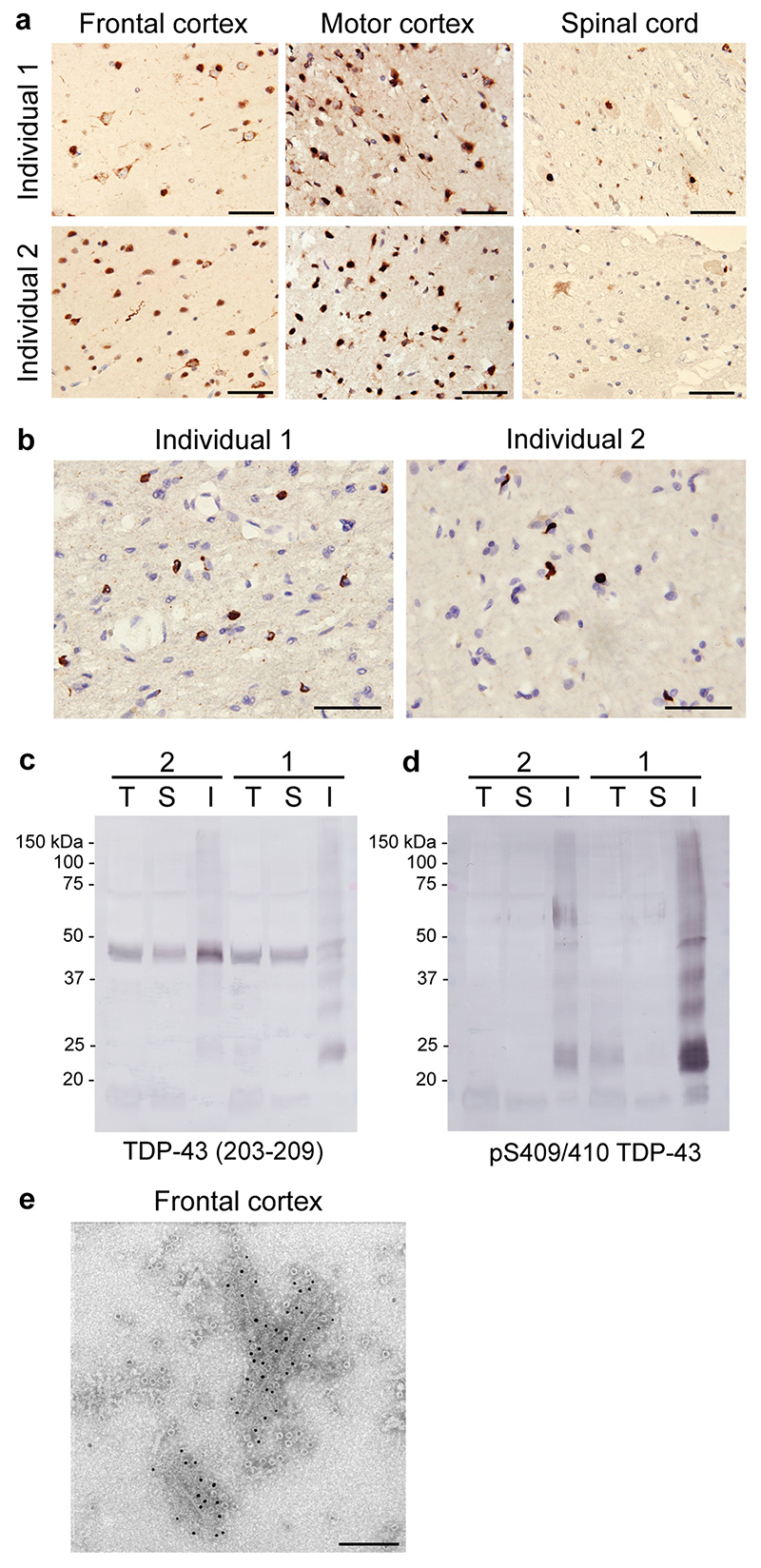
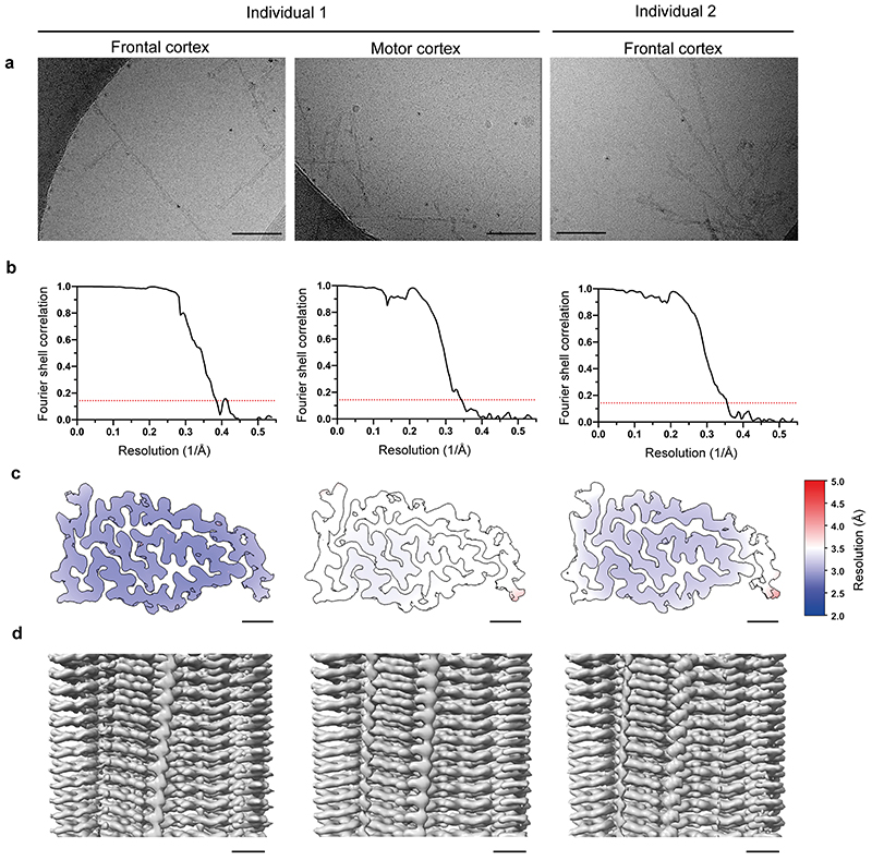
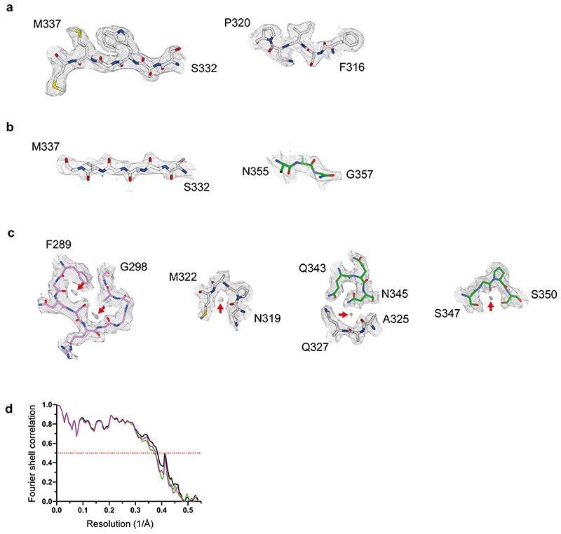
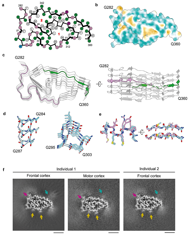
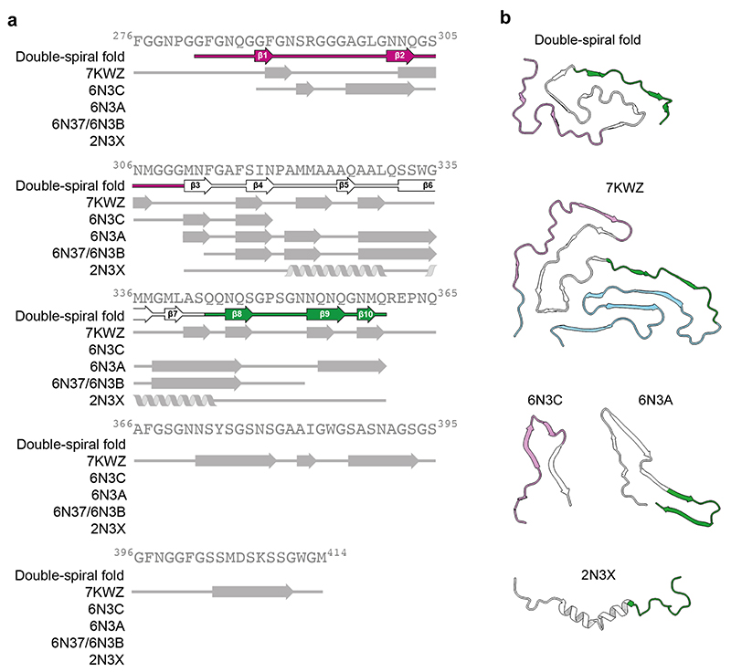
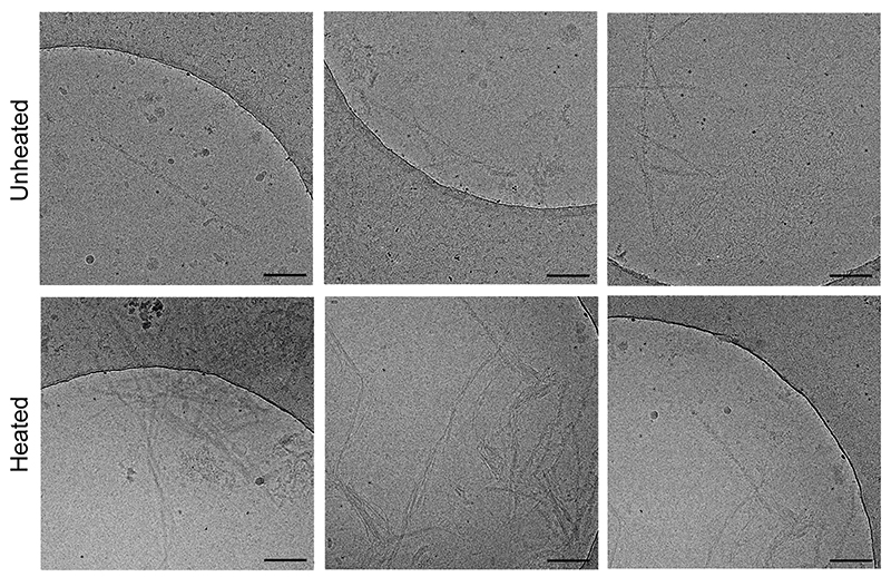
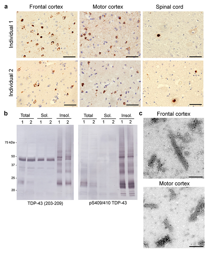
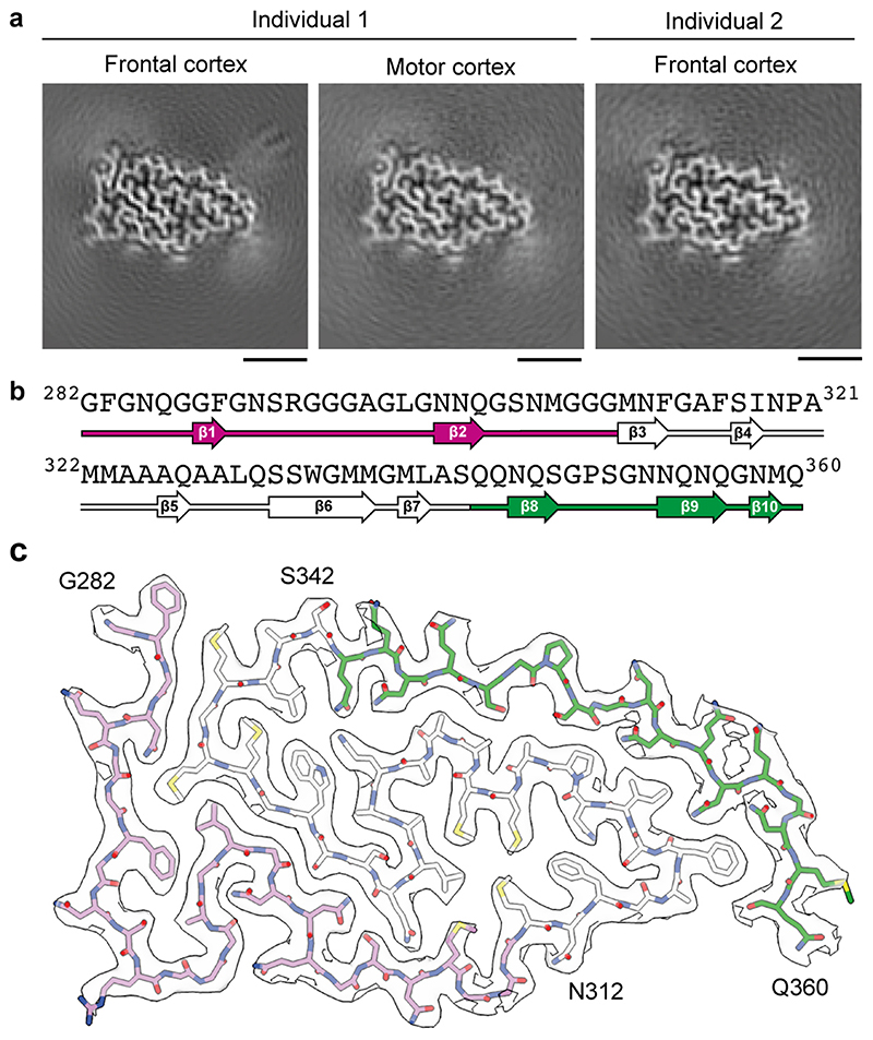
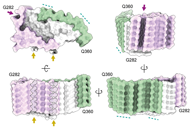
The abnormal aggregation of TAR DNA-binding protein 43 kDa (TDP-43) in neurons and glia is the defining pathological hallmark of the neurodegenerative disease amyotrophic lateral sclerosis (ALS) and multiple forms of frontotemporal lobar degeneration (FTLD)1,2. It is also common in other diseases, including Alzheimer's and Parkinson's. No disease-modifying therapies exist for these conditions and early diagnosis is not possible. The structures of pathological TDP-43 aggregates are unknown. Here we used cryo-electron microscopy to determine the structures of aggregated TDP-43 in the frontal and motor cortices of an individual who had ALS with FTLD and from the frontal cortex of a second individual with the same diagnosis. An identical amyloid-like filament structure comprising a single protofilament was found in both brain regions and individuals. The ordered filament core spans residues 282-360 in the TDP-43 low-complexity domain and adopts a previously undescribed double-spiral-shaped fold, which shows no similarity to those of TDP-43 filaments formed in vitro3,4. An abundance of glycine and neutral polar residues facilitates numerous turns and restricts β-strand length, which results in an absence of β-sheet stacking that is associated with cross-β amyloid structure. An uneven distribution of residues gives rise to structurally and chemically distinct surfaces that face external densities and suggest possible ligand-binding sites. This work enhances our understanding of the molecular pathogenesis of ALS and FTLD and informs the development of diagnostic and therapeutic agents that target aggregated TDP-43.


Conflict of interest statement

Competing interests

The authors declare no competing interests.

Figures

ED Fig. 1
ED Fig. 1. TDP-43 pathology in individuals that succumbed to amyotrophic lateral sclerosis with frontotemporal lobar degeneration continued.
a, Staining of TDP-43 (brown) in the frontal and motor cortices and spinal cords of individuals 1 and 2 with anti-TDP-43 (amino acids 203−209) antibody. Nuclei were counterstained in blue. Scale bars, 50 μm. b, Staining of TDP-43 glial cytoplasmic inclusions (brown) in the motor cortices of individuals 1 and 2 with anti-phosphorylated S409 and S410 TDP-43 antibody. Nuclei were counterstained in blue. Scale bars, 50 μm. c and d, Immunoblots of the total homogenate (T), Sarkosyl-soluble fraction (S) and Sarkosyl-insoluble fraction (I) from the motor cortices of individuals 1 and 2 with anti-TDP-43 (amino acids 203−209) (c) and anti-phosphorylated S409 and S410 TDP-43 (d) antibodies. The original, uncropped blots are shown in Supplementary Fig. 1. e, Immuno-electron microscopy of the Sarkosyl-insoluble fraction from the frontal cortex of individual 2 using anti-phosphorylated S409 and S410 TDP-43 antibody. Scale bar, 100 nm. a−e, Similar results were obtained in at least three independent experiments.
ED Fig. 2
ED Fig. 2. Cryo-EM and helical reconstruction.
a, Representative cryo-EM images of TDP-43 filaments from the frontal and motor cortices of individual 1 and from the frontal cortex of individual 2. Scale bars, 100 nm. Similar results were obtained in at least three independent experiments. b, Fourier shell correlation (FSC) curves for two independently-refined cryo-EM half-maps. The FSC threshold of 0.143 is shown with red dashed lines. c, Local resolution estimates for the Cryo-EM 3D reconstructions. Scale bars, 10 Å. d, Cryo-EM density maps viewed along the helical axis. Scale bars, 10 Å.
ED Fig. 3
ED Fig. 3. Cryo-EM density map and atomic model comparisons.
a−c, Views of the cryo-EM density map of TDP-43 filaments from the frontal cortex of individual 1 and the corresponding atomic model showing representative densities for amino acid side chains (a), peptide group oxygen atoms (b) and ordered solvent molecules (red arrows) (c). d, Fourier shell correlation (FSC) curves for the refined atomic model against the cryo-EM density map (black); for the atomic model shaken and refined using the first half-map against the first half-map (magenta); and for the same atomic model against the second half-map (green). The FSC threshold of 0.5 is shown with a red dashed line.
ED Fig. 4
ED Fig. 4. Double-spiral-shaped fold of TDP-43 filaments from amyotrophic lateral sclerosis with frontotemporal lobar degeneration.
a, Schematic view of the double-spiral fold. The two hydrophobic clusters in the hydrophobic nucleus, composed of the side chains of M307, M311, F313, M322, M323, A326, A329 and L330 (cluster 1); and A328, W334, M336 and L340 (cluster 2), are numbered. b, Hydrophobicity of the double-spiral fold from most hydrophilic (cyan) to most hydrophobic (yellow). c, Secondary structure of the double-spiral fold, depicted as five successive rungs. The glycine-rich (G282-G310, magenta), hydrophobic (M311-S342, white) and Q/N-rich (Q343-Q360, green) regions are highlighted. d, Views of the double-spiral fold, depicted as three successive rungs, showing hydrogen bonds (blue dashed lines) between buried polar side chains and main chain peptide groups. e, Superposition of residues N319-A326 of the double-spiral fold (cyan), depicted as two successive rungs, with residues G88-M95 and G107-A114 of the β-helix domain of glutamate synthase (magenta, PDB ID 1EA0). f, Unmasked cryo-EM 3D reconstructions of TDP-43 filaments from the frontal and motor cortices of individual 1 and from the frontal cortex of individual 2, shown as central slices perpendicular to the helical axis. Additional density within the prominent groove on the filament surface formed by the main chain of G282−Q286 and the side chain of Q286 is indicated with a magenta arrow. Additional densities adjacent to flat strips on the surface of the glycine-rich spiral branch between R293 and A315 are indicated with yellow arrows. Additional less well-resolved density projecting from a polar patch formed by the side chains of N352, Q354 and Q356 on the surface of the Q/N-rich spiral branch is indicated with a cyan arrow. Scale bars, 25 Å.
ED Fig. 5
ED Fig. 5. Comparison of the double-spiral fold with recombinant TDP-43 structures.
a, Amino-acid sequence alignment of the secondary structure elements of the double-spiral fold and recombinant TDP-43 structures with the TDP-43 LC domain. PDB IDs are given for the recombinant TDP-43 structures. The glycine-rich (G282-G310, magenta), hydrophobic (M311-S342, white) and Q/N-rich (Q343-Q360, green) regions are highlighted on the double-spiral fold secondary structure elements. b, Secondary structure of the double-spiral fold and recombinant TDP-43. PDB IDs are given for the recombinant TDP-43 structures. The glycine-rich (G282-G310, magenta), hydrophobic (M311-S342, white) and Q/N-rich (Q343-Q360, green) regions are highlighted.
ED Fig. 6
ED Fig. 6. Heat stability of TDP-43 filaments from amyotrophic lateral sclerosis with frontotemporal lobar degeneration.
Representative cryo-EM images of TDP-43 filaments from the frontal cortex of individual 1 with and without heating at 65°C for 10 min. Scale bars, 100 nm. Similar results were obtained in at least three independent experiments.
Fig. 1
Fig. 1. TDP-43 pathology of amyotrophic lateral sclerosis with frontotemporal lobar degeneration.
a, Staining of TDP-43 neuronal cytoplasmic inclusions (brown) in the frontal and motor cortices and spinal cords of individuals 1 and 2 with anti-phosphorylated S409 and S410 TDP-43 antibody. Nuclei were counterstained in blue. Scale bars, 50 μm. b, Immunoblots of the total homogenate, Sarkosyl-soluble fraction and Sarkosyl-insoluble fraction from the frontal cortices of individuals 1 and 2 with anti-TDP-43 (amino acids 203− 209) and anti-phosphorylated S409 and S410 TDP-43 antibodies. The original, uncropped blots are shown in Supplementary Fig. 1. c, Immuno-electron microscopy of the Sarkosyl-insoluble fractions from the frontal and motor cortices of individual 1 using anti-phosphorylated S409 and S410 TDP-43 antibody. Scale bars, 200 nm. a−c, Similar results were obtained in at least three independent experiments.
Fig. 2
Fig. 2. Cryo-EM maps and atomic model of TDP-43 filaments from amyotrophic lateral sclerosis with frontotemporal lobar degeneration.
a, Cryo-EM maps of TDP-43 filaments from the frontal and motor cortices of individual 1 and from the frontal cortex of individual 2, shown as central slices perpendicular to the helical axis. Scale bars, 25 Å. b, Amino-acid sequence alignment of the secondary structure elements of the double-spiral fold. c, Cryo-EM map (transparent grey) and atomic model of TDP-43 filaments from the frontal cortex of individual 1, shown for a single TDP-43 molecule perpendicular to the helical axis. b and c, The glycine-rich (G282-G310, magenta), hydrophobic (M311-S342, white) and Q/N-rich (Q343-Q360, green) regions are highlighted.
Fig. 3
Fig. 3. Surfaces of TDP-43 filaments from amyotrophic lateral sclerosis with frontotemporal lobar degeneration.
Surface representations of the atomic model of TDP-43 filaments, shown for six molecules. The glycine-rich (G282-G310, magenta), hydrophobic (M311-S342, white) and Q/N-rich (Q343-Q360, green) regions are highlighted. Additional densities are coloured in transparent grey. Additional densities within the prominent groove formed by the main chain of G282-Q286 and the side chain of Q286 are indicated with magenta arrows. Additional planar densities adjacent to flat strips on the surface of the glycine-rich region are indicated with yellow arrows. Polar patches on the surface of the Q/N-rich region are indicated with cyan dashed lines.

Comment in

References

    1. Neumann M, et al. Ubiquitinated TDP-43 in Frontotemporal Lobar Degeneration and Amyotrophic Lateral Sclerosis. Science. 2006;314:130–133. - PubMed
    1. Arai T, et al. TDP-43 is a component of ubiquitin-positive tau-negative inclusions in frontotemporal lobar degeneration and amyotrophic lateral sclerosis. Biochem Biophys Res Commun. 2006;351:602–611. - PubMed
    1. Cao Q, Boyer DR, Sawaya MR, Ge P, Eisenberg DS. Cryo-EM structures of four polymorphic TDP-43 amyloid cores. Nature Struct Mol Biol. 2019;26:619–627. - PMC - PubMed
    1. Li Q, Babinchak WM, Surewicz WK. Cryo-EM structure of amyloid fibrils formed by the entire low complexity domain of TDP-43. Nature Commun. 2021;12:1620. - PMC - PubMed
    1. Brettschneider J, et al. Stages of pTDP-43 pathology in amyotrophic lateral sclerosis. Ann Neurol. 2013;74:20–38. - PMC - PubMed

Publication types

MeSH terms